Jump to content
In this section we have spot diagnoses posted on a daily basis since June 2010, now over 4000! You can review the archived cases and read the suggested diagnoses by users and the final comment by the contributors.
Case are uploaded each week day by 10 am UK time with the correct diagnosis will generally be posted at 8 pm UK time. Why not view the most recent spot diagnosis and proffer a diagnosis?

Case Number : Case 2621 - 23 July 2020 Posted By: Saleem Taibjee

Please read the clinical history and view the images by clicking on them before you proffer your diagnosis.
Submitted Date :
   (0 reviews)

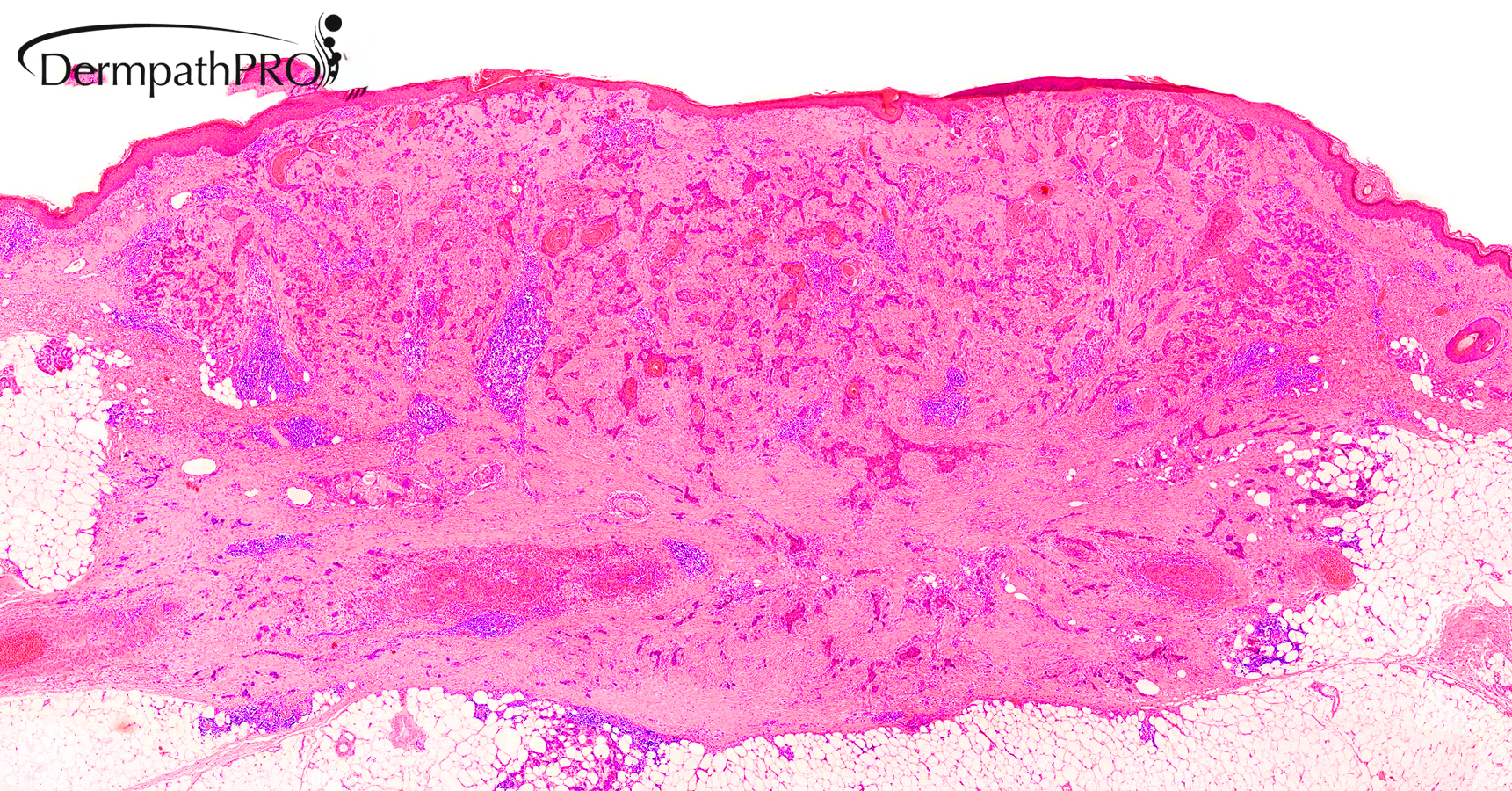
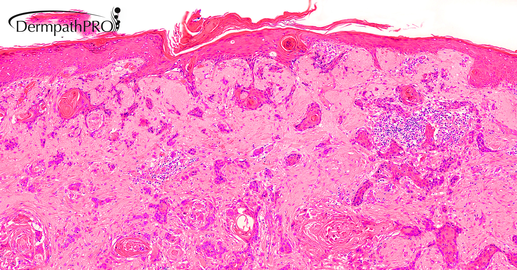
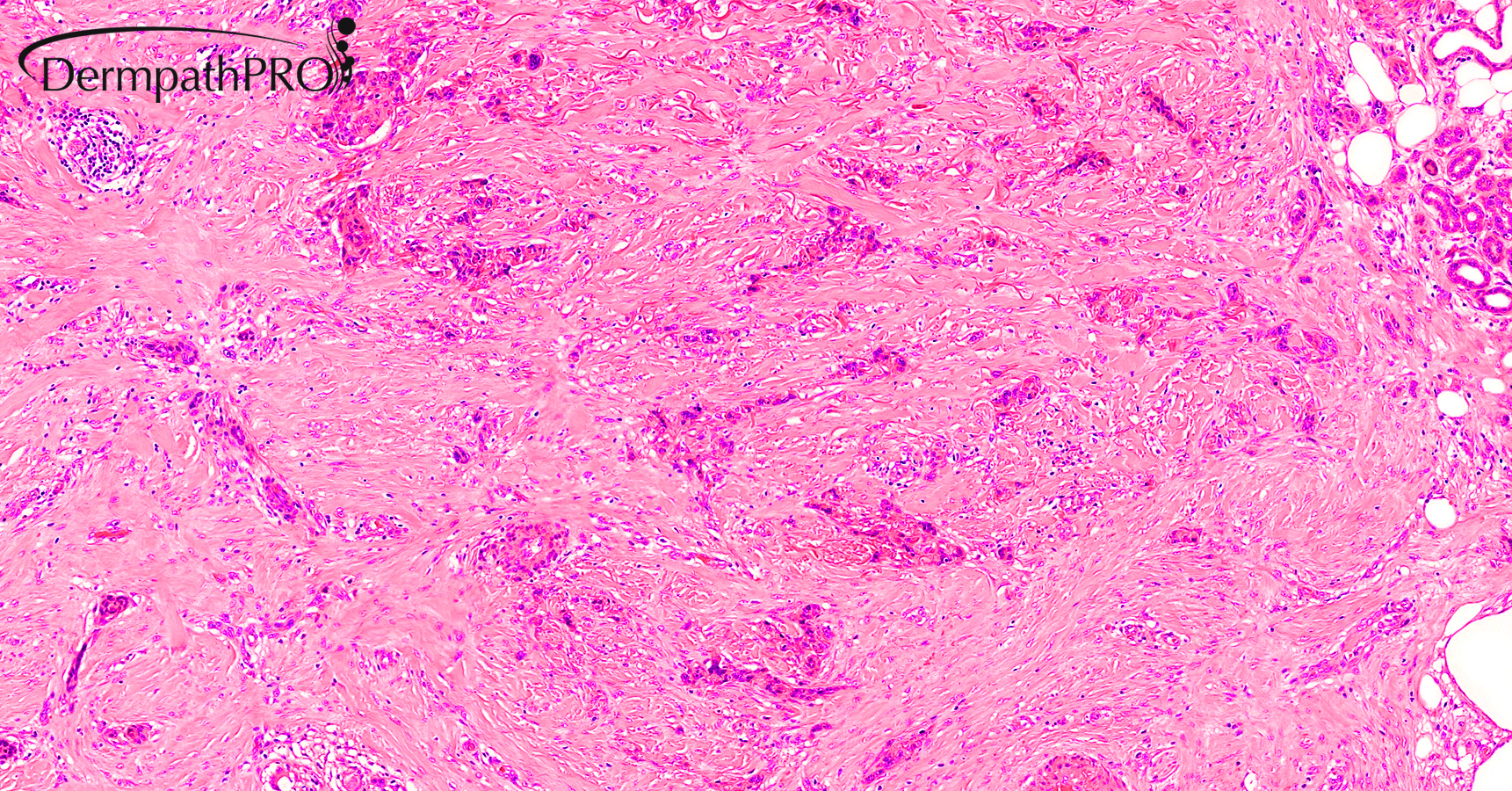
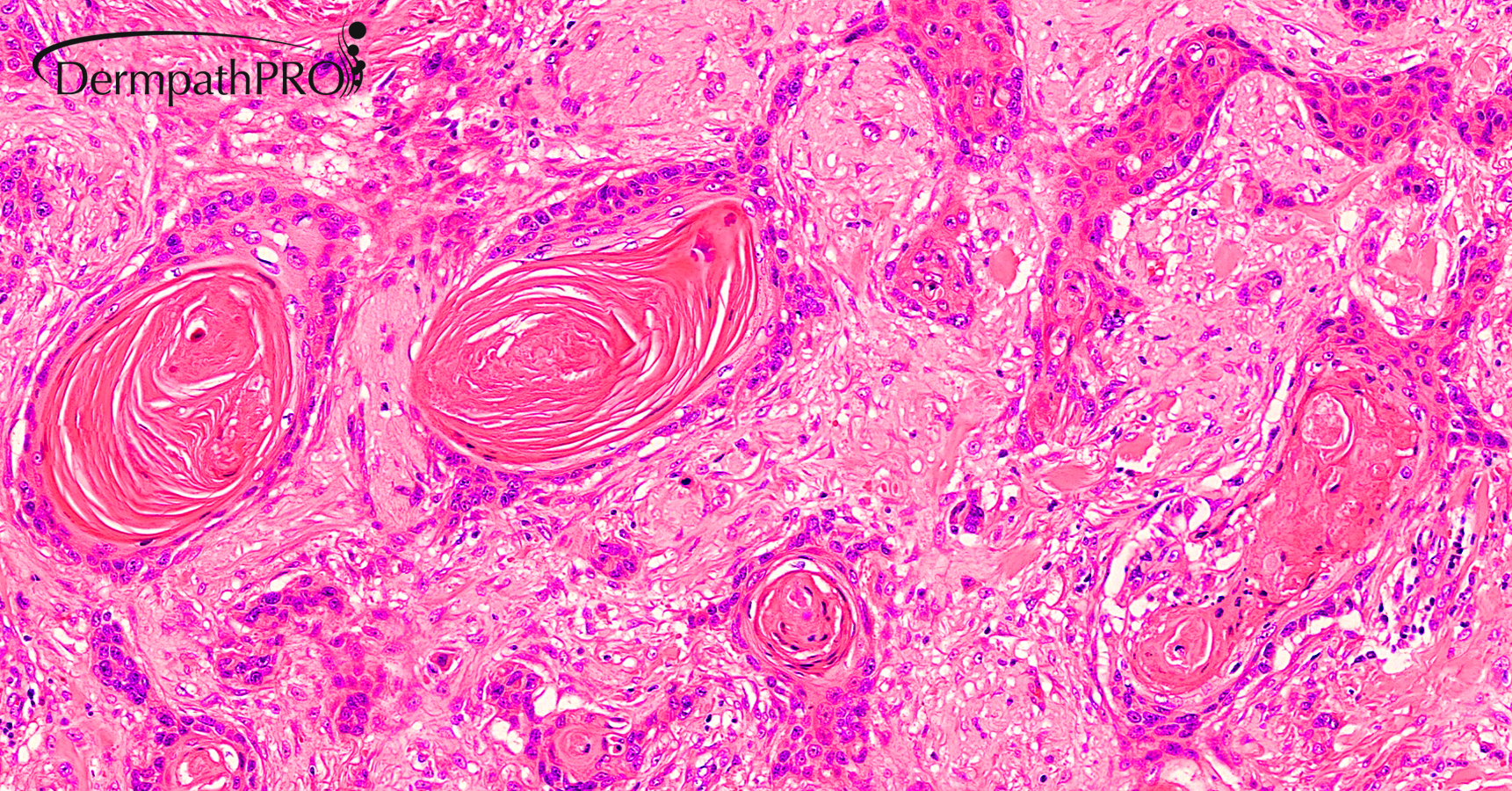
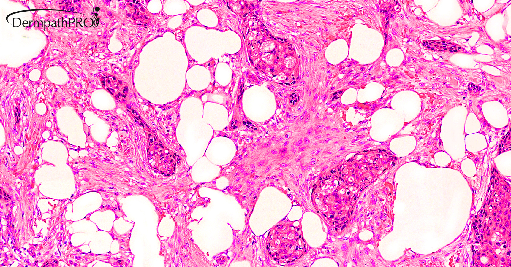
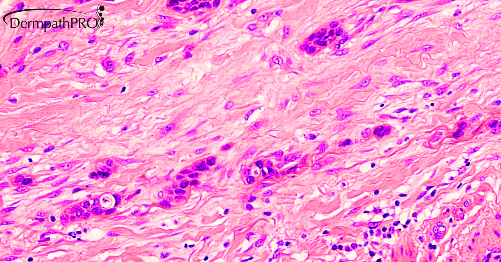
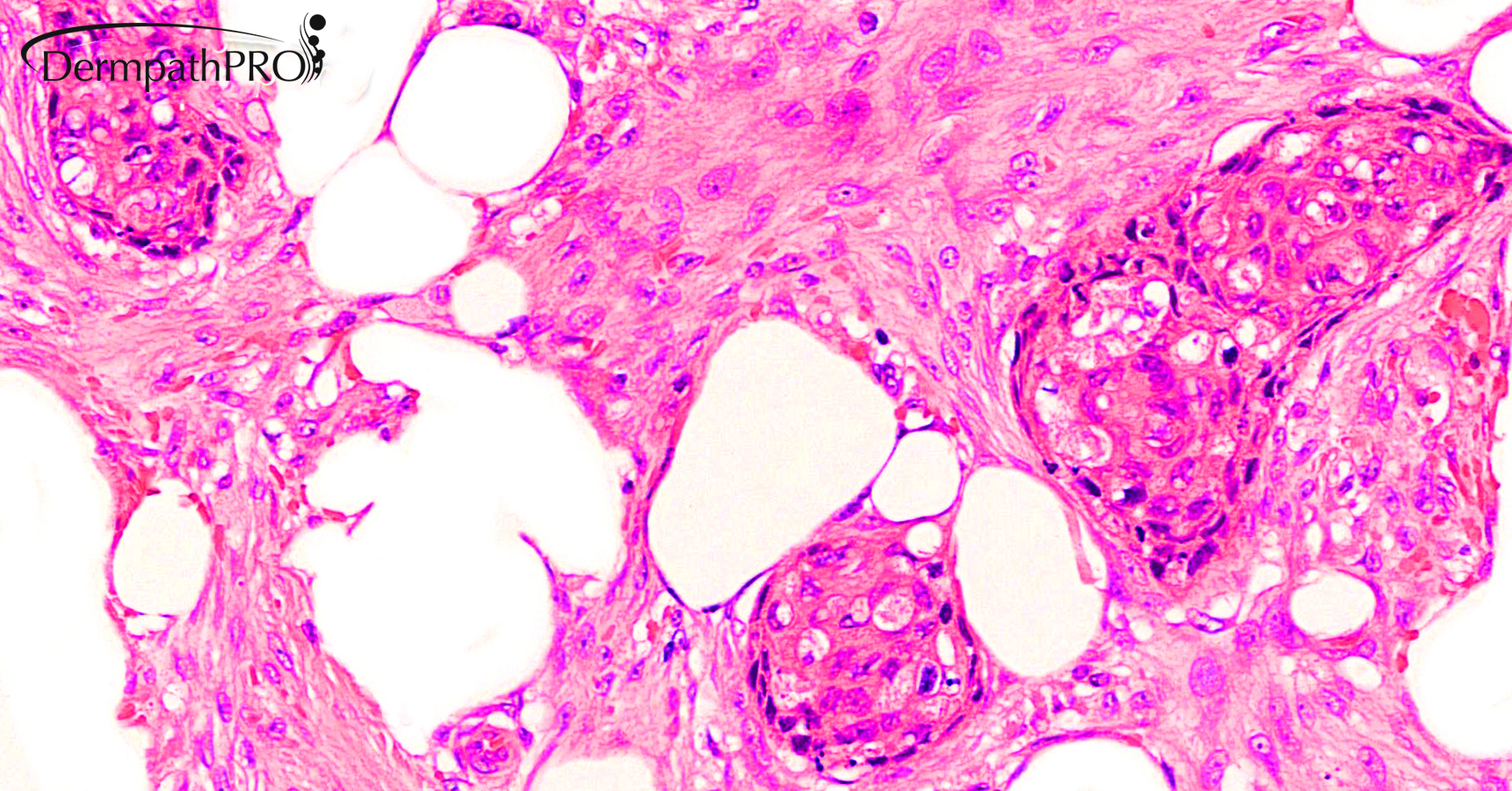
80M, Excision right wrist.


  • Like 1
  Report Record

User Feedback


Richard Logan

Posted

To me this looks like well-differentiated invasive squamous cell carcinoma.  I am not convinced that it has originated locally as connection with the epidermis is tenuous and the overlying epidermis shows neither much in the way of dysplasia nor any sign of ulceration. Perhaps this is intravascular spread from a nearby primary SCC.

Share this comment


Link to comment
share_externally

Cem Leblebici

Posted

I think tumor shows squamoid and ductal differantiation.

My thought is squamoid eccrine ductal carcinoma.

Share this comment


Link to comment
share_externally

Dr. Mona Abdel-Halim

Posted

Squamoid eccrine ductal carcinoma


Share this comment


Link to comment
share_externally

Krishnakumar subramanian

Posted

Squamoid eccrine ductal carcinoma

Share this comment


Link to comment
share_externally

I was thinking of a solid variant of MAC, but this looks like a high grade carcinoma. So favor a Porocarcinoma. 

Share this comment


Link to comment
share_externally

Saleem Taibjee

Posted

Yes, I favoured squamoid eccrine ductal carcinoma. Low power shows a poorly demarcated and infiltrative tumour extending into subcutis. The superficial aspect shows focal connection with the epidermis and infundibula, with prominent squamoid differentiation. At the deep aspect the tumour is more infiltrative with ductal differentiation evident; cuticle-like lumina and intracytoplasmic vacuoles, also highlighted by the CEA. There is variable cytological atypia and mitotic activity.

Share this comment


Link to comment
share_externally


Join the conversation

You can post now and register later. If you have an account, sign in now to post with your account.

Guest
Add a comment...

×   Pasted as rich text.   Paste as plain text instead

  Only 75 emoji are allowed.

×   Your link has been automatically embedded.   Display as a link instead

×   Your previous content has been restored.   Clear editor

×   You cannot paste images directly. Upload or insert images from URL.

Loading...

×
×
  • Create New...