Jump to content
In this section we have spot diagnoses posted on a daily basis since June 2010, now over 4000! You can review the archived cases and read the suggested diagnoses by users and the final comment by the contributors.
Case are uploaded each week day by 10 am UK time with the correct diagnosis will generally be posted at 8 pm UK time. Why not view the most recent spot diagnosis and proffer a diagnosis?

Case Number : Case 2622 - 24 July 2020 Posted By: Dr. Richard Carr

Please read the clinical history and view the images by clicking on them before you proffer your diagnosis.
Submitted Date :
   (0 reviews)

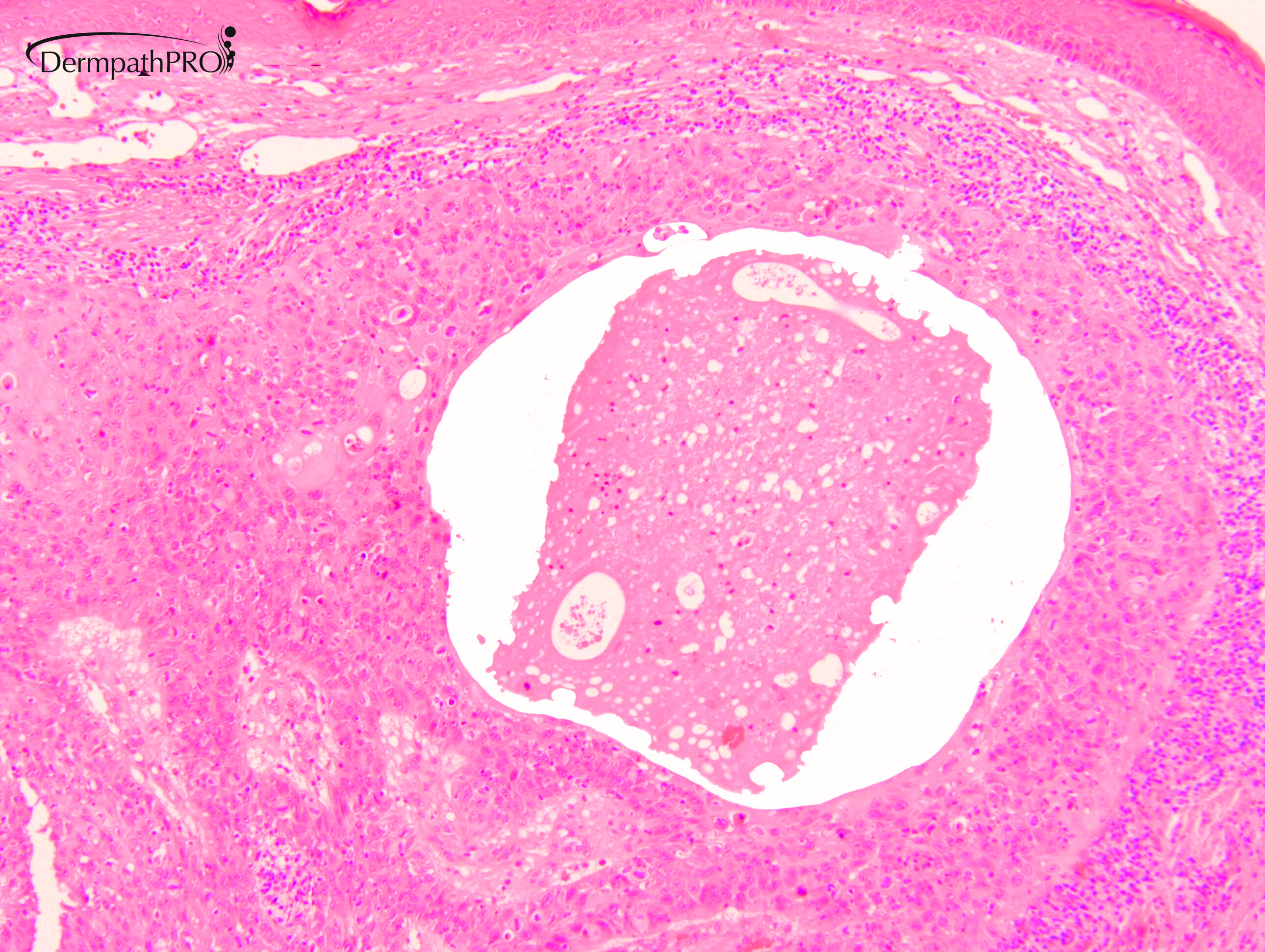
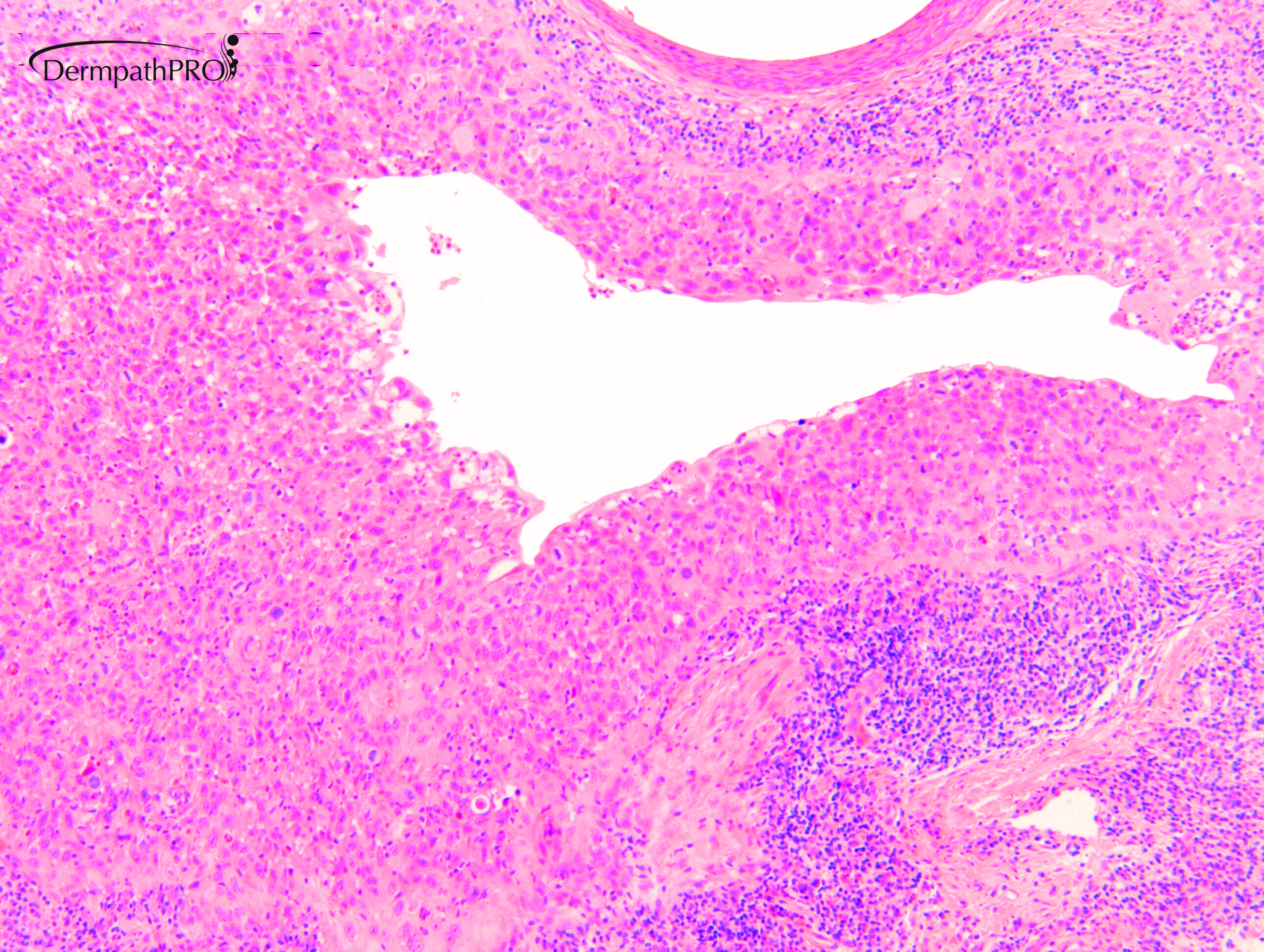
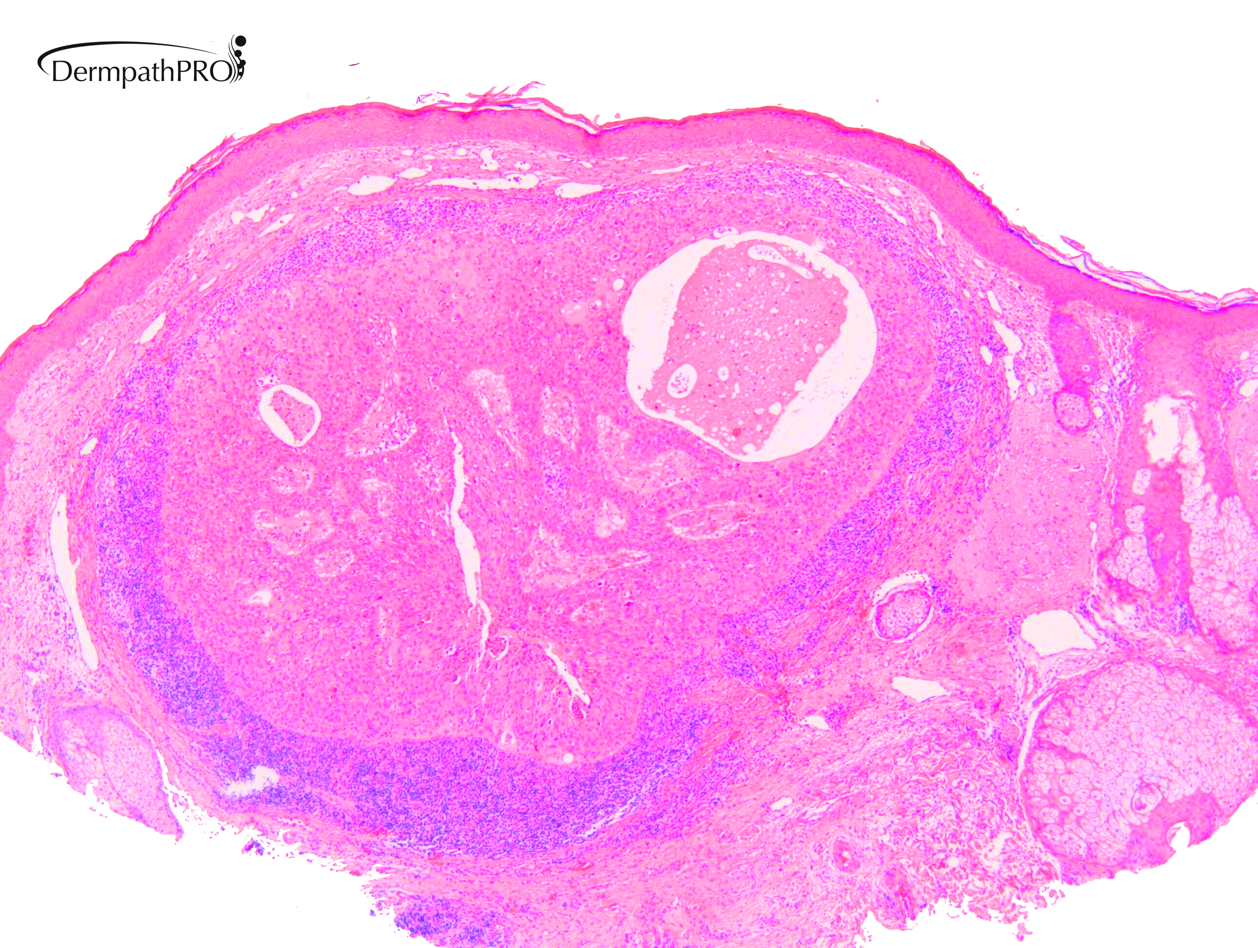
M80. Cheek. Initially 8 x 8mm lesion (3 months) but at 4 weeks partially regressed to a 3 x 4mm papule. ?BCC ?BAK ?SCC

  Report Record

User Feedback


Cem Leblebici

Posted

Atypical hidradenoma vs hidradenocarcinoma. It may be fit atypical hidradenoma, but I prefer the last one. 

Share this comment


Link to comment
share_externally

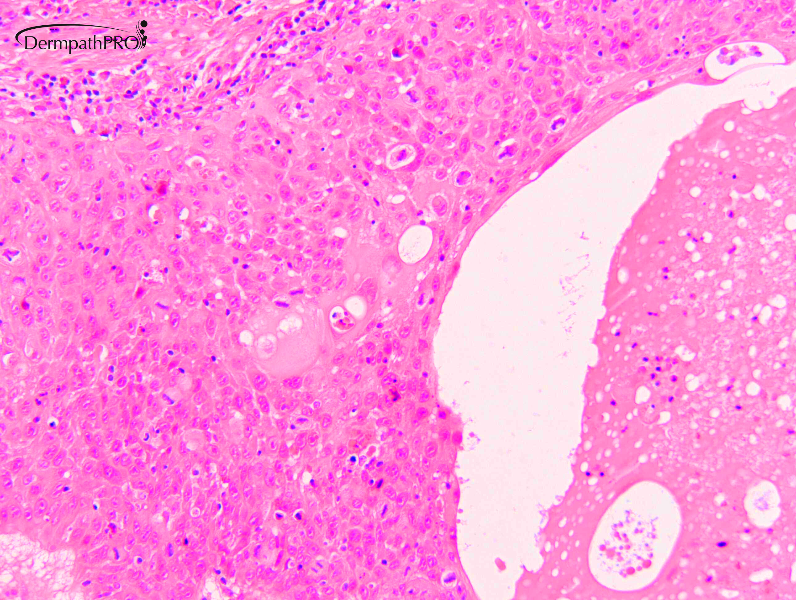
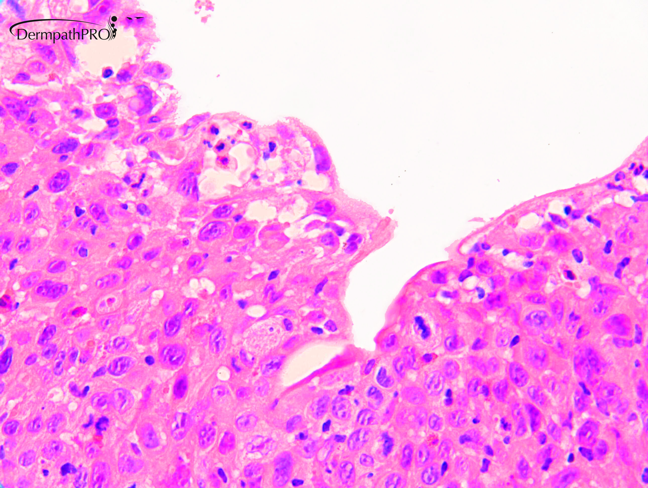
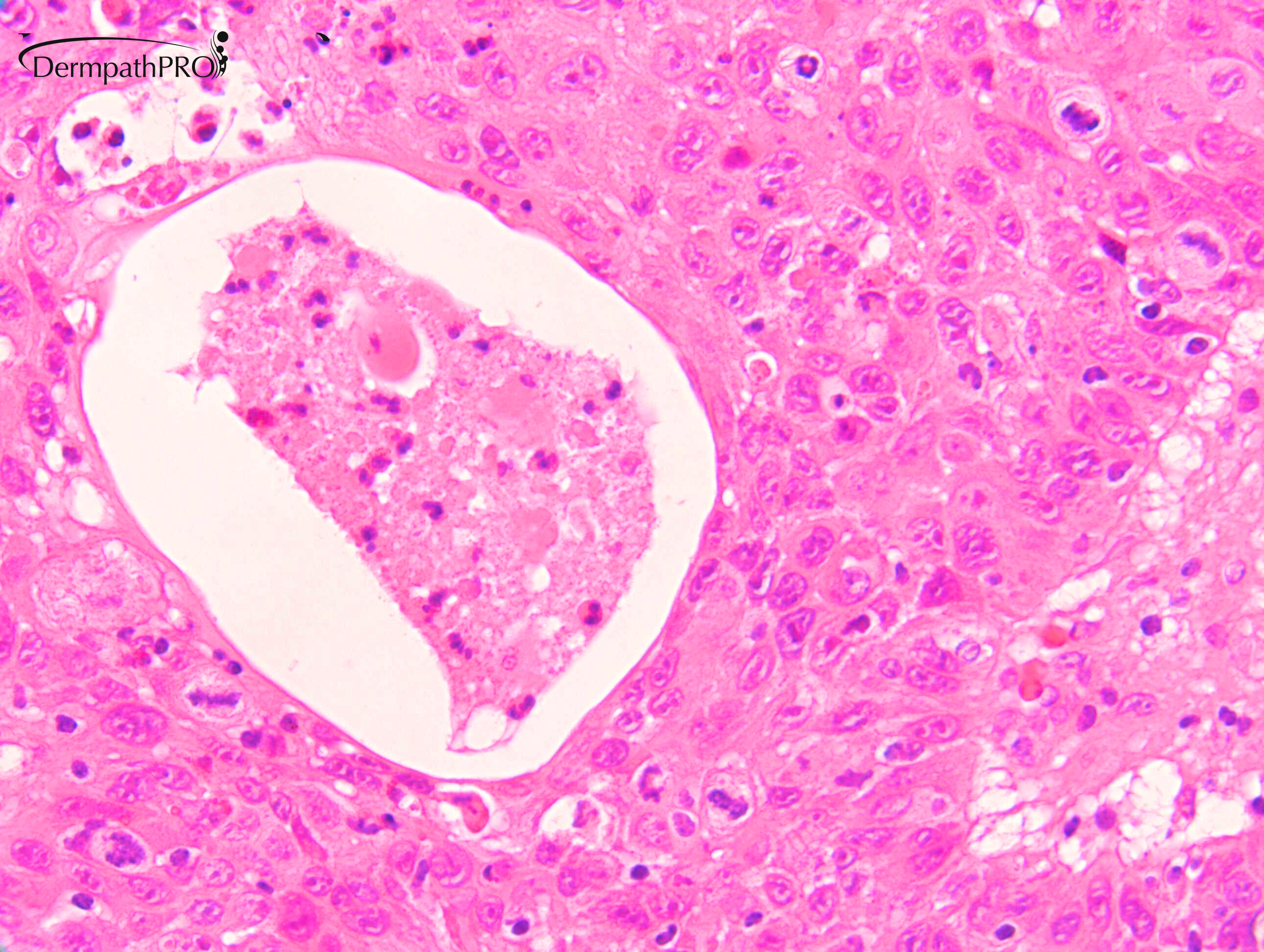
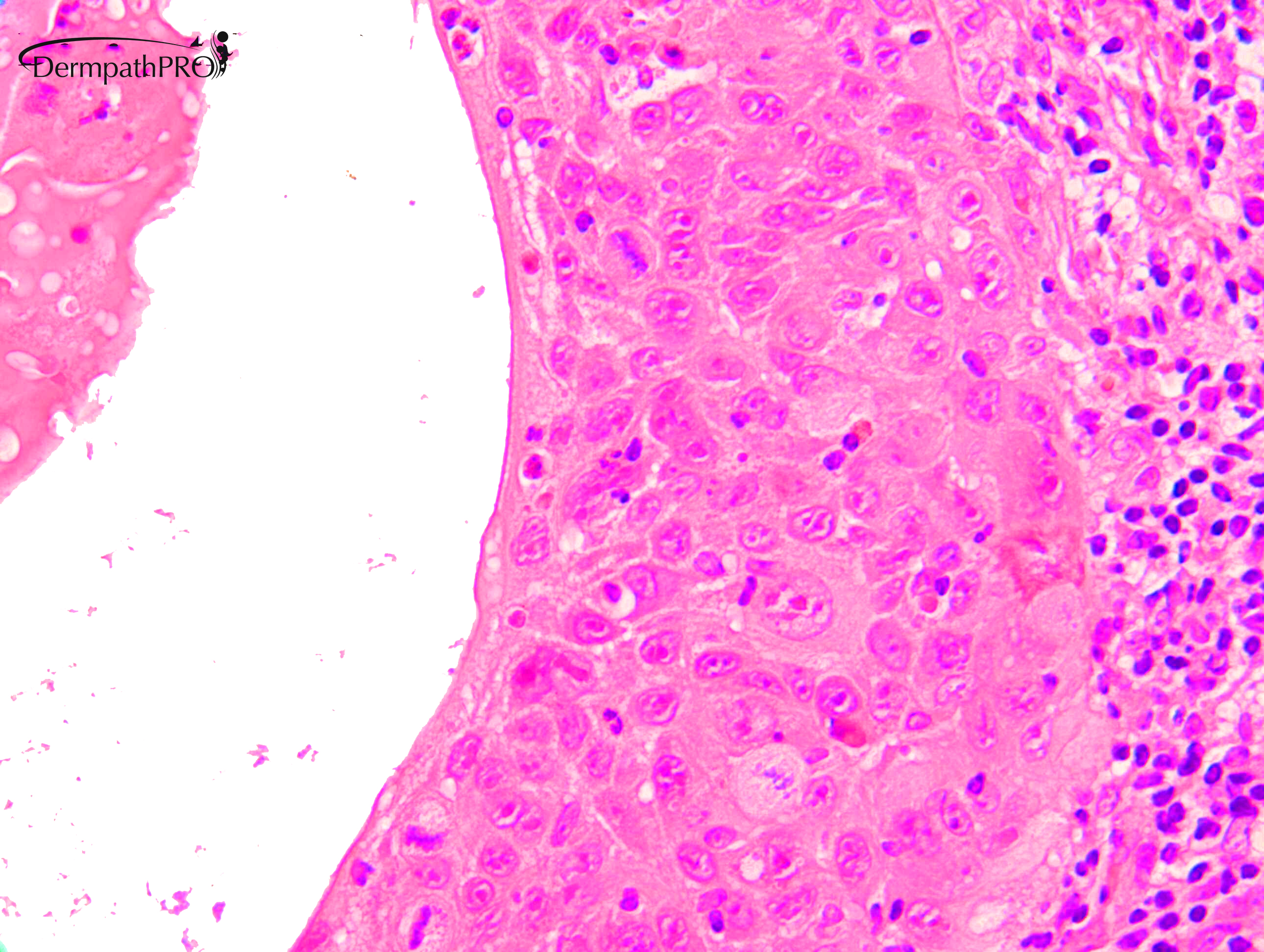
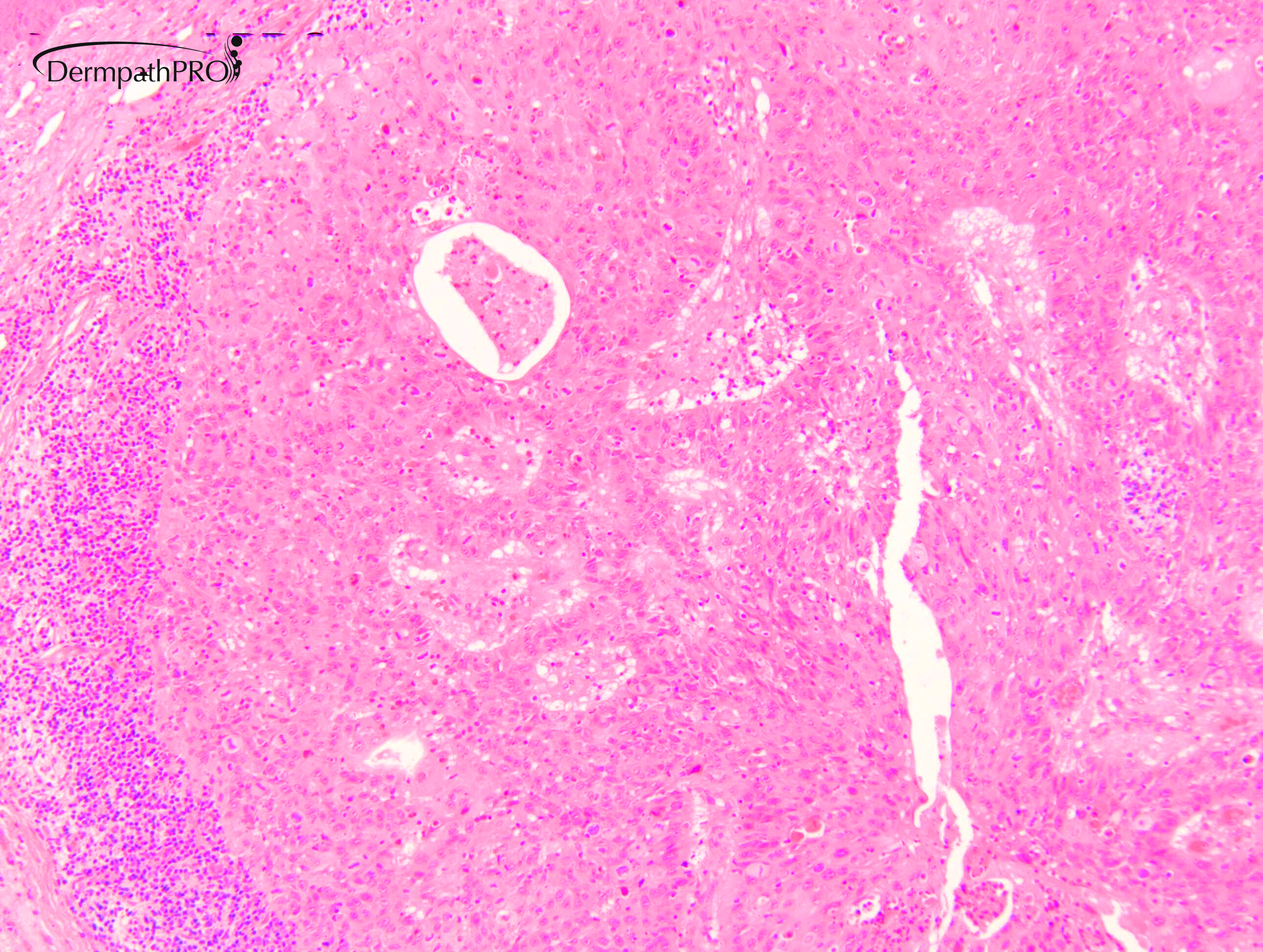
There are intercellular bridges between the neoplastic cells so it is a tumor with squamous differentiation. Quite mitotically active and cellular atypia. I think it is best classified as a squamous cell carcinoma. But I do not an in situ component and the history of regression is unusual. Was it treated with some topical?

Share this comment


Link to comment
share_externally

Richard Logan

Posted

I agree that this looks like a malignant lesion with squamous differentiation.  The morphology looks like a metastasis which has undergone cystic degeneration and there is a host inflammatory response, both of which might account for the apparent shrinkage.  It is unlikely that a topical treatment would have been used in this clinical situation and there is no epidermal change that would usually be seen if a topical such as 5-fluorouracil or imiquimod had been used.

Share this comment


Link to comment
share_externally

Dr. Mona Abdel-Halim

Posted

I think it is SCC (mostly fSCC, may be tangentially cut?) undergoing regression?

Share this comment


Link to comment
share_externally

Dr. Richard Carr

Posted

I've asked for some IHC to be posted.

Share this comment


Link to comment
share_externally

?salivary duct carcinoma of parotid gland...

Share this comment


Link to comment
share_externally

Dr. Richard Carr

Posted

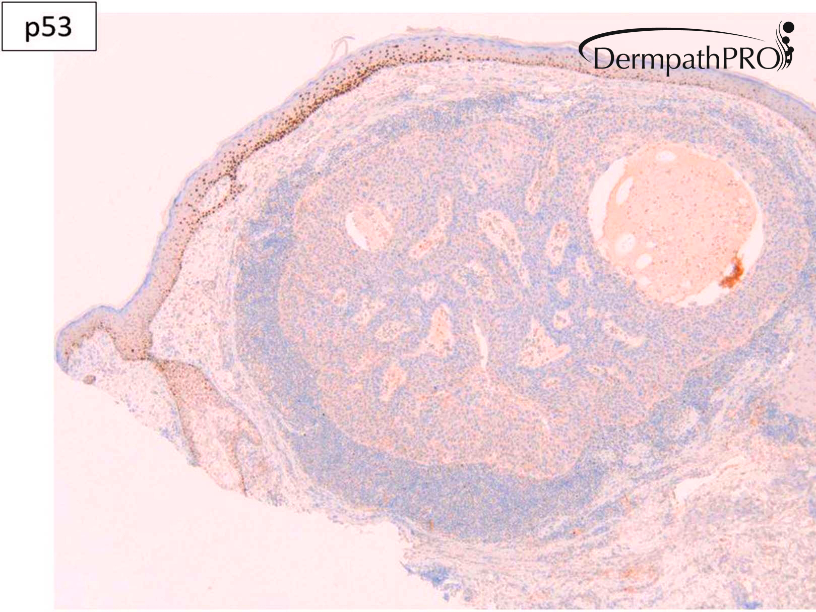
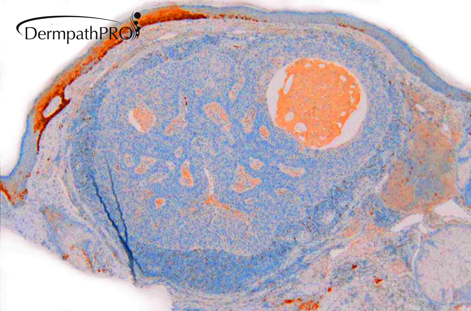
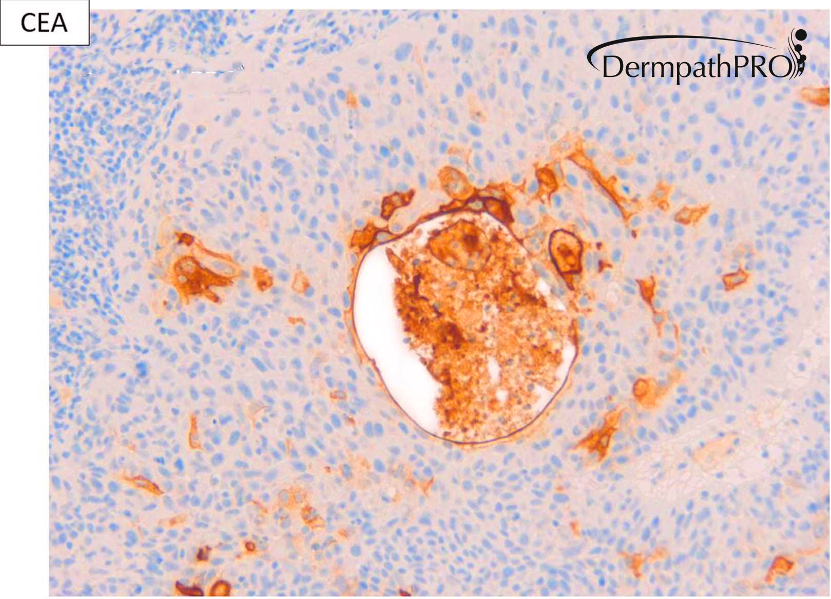
See if you can guess the unlabelled IHC. It's showing a patchy epidermal staining and modoerate staining of macrophages in dermis but is NULL in the tumour.

Share this comment


Link to comment
share_externally

MITF-1?

SCC with ductal differentiation? Adenosquamous carcinoma? But still does not explain why the tumor regressed.

Share this comment


Link to comment
share_externally

Richard Logan

Posted

Has the patient been treated with systemic chemotherapy for their primary mailgnancy?

Share this comment


Link to comment
share_externally

Dr. Richard Carr

Posted

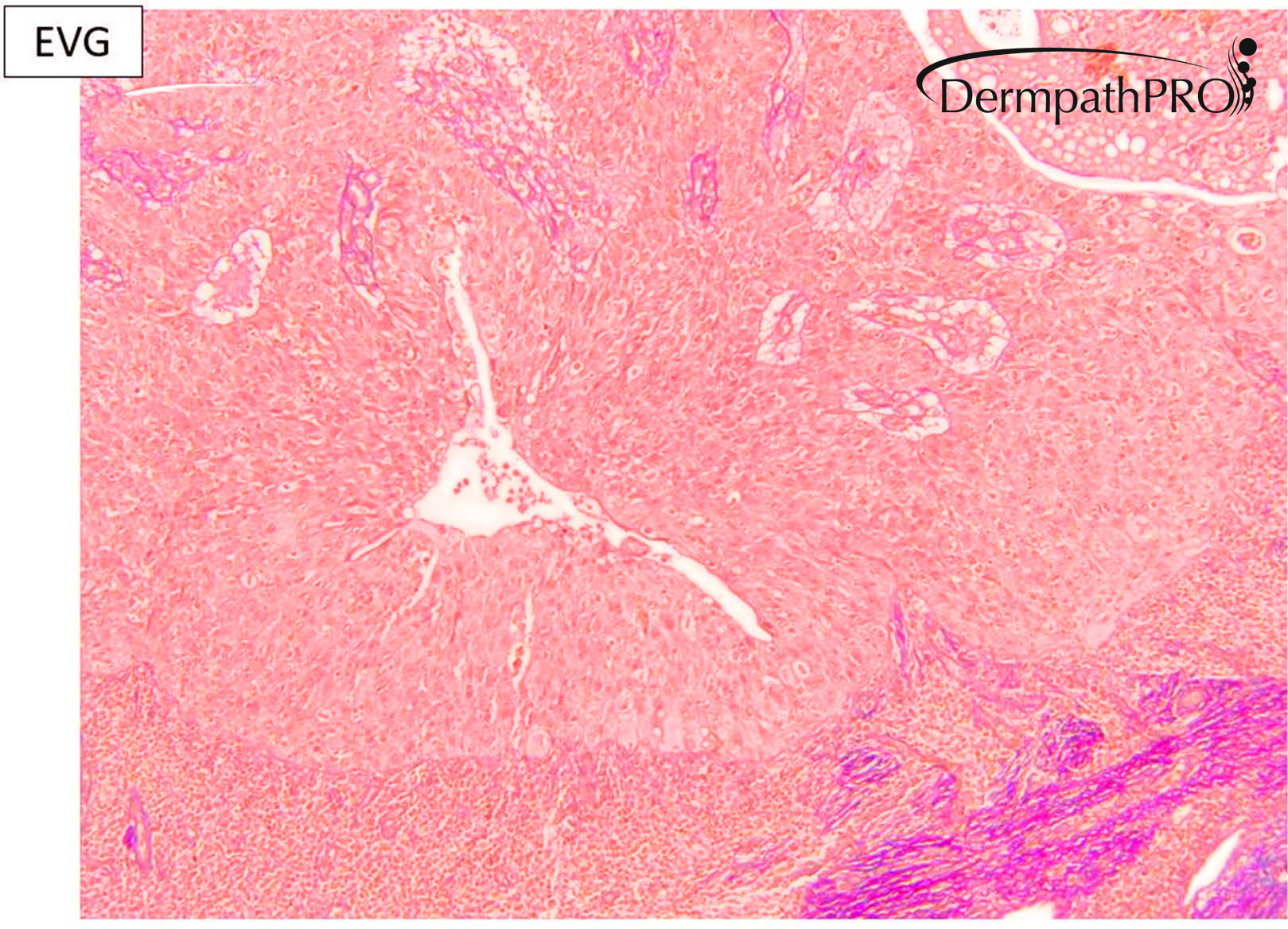
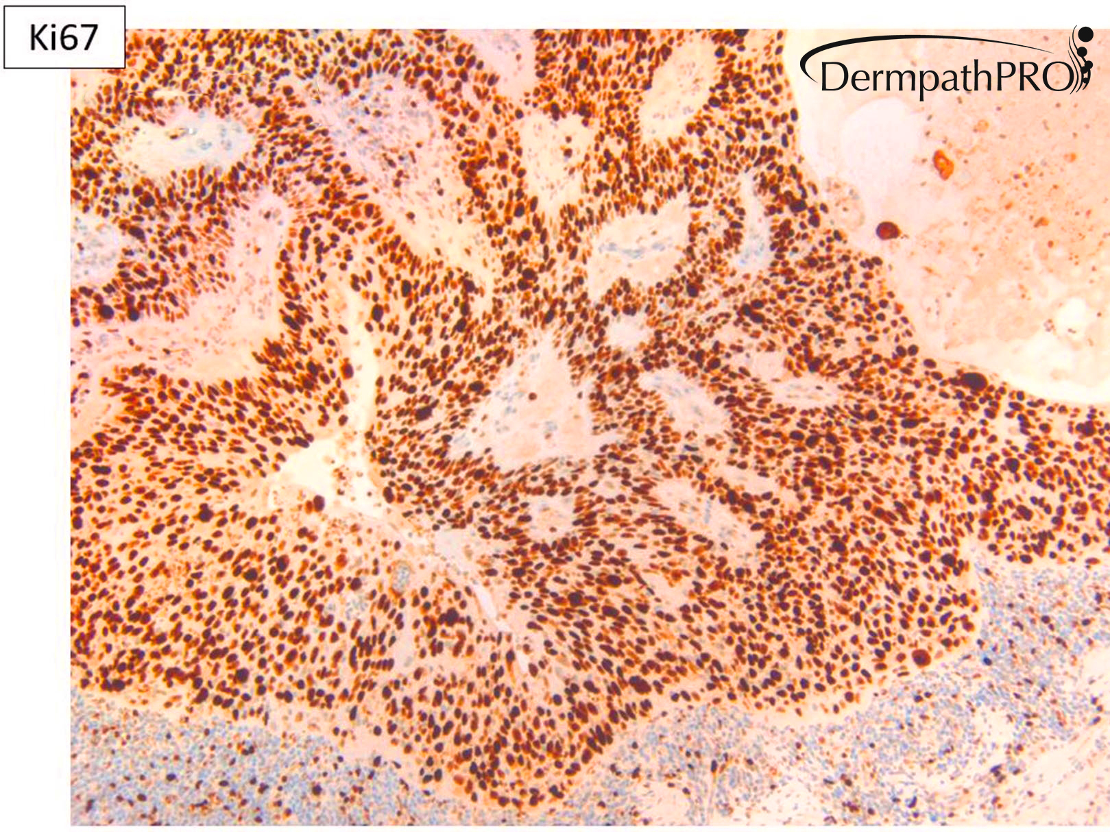
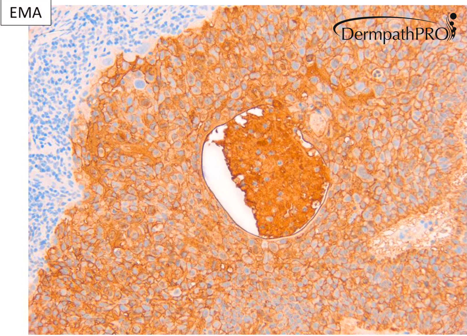
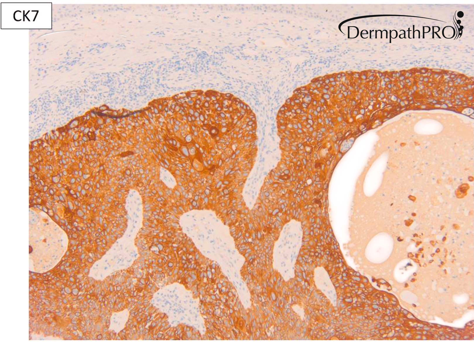
I think it's always good to consider a metastasis especially for a large rounded lump centred in the deep dermis and involving subcutis - not the case here. I've been increasingly using EVG to determine if a tumour is invasive. In this case the tumour is completely circumscribed and there is no entrapment of elastic / collagen which would tend to indicate a non-invasive lesion so I think this is an in situ adnexal primary (I'm sure this is under-recognised in cutaneous adnexal tumours inclulding the in situ variants of follicular SCC). Given that there is clear-cut ductal differentiation including squamoid ductal lining cells I'd label it a hidradenocarcinoma "in situ". I have no problem with designating in "adenosquamous" carcinoma. Many porocarcinomas, sebaceous carcinomas have mixed squamous ductal - sebaceous differentiation and no doubt some arise from the follicular-sebaceous-apocrine ductal unit.

The p53 shows a complete NULL which is interpreted as a mutant pattern. I'm not too sure if I've mentioned previously but we are finding p16 NULL is a common finding in follicular SCC (arouund 50% of cases) and it appears to be seen in other cutaneous malignancies associated with severe sun-damage - superficial adnexal carcinomas, AFX, melanoma to name three. So this is an example of a NULL p16. I will be illustrating many examples later in the year of the combinations of Ki67, p16, p53 and EVG in challenging squamoproliferative lesions including a digitised series for the UK National Skin EQA in November. The staining patterns can be complex to interpret but I think their use will become a new paradigm in the assessment of KA and KA-like lesions (to help differentiate KA from KA-like follicular SCC). For those interested send me an e-mail and i'll send you a link to my lectures and you can see my taster lecture from the ISDP meeting in Lisbon last year although we now have over 400 cases in our audit database and we've learn't some new patterns since then.

Share this comment


Link to comment
share_externally

Dr. Mona Abdel-Halim

Posted

14 hours ago, Dr. Richard Carr said:

I think it's always good to consider a metastasis especially for a large rounded lump centred in the deep dermis and involving subcutis - not the case here. I've been increasingly using EVG to determine if a tumour is invasive. In this case the tumour is completely circumscribed and there is no entrapment of elastic / collagen which would tend to indicate a non-invasive lesion so I think this is an in situ adnexal primary (I'm sure this is under-recognised in cutaneous adnexal tumours inclulding the in situ variants of follicular SCC). Given that there is clear-cut ductal differentiation including squamoid ductal lining cells I'd label it a hidradenocarcinoma "in situ". I have no problem with designating in "adenosquamous" carcinoma. Many porocarcinomas, sebaceous carcinomas have mixed squamous ductal - sebaceous differentiation and no doubt some arise from the follicular-sebaceous-apocrine ductal unit.

The p53 shows a complete NULL which is interpreted as a mutant pattern. I'm not too sure if I've mentioned previously but we are finding p16 NULL is a common finding in follicular SCC (arouund 50% of cases) and it appears to be seen in other cutaneous malignancies associated with severe sun-damage - superficial adnexal carcinomas, AFX, melanoma to name three. So this is an example of a NULL p16. I will be illustrating many examples later in the year of the combinations of Ki67, p16, p53 and EVG in challenging squamoproliferative lesions including a digitised series for the UK National Skin EQA in November. The staining patterns can be complex to interpret but I think their use will become a new paradigm in the assessment of KA and KA-like lesions (to help differentiate KA from KA-like follicular SCC). For those interested send me an e-mail and i'll send you a link to my lectures and you can see my taster lecture from the ISDP meeting in Lisbon last year although we now have over 400 cases in our audit database and we've learn't some new patterns since then.

Thank u Dr. Carr for always fascinating me :-))

Share this comment


Link to comment
share_externally

Dr. Carr,


Interesting concept. But just for the sake of academic discussion, I have 2 questions:

1. In situ carcinoma per definition is a proliferative malignant neoplastic process confined within a preexisting compartment or structure. For example, SCC in situ of the skin is confined within the epidermis or in ductal carcinoma in situ of the breast, the tumor cells proliferate in a preexisting breast duct. In this case, the tumor just sits in the dermis so it's difficult to visualize what the preexisting structure is. Of course it doesn't matter so much if the lesion is completely excised (unless it recurs with a nodal or pulmonary metastasis, then it becomes problematic).


2. I never heard about using elastic stain to show invasion but conceptually it makes completely sense in the majority of the cases where the invasive pattern is infiltrative. But we all know that some tumors invade in a pushing manner. In such tumors, what is your experience with elastic stain?


Anh 

Share this comment


Link to comment
share_externally

Dr. Richard Carr

Posted

Thanks Anh for you very considered thoughts.

We have to be very very careful with this concept. I have seen cases of thick bowenoid lesions with NEARLY pushing only pattern but clear focal evidence of elastic & collagen entrapment that also had extensive lymphovascular invasion and another similar case on punch biopsy reported as Bowen's that had subtle pushing invasion with entrapment (EVG) on review of a recurrent lesion with extensive invasion & lymphovascular invasion in a recurrence nine months later. The patient had been under-treated. So in my experience I'd have a low threshold for doing an EVG on Bowenoid lesions with deep pushing extensions or any blurred margins and report any lesions in which there is elastic and collagen entrapment as favouring poorly differentiated invasive carcinoma with appropriate management & follow-up (I think I illustrate this in my Lisbon & RCPath lectures). As you know I believe many more well differentiated follicular SCC with COMPLETELY CIRCUMSCRIPT borders, supported by complete absence of elastic & collagen entrapment are IN SITU FOR PRACTICAL PURPOSES. So I think I agree with your caution about the above concepts nevertheless I do think IN SITU FOR PRACTICAL PURPOSES is under-recognised in SCC, particularly the follicular variant, and adnexal carcinomas too. On the other had one can both under-recognise or over-recognise invasion in the setting of thick bowenoid lesions and I'm finding EVG a very useful adjunct to the H&E assessment. Another helpful clue is the preservation of small reactive p53 stained cells in the peripheral lining of Bowenoid (non-invasive) compared with loss of these reactive cells in pushing invasive lesions (pseudo-in situ). The latter is helped when the lesion is p53 null - not uncommon in frank bowen's). My audit database of EVG, Ki67, p16, p53 has 433 cases (as of yesterday) and we're still learning but this panel will become the new paradigm in challenging cutaneous epithelial neoplasms. Warm regards Richard

Share this comment


Link to comment
share_externally

Great. Thanks for the comments.

I will try elastic stain in difficult keratinocytic lesions to learn how it works.

Share this comment


Link to comment
share_externally


Join the conversation

You can post now and register later. If you have an account, sign in now to post with your account.

Guest
Add a comment...

×   Pasted as rich text.   Paste as plain text instead

  Only 75 emoji are allowed.

×   Your link has been automatically embedded.   Display as a link instead

×   Your previous content has been restored.   Clear editor

×   You cannot paste images directly. Upload or insert images from URL.

Loading...

×
×
  • Create New...