Jump to content
In this section we have spot diagnoses posted on a daily basis since June 2010, now over 4000! You can review the archived cases and read the suggested diagnoses by users and the final comment by the contributors.
Case are uploaded each week day by 10 am UK time with the correct diagnosis will generally be posted at 8 pm UK time. Why not view the most recent spot diagnosis and proffer a diagnosis?

Case Number : Case 2586 - 04 June 2020 Posted By: Saleem Taibjee

Please read the clinical history and view the images by clicking on them before you proffer your diagnosis.
Submitted Date :
   (0 reviews)

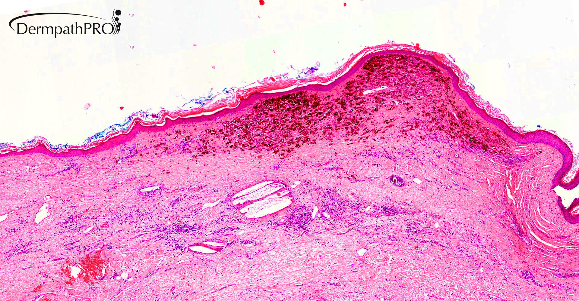
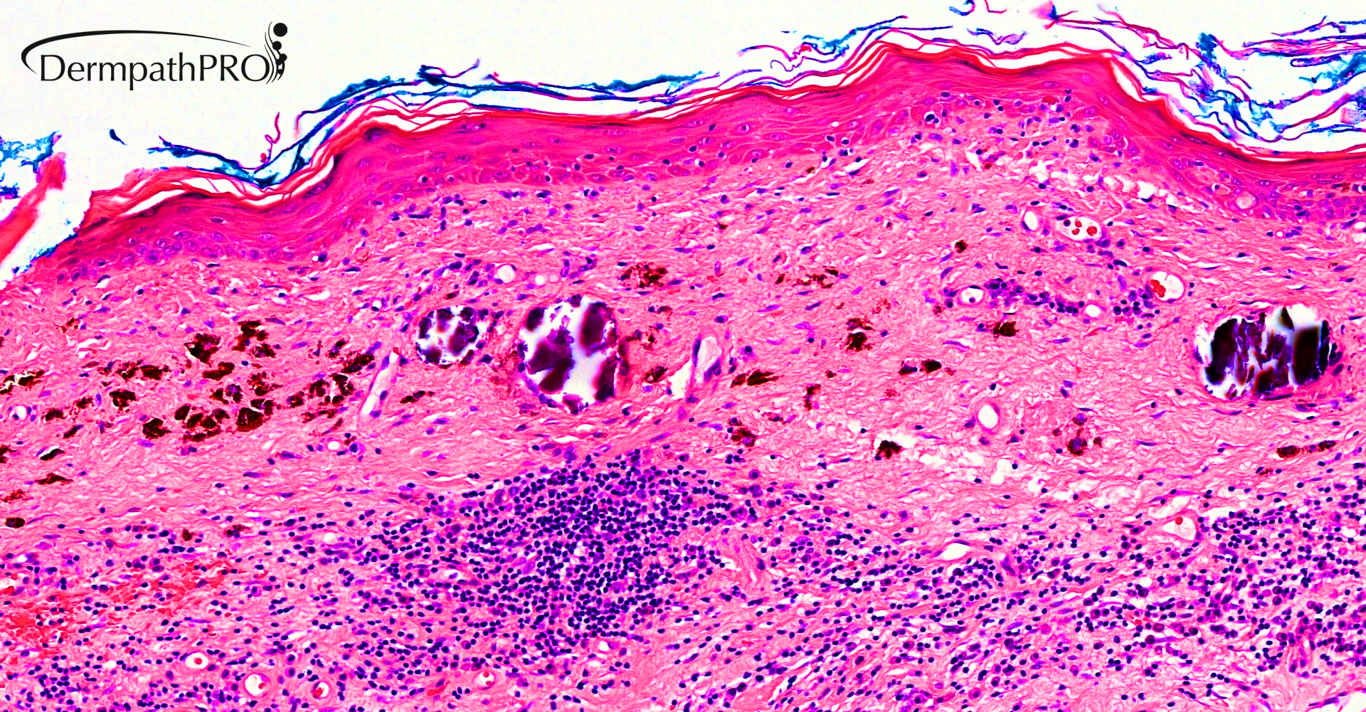
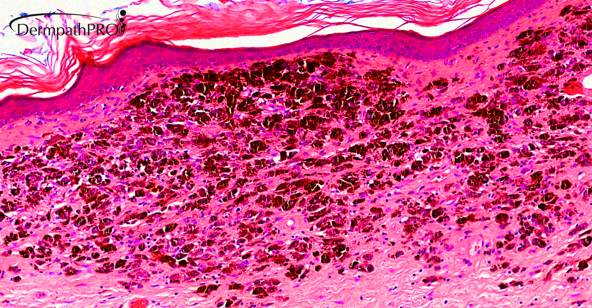
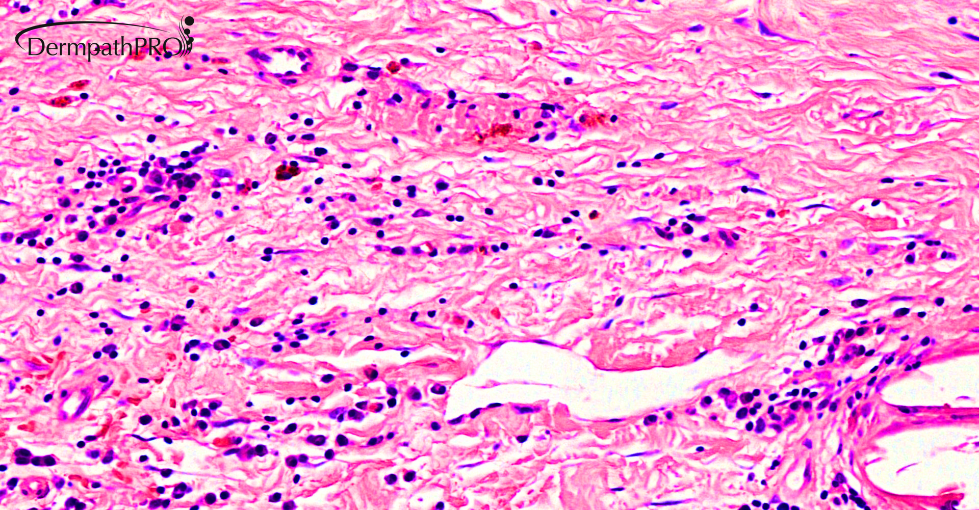
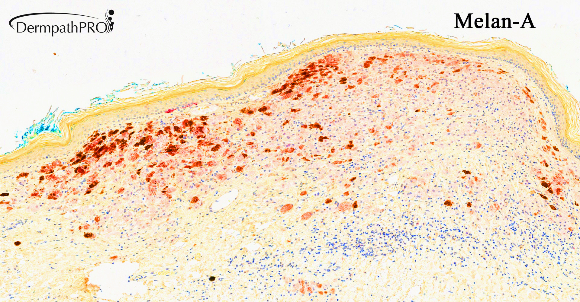
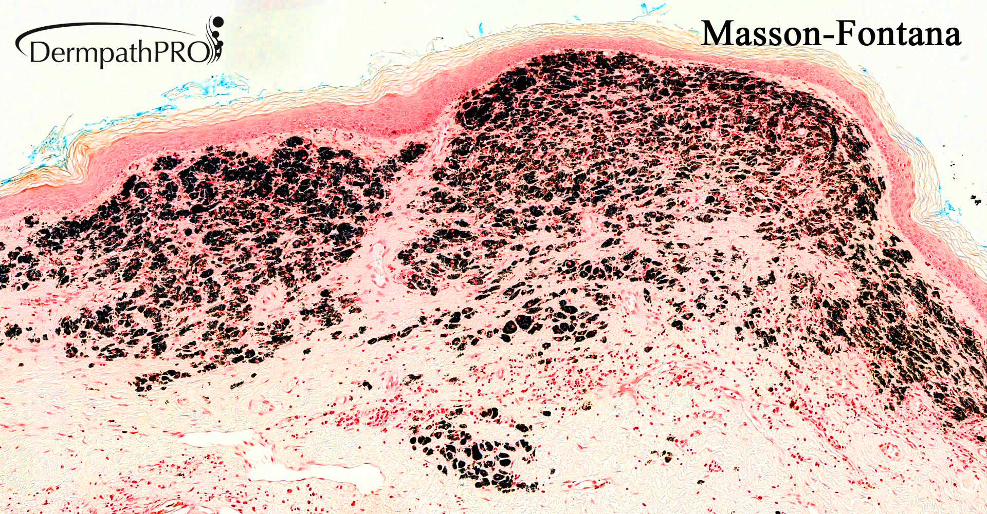
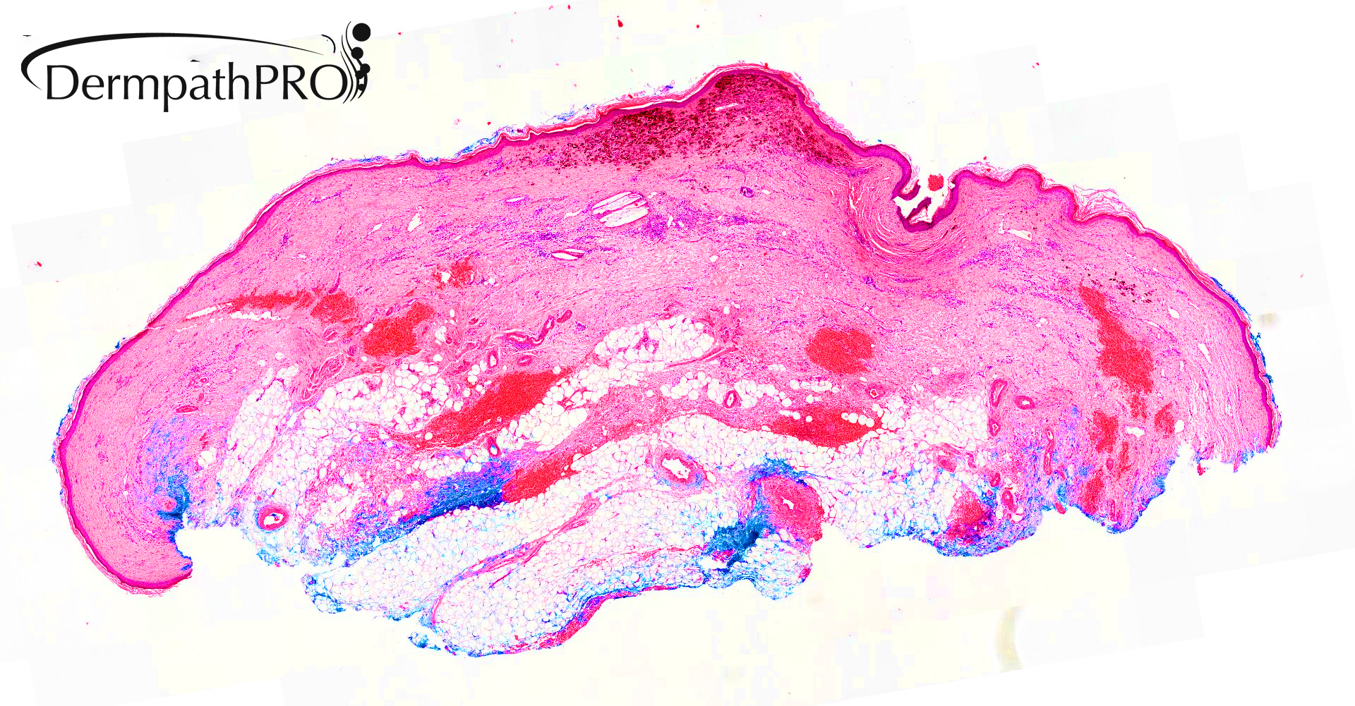
79M: Incisional biopsy left shoulder. Pigmented lesion noted within the field of previous radiotherapy for basal cell carcinoma.

  Report Record

User Feedback


vincenzo

Posted

59 minutes ago, Cem Leblebici said:

Tumoral melanosis

Agree. 

A question: Was the previous bcc histologically assessed?

Share this comment


Link to comment
share_externally

Saleem Taibjee

Posted

Some interim points/food for thought:

1. Yes, the BCC was histologically confirmed.

2. If tumoral melanosis, what are the implications / cause / management?

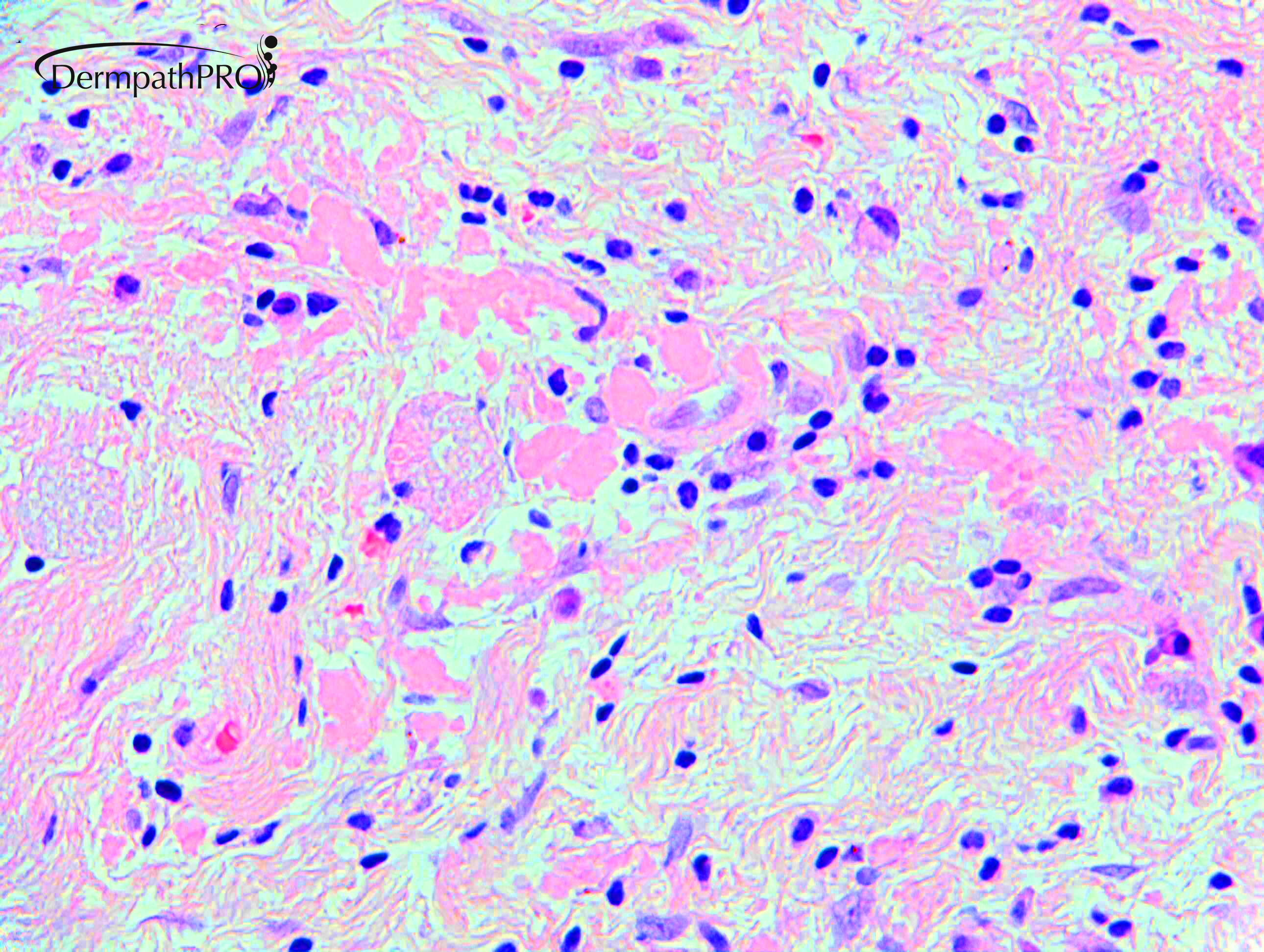
3. The significance of amyloid?

Share this comment


Link to comment
share_externally

vincenzo

Posted

I studied that there is a correlation between BCC and amyloid reactive stroma, but I don’t know much about this. 

However, I’m not sure we can rule out melanoma. 

Share this comment


Link to comment
share_externally

Saleem Taibjee

Posted

This is an interesting case to demonstrate the pitfall that impressive tumoral melanosis can also occasionally arise as a result of regression of pigmented epithelial lesions.

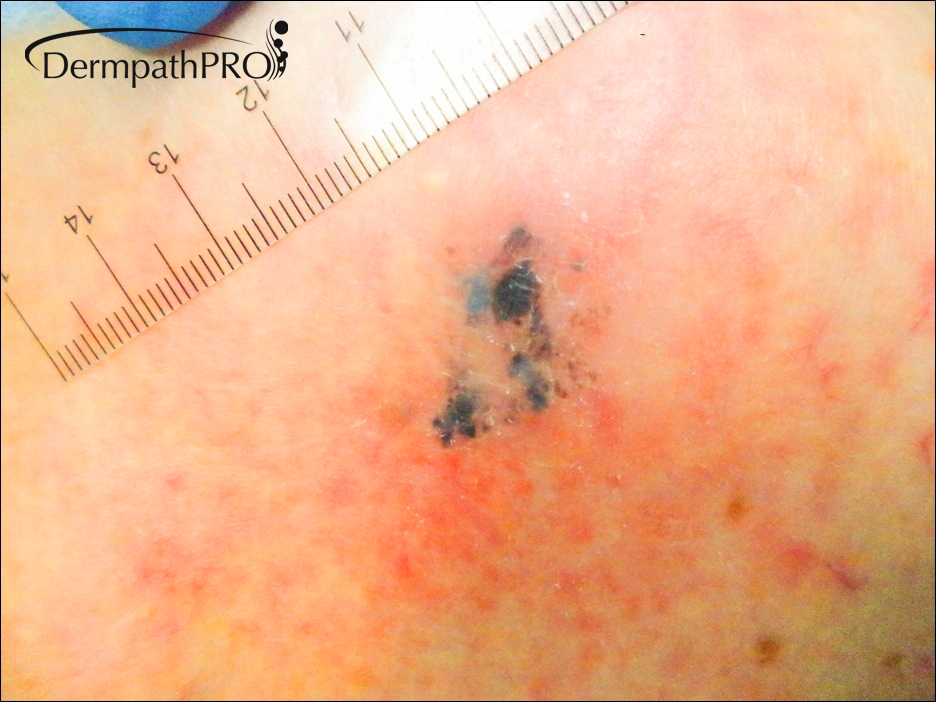
I reviewed the original clinical images of the BCC which was treated by radiotherapy the preceding year, and as you can see below, it was a pigmented variant of BCC. The histology was clearly BCC, with no evidence of any melanocytic lesion in the original biopsy prior to radiotherapy.

There is a pertinent publication in this regard.

Flax et al. Nodular melanosis due to epithelial neoplasms: A finding not restricted to regressed melanomas.  Am J Dermatopathol 1998;20:118-22

The table from the paper is shown below, showing the range of epithelial lesions which were described in their series.

This means that we do sometimes need to be wary of automatically inferring a regressed melanocytic lesion or melanoma when encountering tumoral melanosis, even though that remains the most likely cause.

clinical photo 2.jpg

Table.jpg

Share this comment


Link to comment
share_externally

Saleem Taibjee

Posted

As Vincenzo correctly points out, epithelial tumours, in particular BCC, not infrequently result in residual (keratin-derived) amyloid when regressing. The amyloid can be highlighted by a pankeratin stain if necessary. This is a further clue in this case as to the origin of the tumoral melanosis.

Share this comment


Link to comment
share_externally

Thanks so much, for the many learning points. 

Share this comment


Link to comment
share_externally

Dr. Mona Abdel-Halim

Posted

very informative case!

Share this comment


Link to comment
share_externally


Join the conversation

You can post now and register later. If you have an account, sign in now to post with your account.

Guest
Add a comment...

×   Pasted as rich text.   Paste as plain text instead

  Only 75 emoji are allowed.

×   Your link has been automatically embedded.   Display as a link instead

×   Your previous content has been restored.   Clear editor

×   You cannot paste images directly. Upload or insert images from URL.

Loading...

×
×
  • Create New...