Jump to content
In this section we have spot diagnoses posted on a daily basis since June 2010, now over 4000! You can review the archived cases and read the suggested diagnoses by users and the final comment by the contributors.
Case are uploaded each week day by 10 am UK time with the correct diagnosis will generally be posted at 8 pm UK time. Why not view the most recent spot diagnosis and proffer a diagnosis?

Case Number : Case 2587 - 05 June 2020 Posted By: Dr. Richard Carr

Please read the clinical history and view the images by clicking on them before you proffer your diagnosis.
Submitted Date :
   (0 reviews)

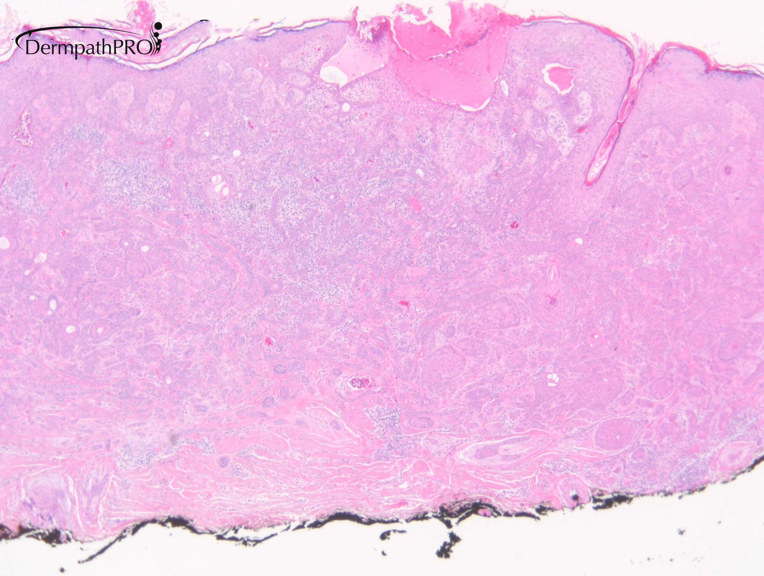
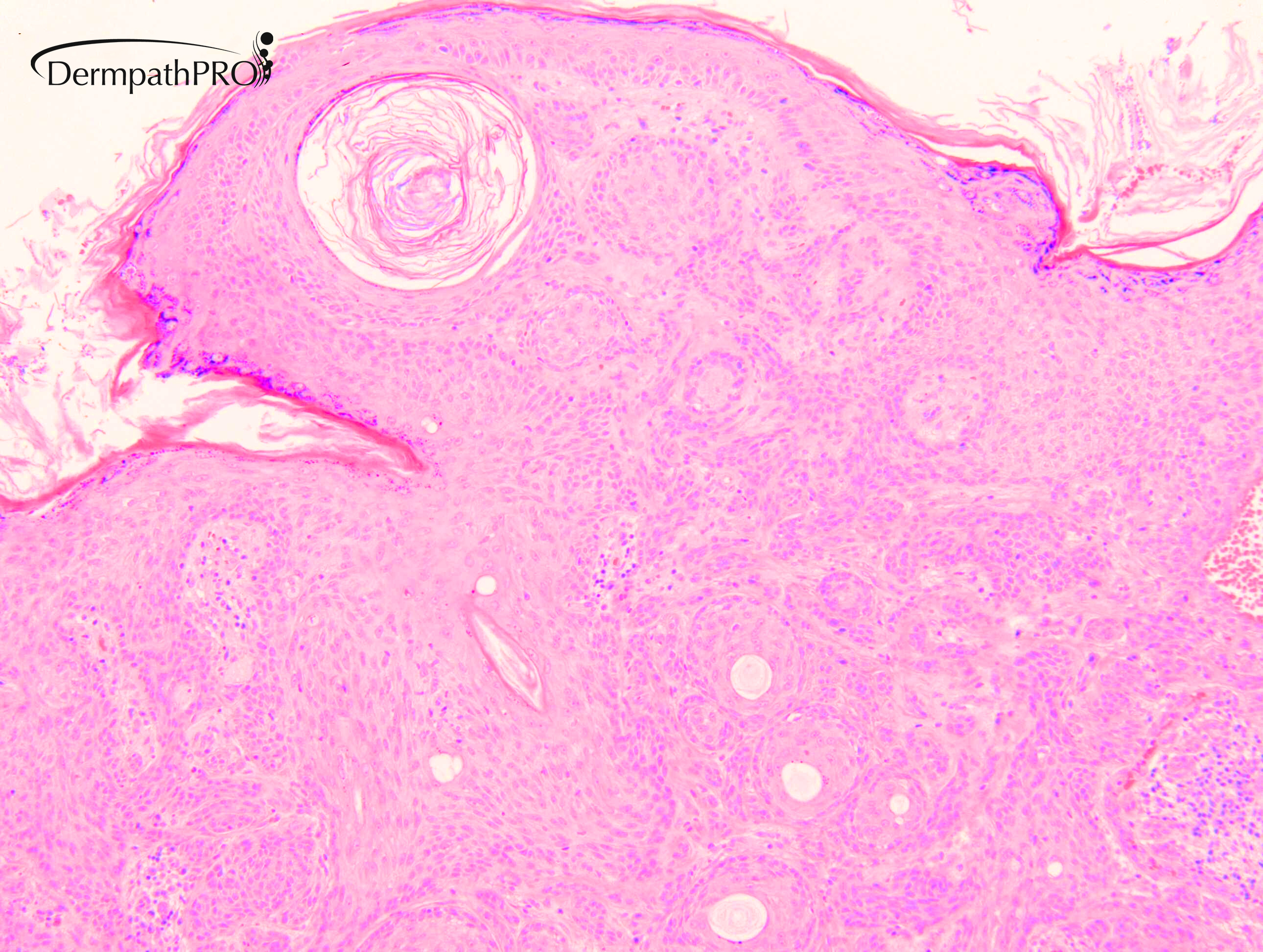
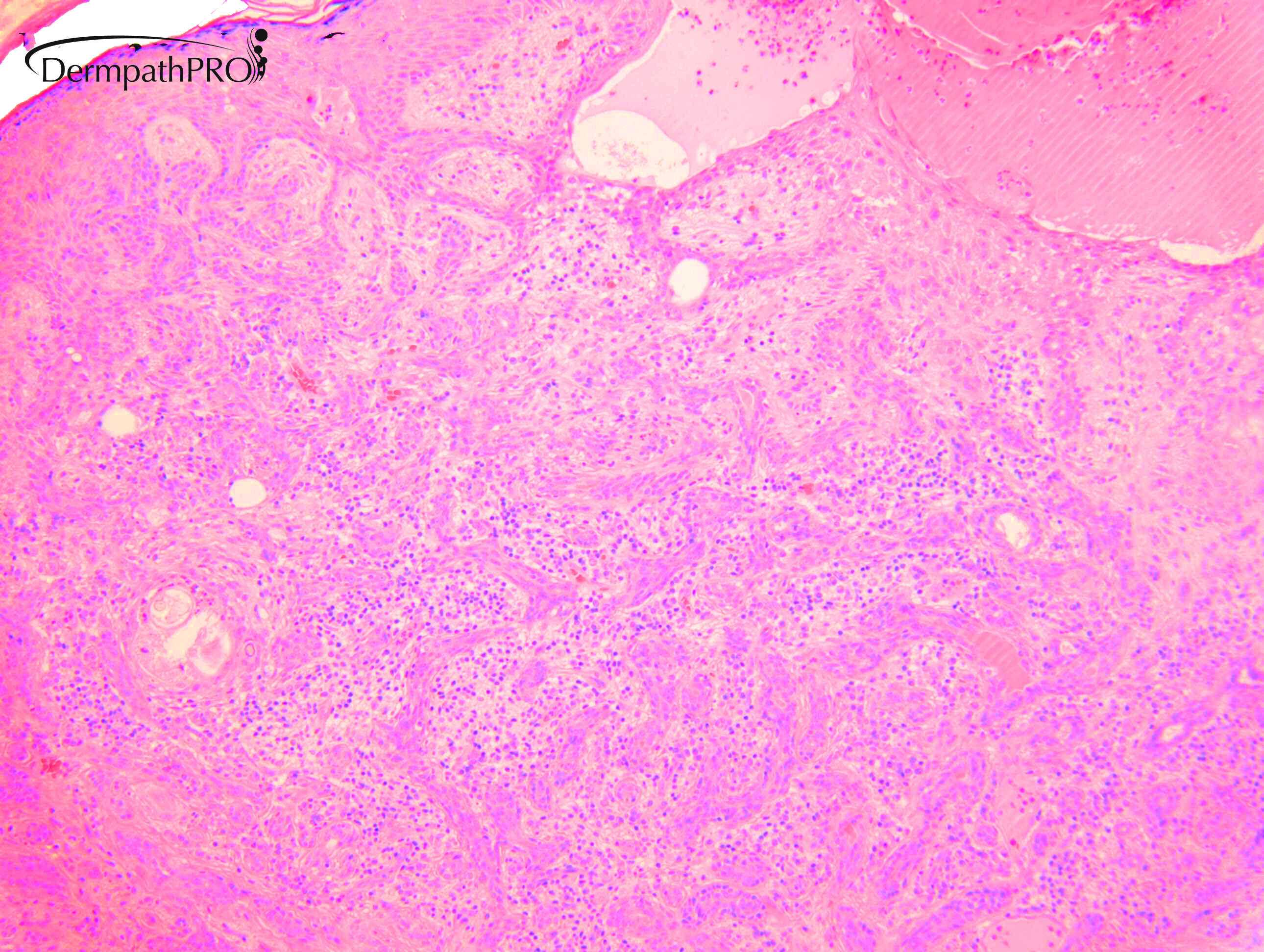
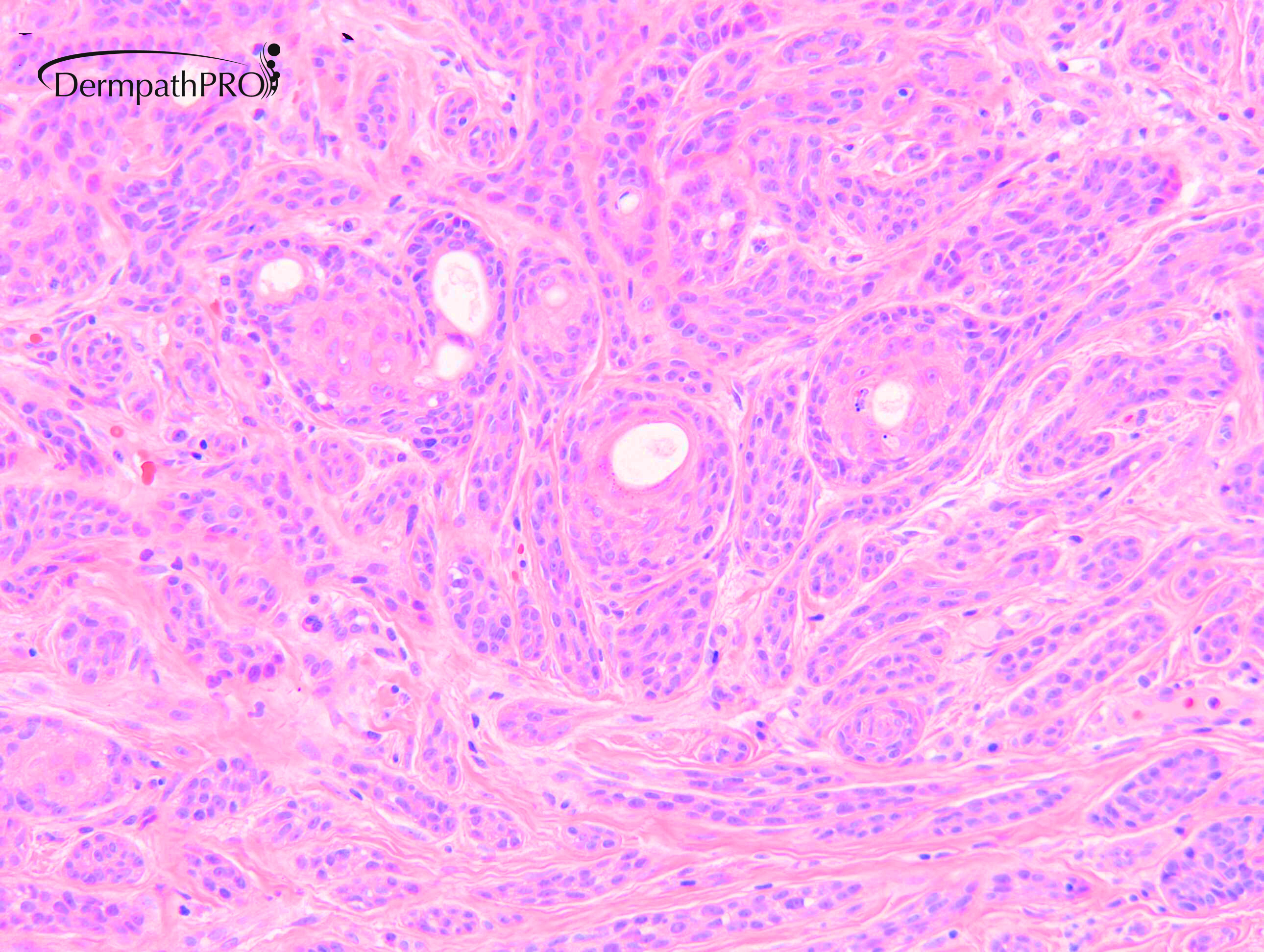
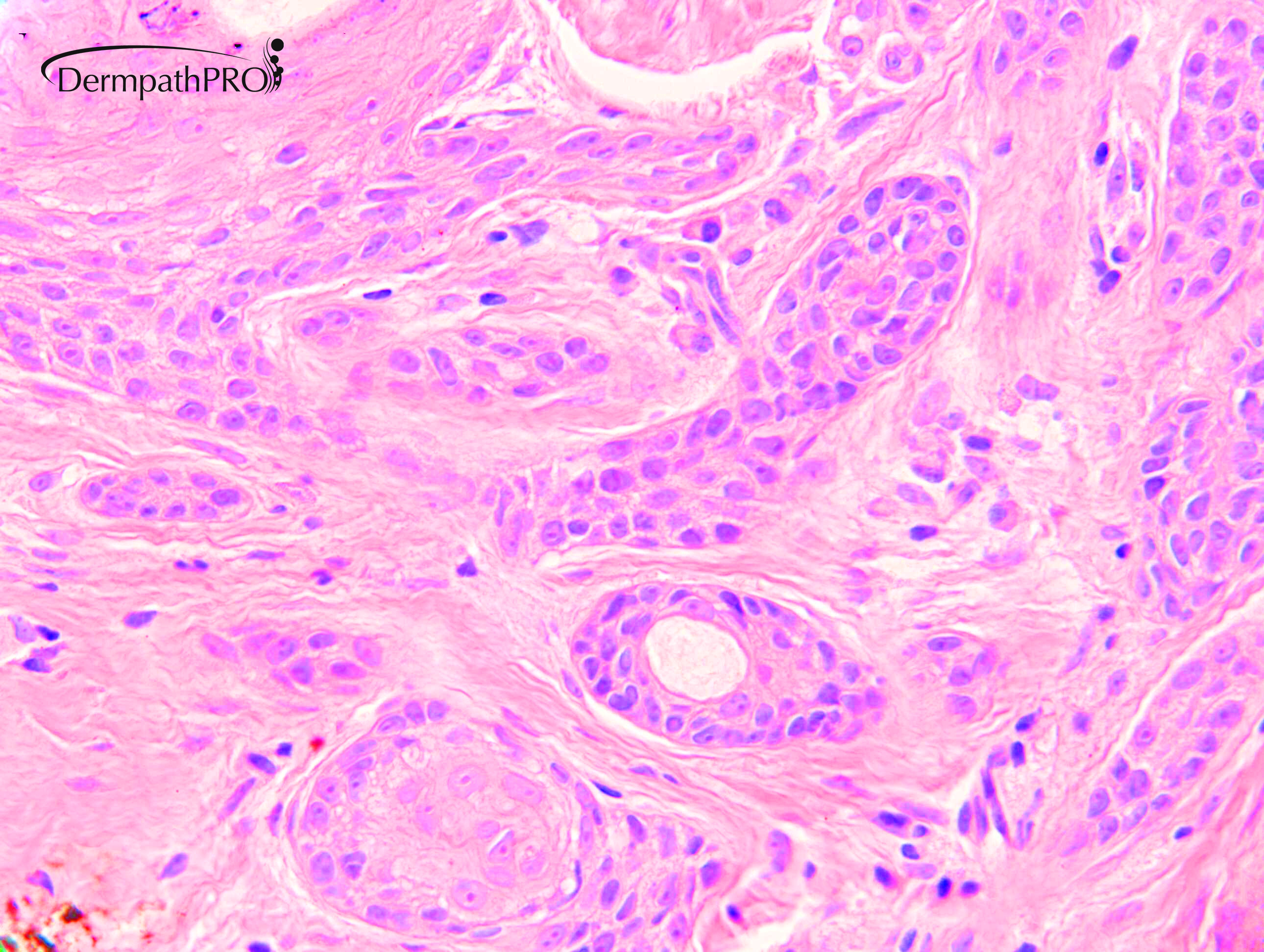
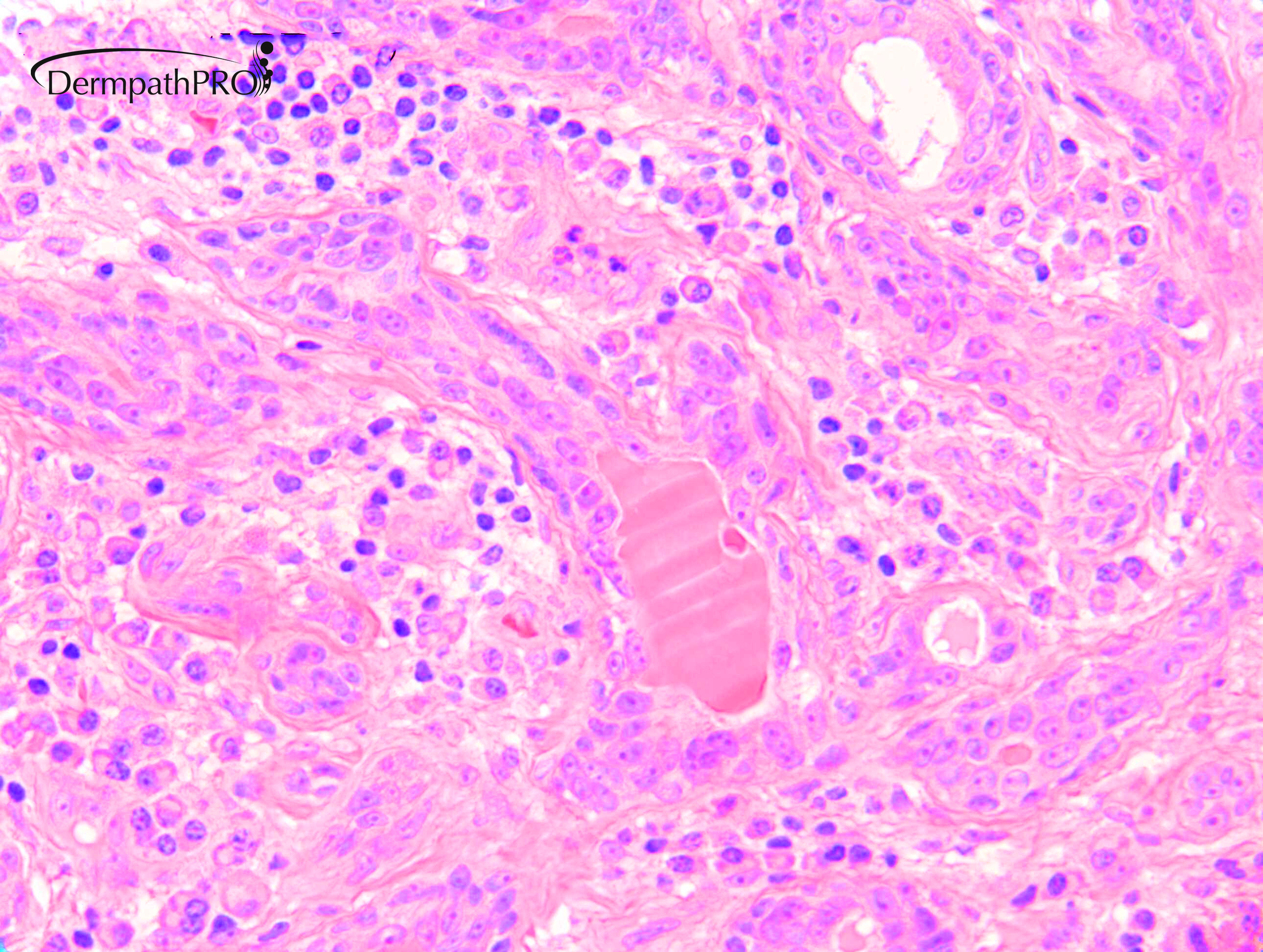
M75. Cheek. 2 years. Raised lesion ?SEBK. Case c/o Dr S. Littleford


  • Thanks 1
  Report Record

User Feedback


Victor Delgado

Posted

Inverted Follicular Keratosis

Share this comment


Link to comment
share_externally

Meenakshi Batrani

Posted

Reticulated pattern of growth with pale cells. IIt makes me consider tumor of follicular infundibulum but there are also some ductular structures which sometimes may be seen in TFI. But I am not sure if these represent eccrine differentiation, which makes me also suspicious of syrigofibroadenoma/acrosyringeal nevus but cells appear pale for this. 

Share this comment


Link to comment
share_externally

John Zhang

Posted

The lower border of this lesion is somewhat infiltrative. In a patient with this advanced age I am leaning towards maligant. There is a hint of tubular structures, and to me this case shows some similarity to a previous spot diagnosis case. Over I favor microcystic adnexal carcinoma/solid carcinoma and will recommend complete excision/Mohs. Hope I am right!


Share this comment


Link to comment
share_externally

Nivedita

Posted

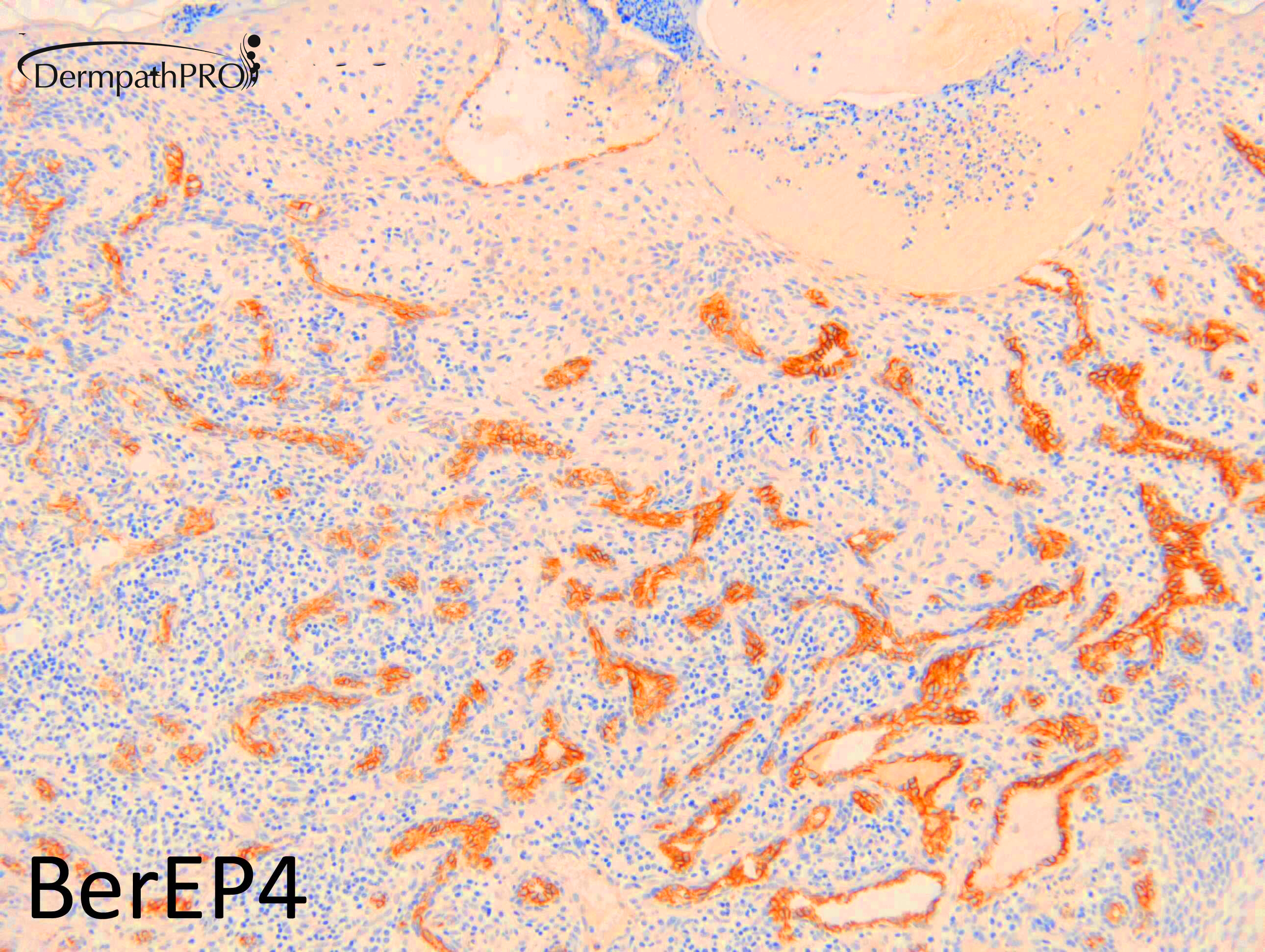
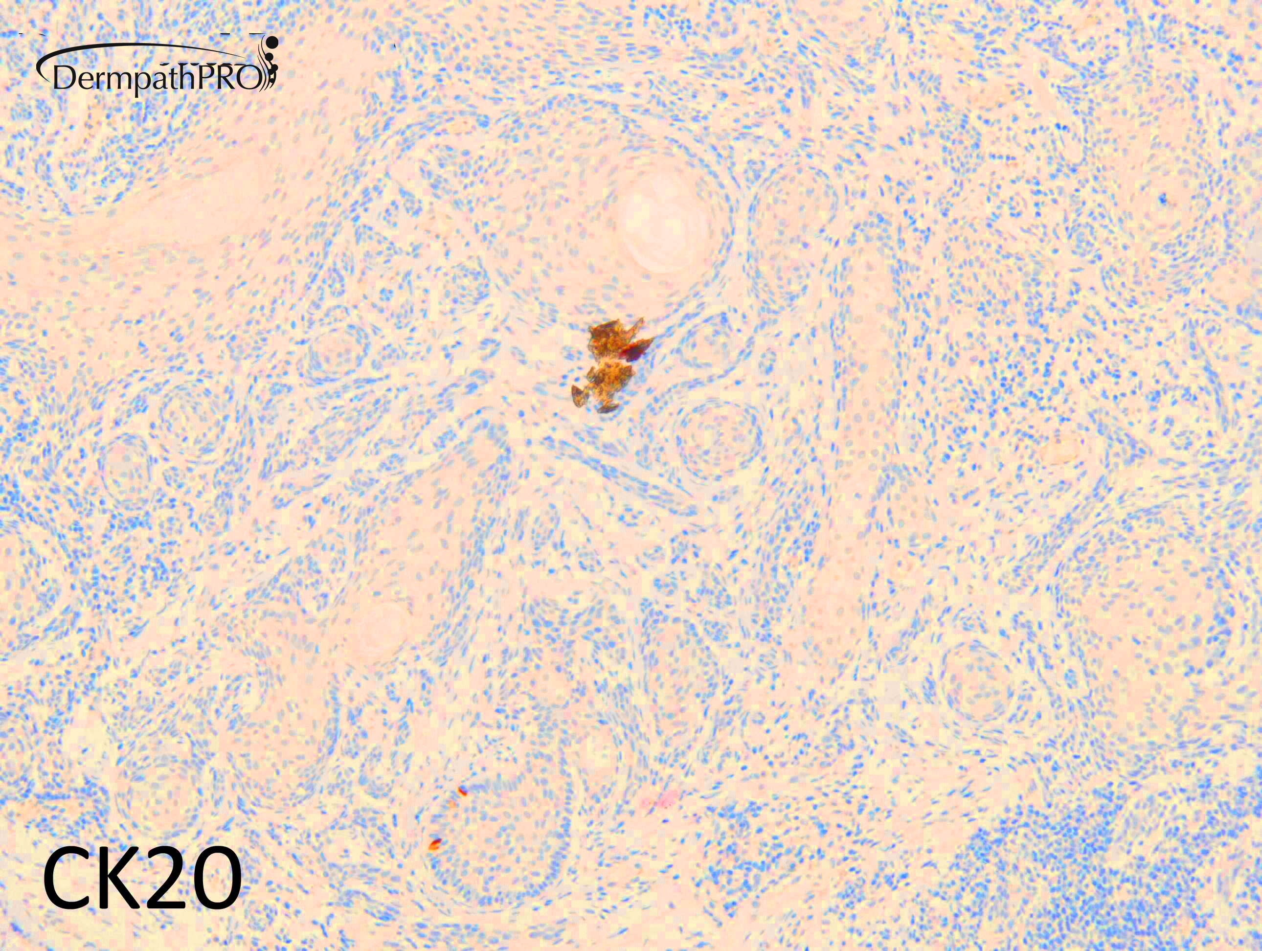
Can we consider bcc since it’s expressing BerEP 4 and absence of CK20 cells? Thanks for sharing this case

Share this comment


Link to comment
share_externally

John Zhang

Posted

Positive BerEP4 argues against MAC. Now I am leaning towards eccrine carcinoma. 

Share this comment


Link to comment
share_externally

vincenzo

Posted

Why not an Infundibulocystic BCC?

Share this comment


Link to comment
share_externally

Eman El-Nabarawy

Posted

Squamoid eccrine ductal carcinoma or Adenosquamous carcinoma porocarcinoma with squamous differentiation.

Share this comment


Link to comment
share_externally

Dr. Richard Carr

Posted

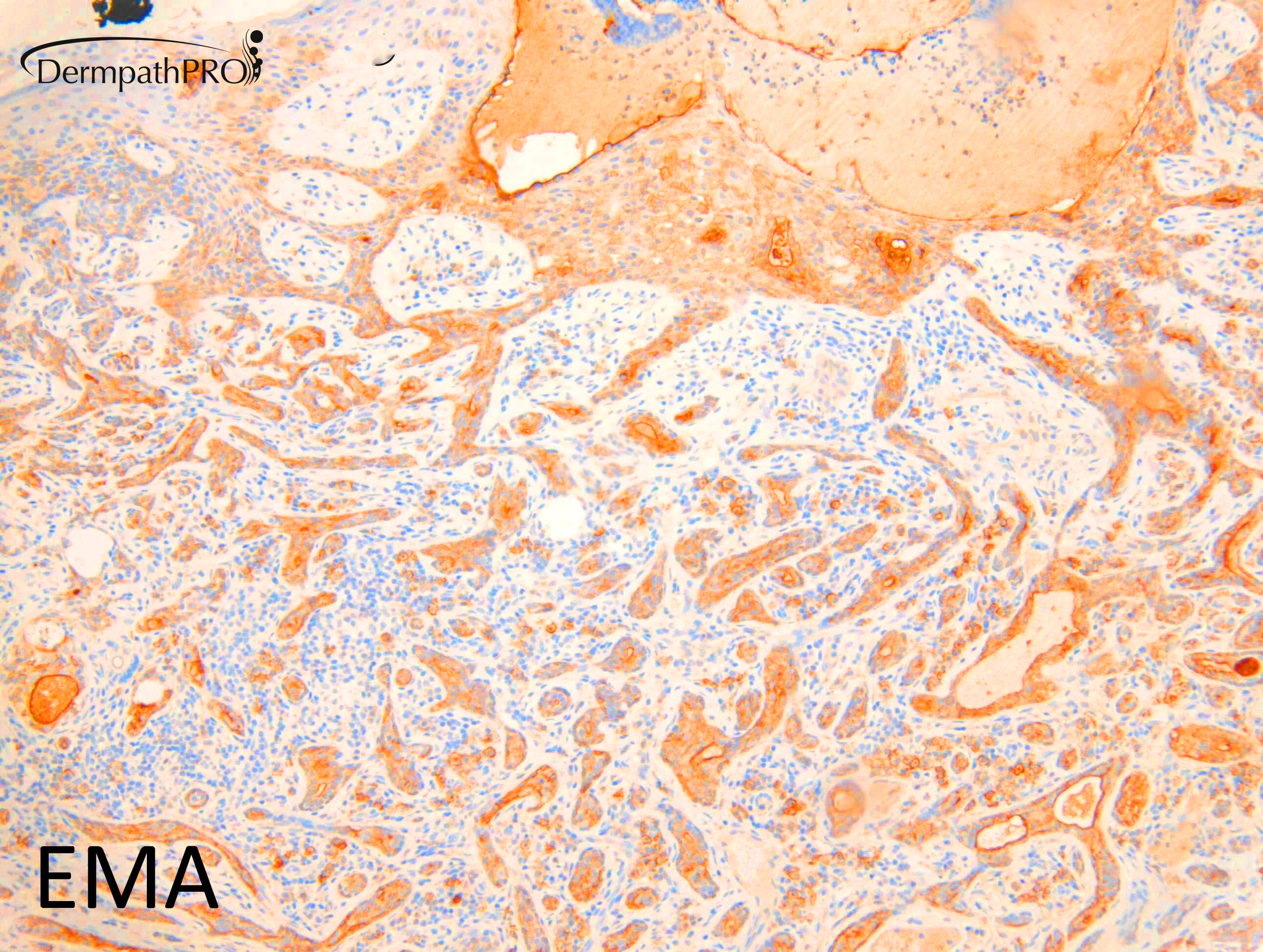
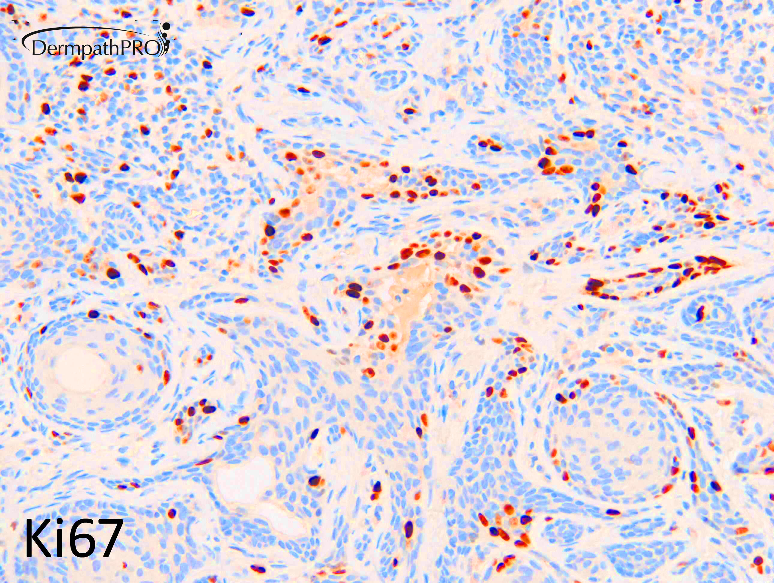
Well done John. These are nearly always highly challenging cases and this is a referred case. I reported it as microcystic adnexal carcinoma (MAC) and advised margin control surgery and commented on the potential for deep and possible extensive perineural invasion. BerEP4 is diffusely strongly positive in infiltrative BCC and EMA is always negative in the basaloid cells of BCC.  BerEP4 and EMA can highlight glandular differentiation in MAC. For me the original reports of squamoid eccrine ductal carcinoma were of a low grade lesion akin to MAC and low grade syringoid and sclerosing eccrine ductal carcinoma. Eccrine epithelioma is an out-dated term and for me and was never precisely defined. If a lesion is "adenosquamous" but intermediate to high grade and not a typical porocarcinoma (can have a benign poromatous component), hidradenocarcinoma (benign hidradenoma component) or eccrine ductal carcinoma (identical to ductal carcinoma of breast) I prefer "adenosquamous" carcinoma NOS. CK20 shows reliably dendritic Merkel cells in desmoplastic trichoepithelioma but as far as I'm aware is otherwise not useful for BCC v's syringoma v's MAC (and allied low grade lesions above). Ultimately it is recognising it does not fit with a benign lesion, and having a high index of suspicion, it's low grade (low mitotic and proliferation, mild atypia only) and can be highly locally infiltrative (if left for years) but rarely if ever metastasises. Some quite horrific surgery may be required to remove these low-grade lesions if left to grow indolently for many years (i.e. similar, in almost every way, to an indolent infiltrative BCC).

Share this comment


Link to comment
share_externally

Nivedita

Posted

Thank you Dr.Richard Carr for this case learning.... many important points to remember 

Share this comment


Link to comment
share_externally

John Zhang

Posted

Thank you so much for your effort of continuously educating our dermpath community. I probably would not have any idea about this case if I didn't study case 806. I benefit a lot from Dermpathpro!

Share this comment


Link to comment
share_externally


Join the conversation

You can post now and register later. If you have an account, sign in now to post with your account.

Guest
Add a comment...

×   Pasted as rich text.   Paste as plain text instead

  Only 75 emoji are allowed.

×   Your link has been automatically embedded.   Display as a link instead

×   Your previous content has been restored.   Clear editor

×   You cannot paste images directly. Upload or insert images from URL.

Loading...

×
×
  • Create New...