Jump to content
In this section we have spot diagnoses posted on a daily basis since June 2010, now over 4000! You can review the archived cases and read the suggested diagnoses by users and the final comment by the contributors.
Case are uploaded each week day by 10 am UK time with the correct diagnosis will generally be posted at 8 pm UK time. Why not view the most recent spot diagnosis and proffer a diagnosis?

Case Number : Case 2588 - 08 June 2020 Posted By: Iskander H. Chaudhry

Please read the clinical history and view the images by clicking on them before you proffer your diagnosis.
Submitted Date :
   (0 reviews)

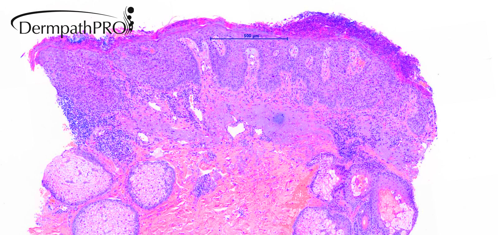
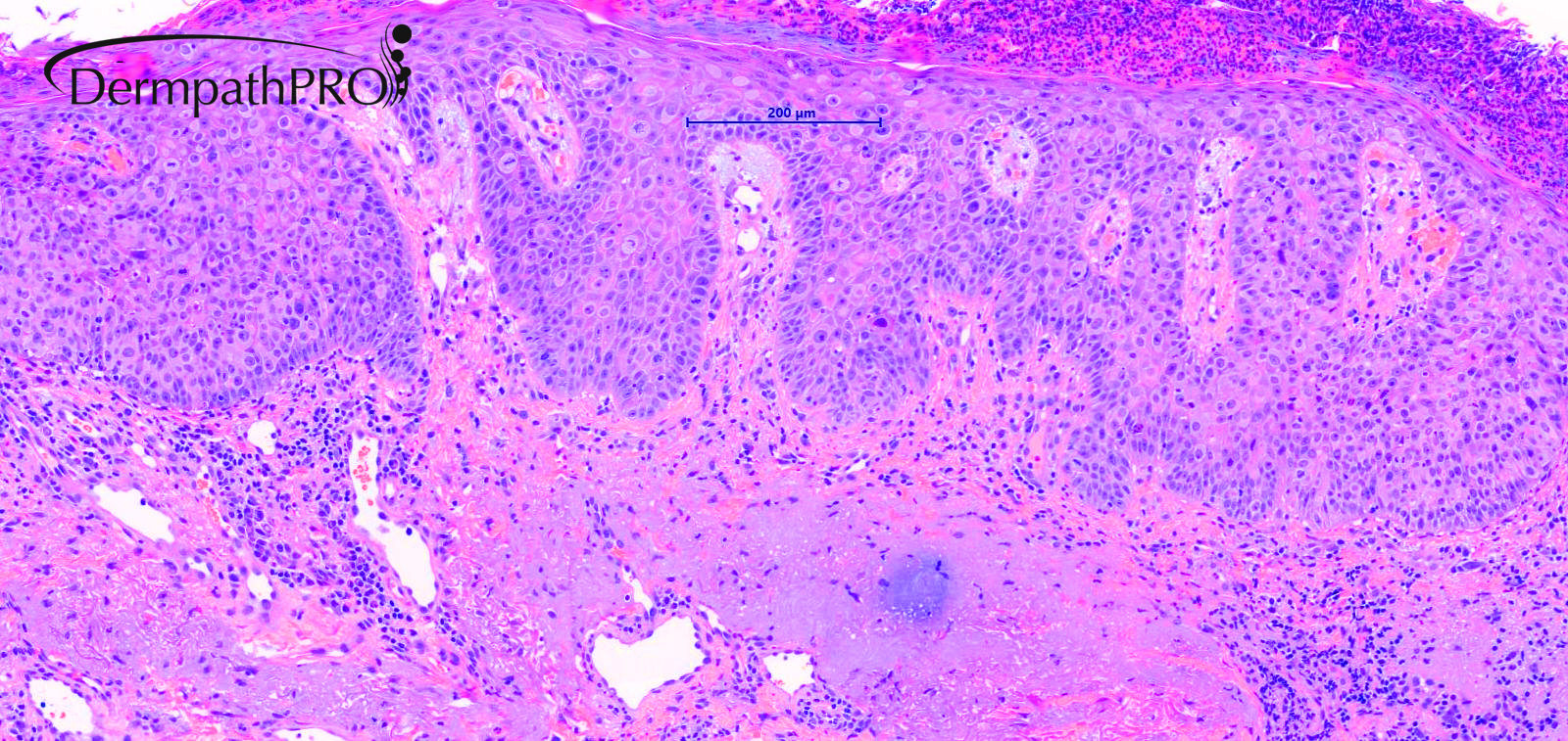
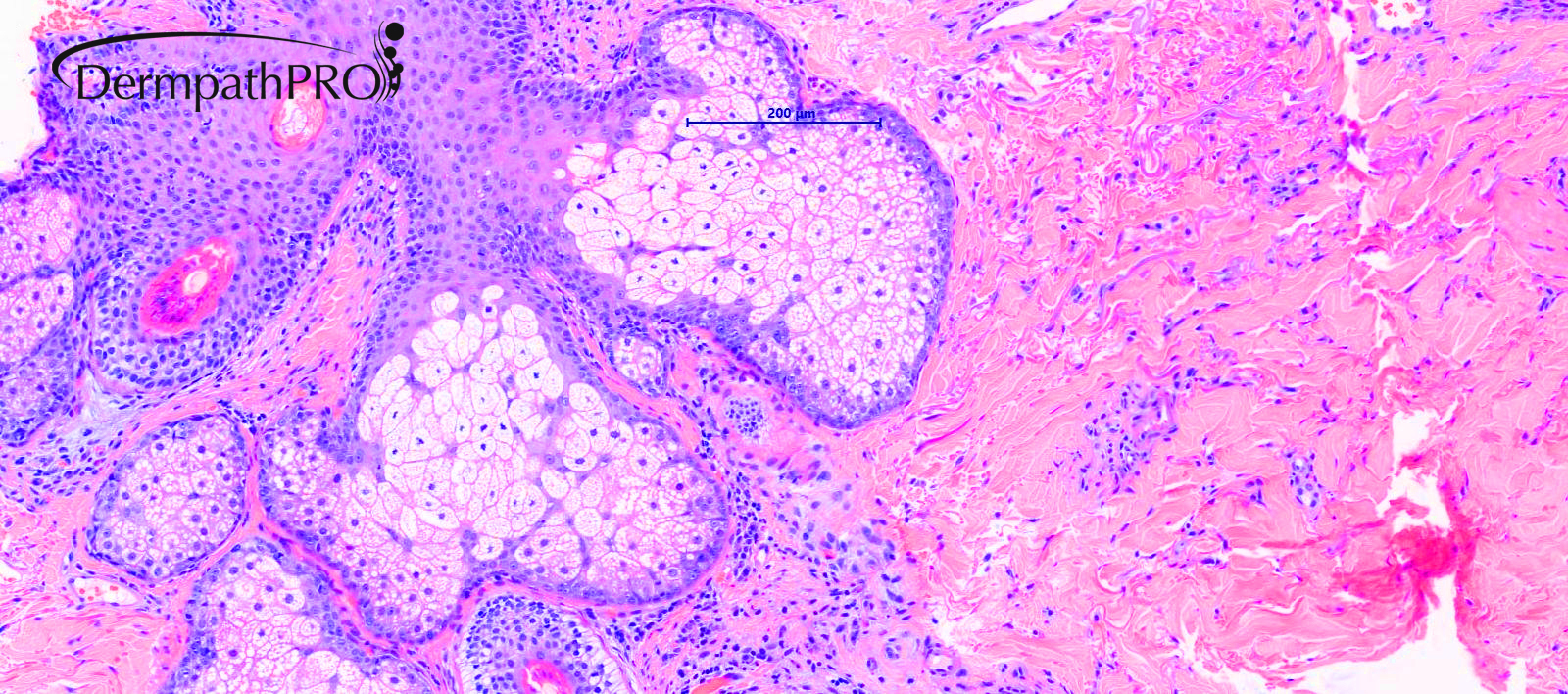
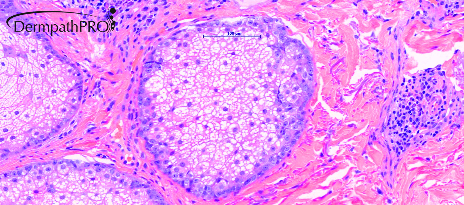
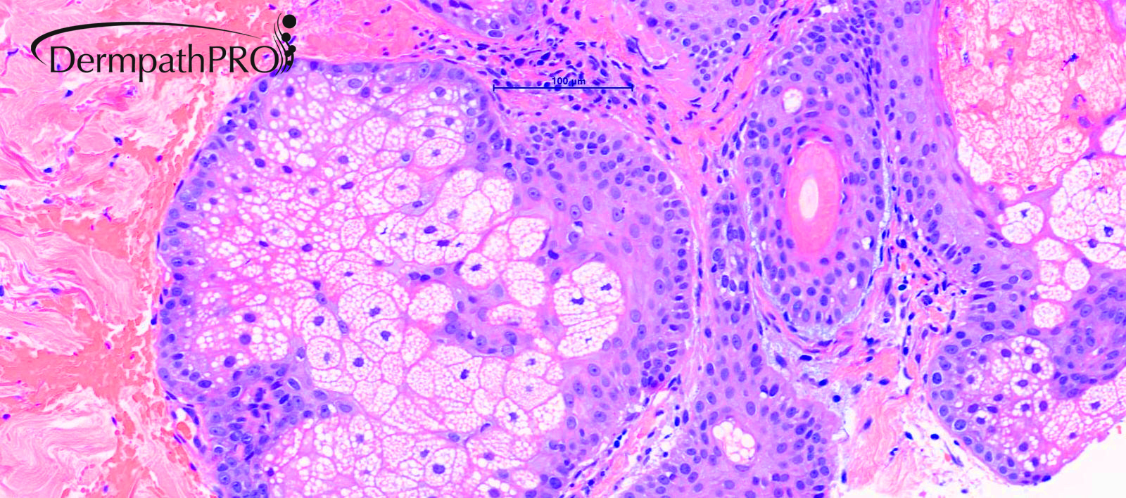
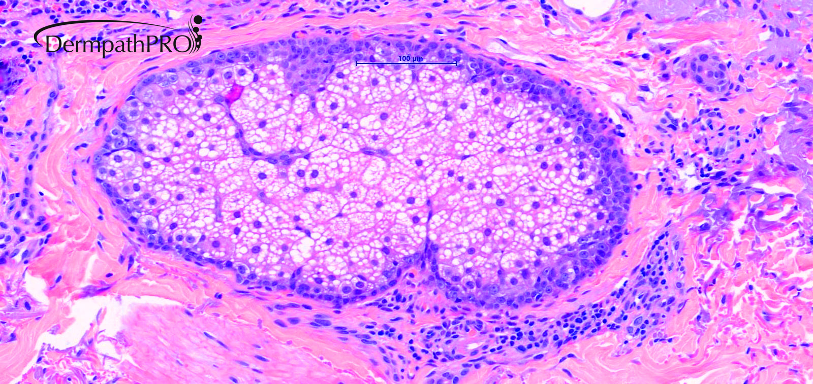
76M, Erythematous - 6cm scaly area not responding to steroid by GP.


  Report Record

User Feedback


vincenzo

Posted

4 hours ago, Hamad said:

SCCIS / Paget’s disease 

Agree.

Share this comment


Link to comment
share_externally

Josie Bisi

Posted

I would do immunohistochemistry to differentiate pagetoid neoplasms such as: Bowen's disease, Paget's disease (I believe to be one of these two), sebaceous carcinoma, among others.

Share this comment


Link to comment
share_externally

Krishnakumar subramanian

Posted

agree could be Bowen's or Paget's disease

Share this comment


Link to comment
share_externally

Dr. Mona Abdel-Halim

Posted

Agree with the above mentioned possibilities, in situ seb carc also

Share this comment


Link to comment
share_externally

Richard Logan

Posted

Hello everyone.  I have been one of the "silent majority" not putting my head above the parapet for the last few years.  Retirement from Dermatological practice and "lockdown" has given me lots of time to study this wonderful website, and I would like to say a collective thank you to all the erudite contributors who have educated and entertained me in equal measure.


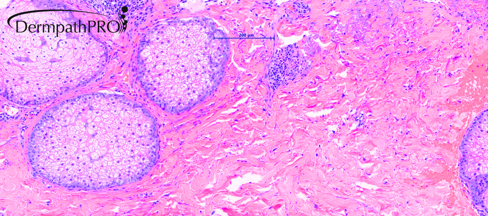
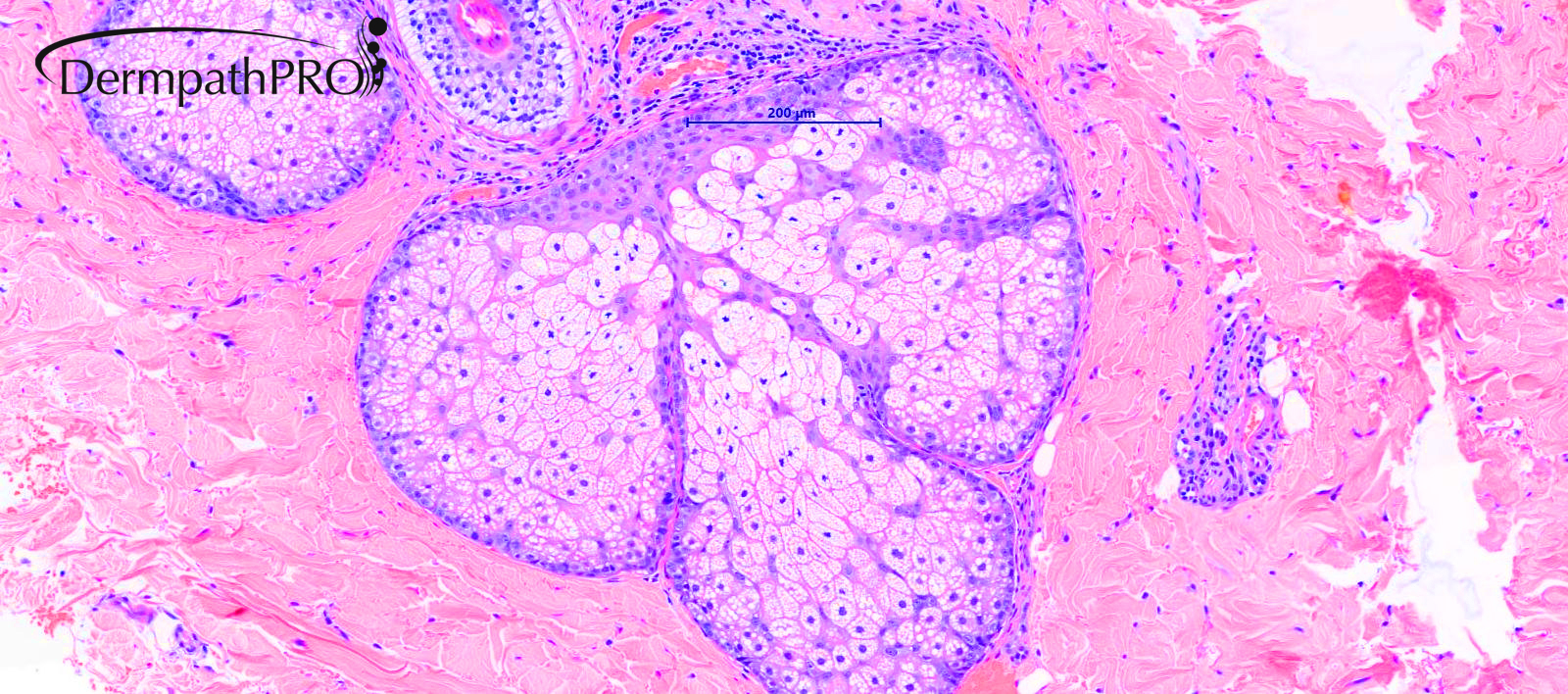
Regarding this case, the action is clearly in the epidermis and I agree that Bowen's or Paget's disease are the most likely diagnoses.  However, Iskander has shown several views of the sebaceous glands, so I wonder what his final thoughts on this case were?

Share this comment


Link to comment
share_externally

Iskander H. Chaudhry

Posted

Dear All 

The final diagnosis is Clear Cell Bowen's based on immunochemistry. I exclude Paget's and also MMIS. The sebaecous gland photomicrographs I added for you to consider sebaceous carcinoma in situ which there was not compelling evidence of on morphology or immunochemistry. A panel of markers  for sebaecous differentiation include: EMA, CK7, Ber-EP4, Factor XIIIA, androgen receptor, p53, adipophilin, progesterone receptor membrane component 1 (PGRMC1), squalene synthase (SQS), and alpha/beta hydrolase domain-containing protein 5 (ABHD5) :). 

Share this comment


Link to comment
share_externally


Join the conversation

You can post now and register later. If you have an account, sign in now to post with your account.

Guest
Add a comment...

×   Pasted as rich text.   Paste as plain text instead

  Only 75 emoji are allowed.

×   Your link has been automatically embedded.   Display as a link instead

×   Your previous content has been restored.   Clear editor

×   You cannot paste images directly. Upload or insert images from URL.

Loading...

×
×
  • Create New...