Jump to content
In this section we have spot diagnoses posted on a daily basis since June 2010, now over 4000! You can review the archived cases and read the suggested diagnoses by users and the final comment by the contributors.
Case are uploaded each week day by 10 am UK time with the correct diagnosis will generally be posted at 8 pm UK time. Why not view the most recent spot diagnosis and proffer a diagnosis?

Case Number : Case 2591 - 11 June 2020 Posted By: Saleem Taibjee

Please read the clinical history and view the images by clicking on them before you proffer your diagnosis.
Submitted Date :
   (0 reviews)

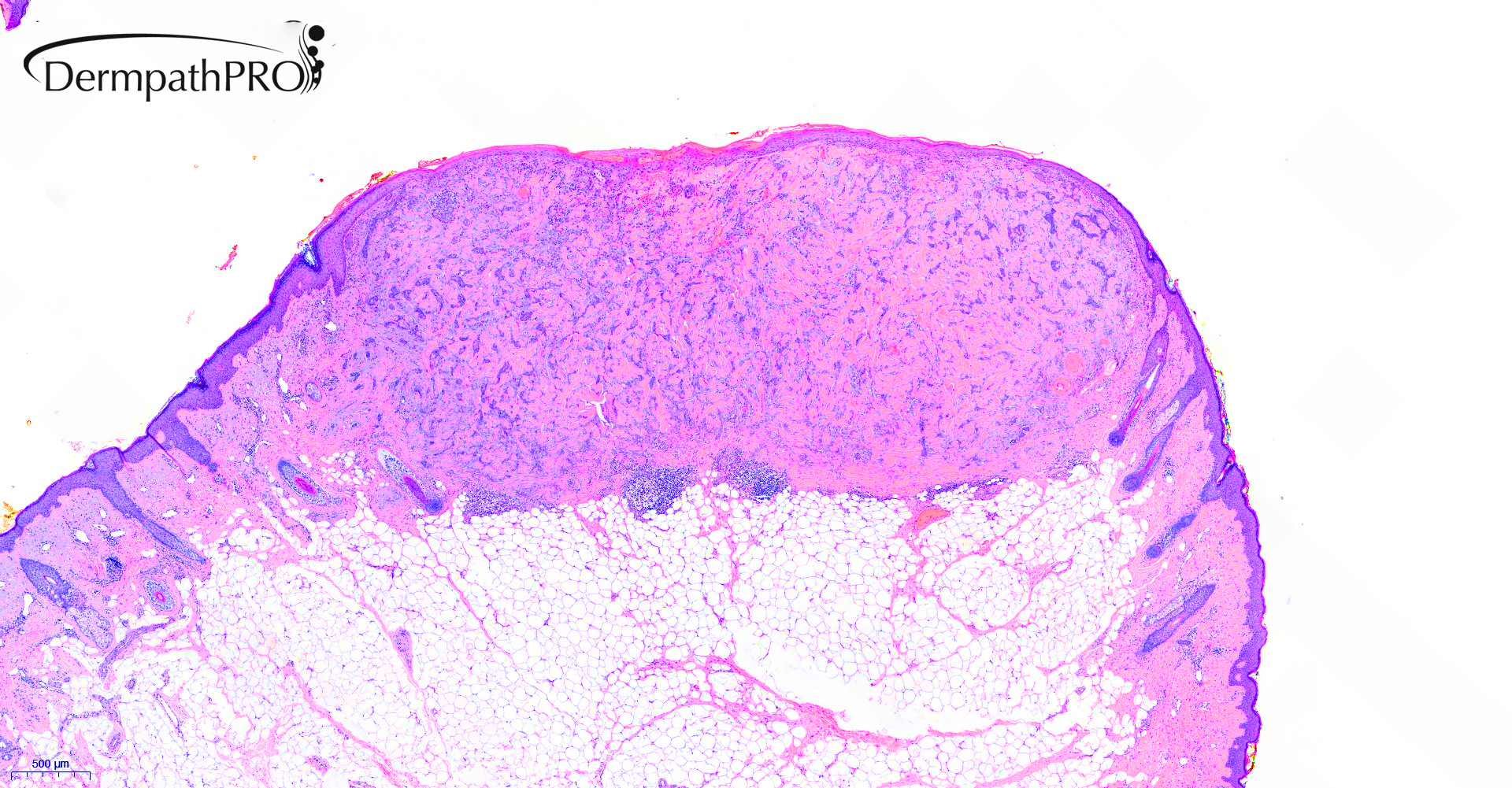
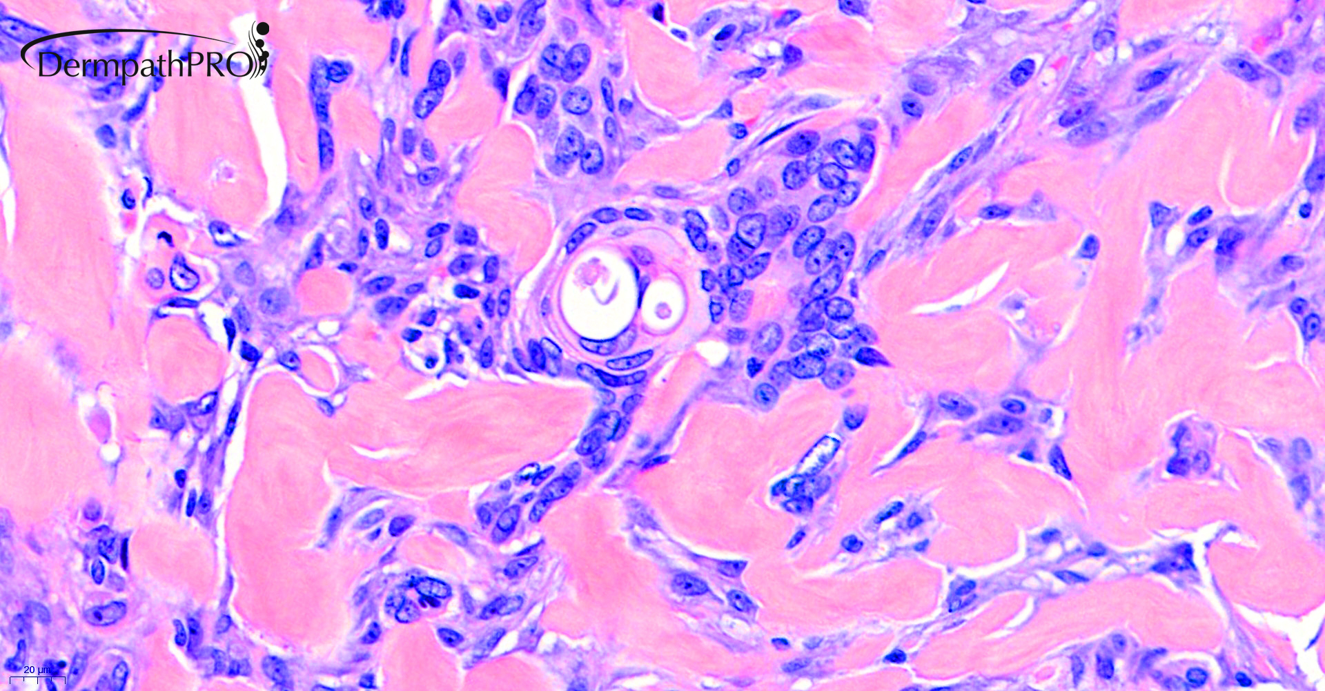
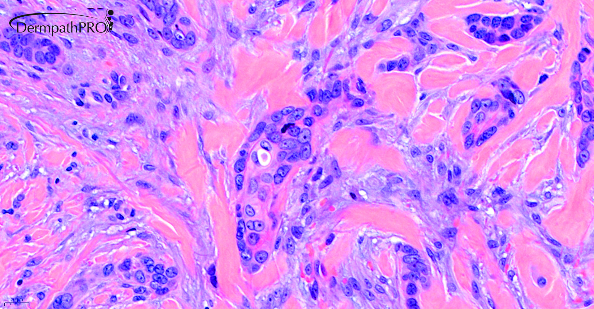
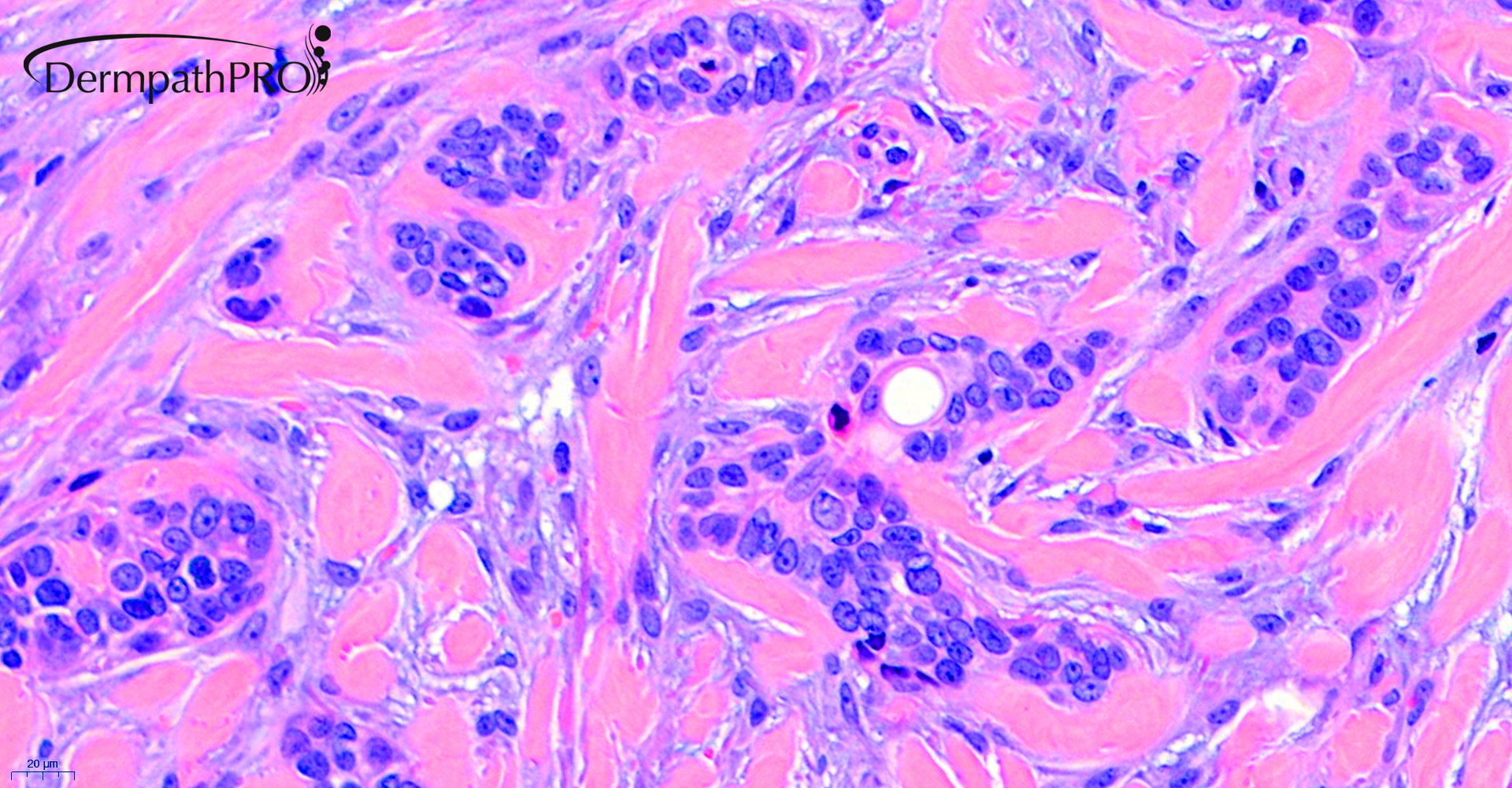
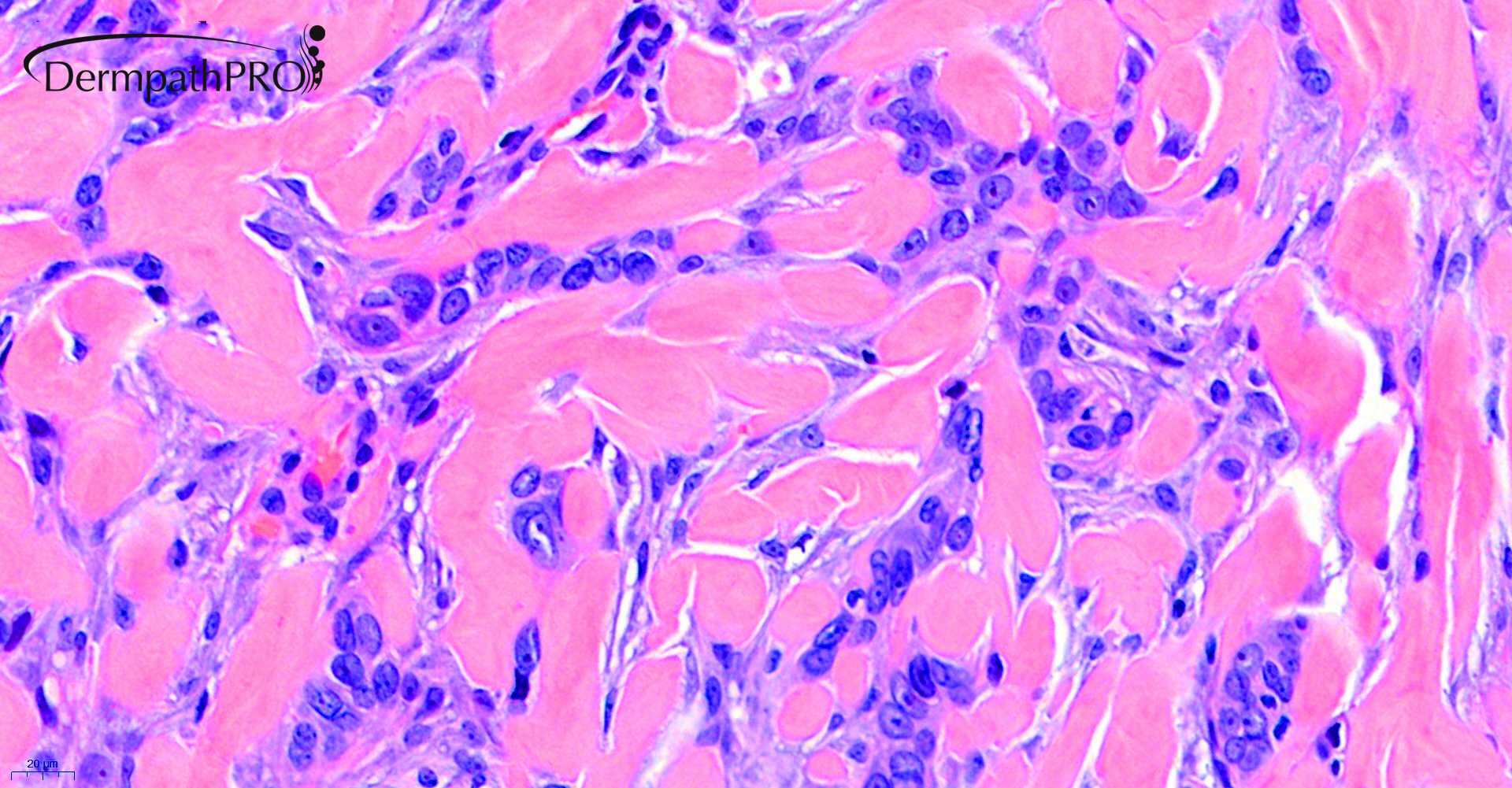
54F, Excision left ear. suspected BCC.


  Report Record

User Feedback


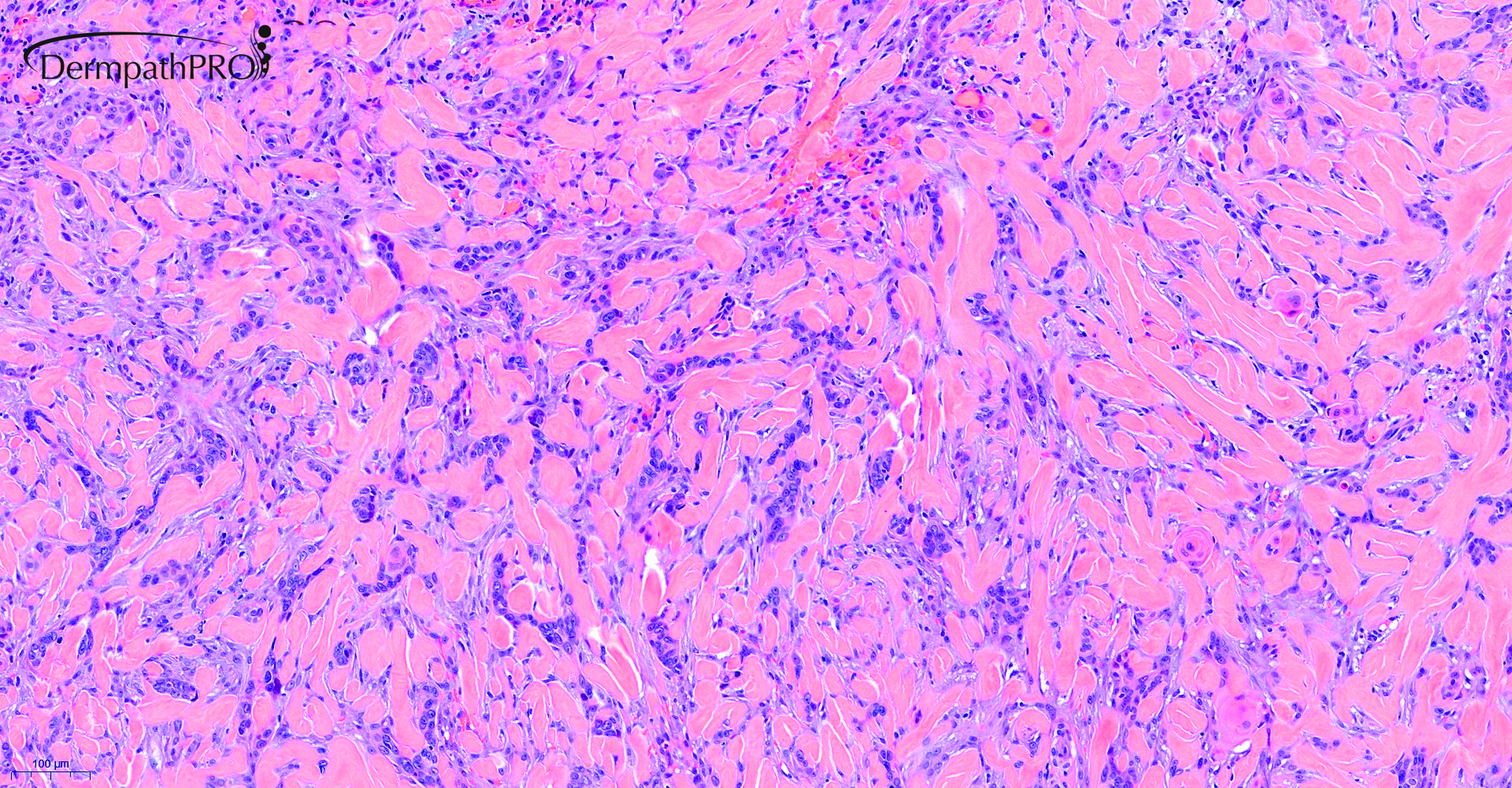
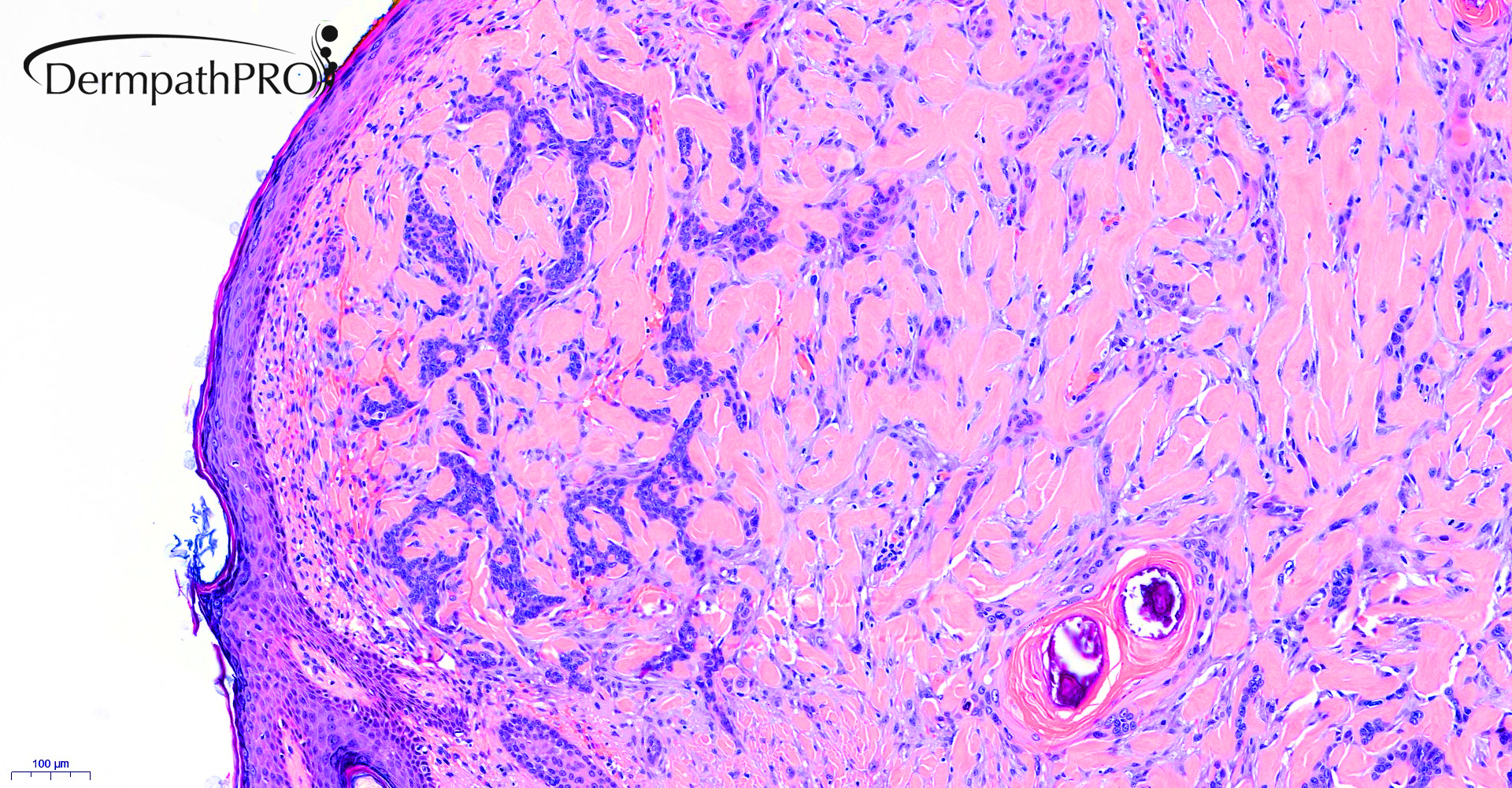
I am thinking of both DT and MAC. There are keratin cysts as well as duct formation and infiltration into the subcutaneous fat with desmoplastic stroma...not able to further differentiate these both. 
There are no obvious BCC like nests.

 Thank you for this case learning.... waiting to learn some important points

Share this comment


Link to comment
share_externally

Favor DTE. I don’t see any perineurial infiltration, and  there are calcifications. 

Share this comment


Link to comment
share_externally

Krishnakumar subramanian

Posted

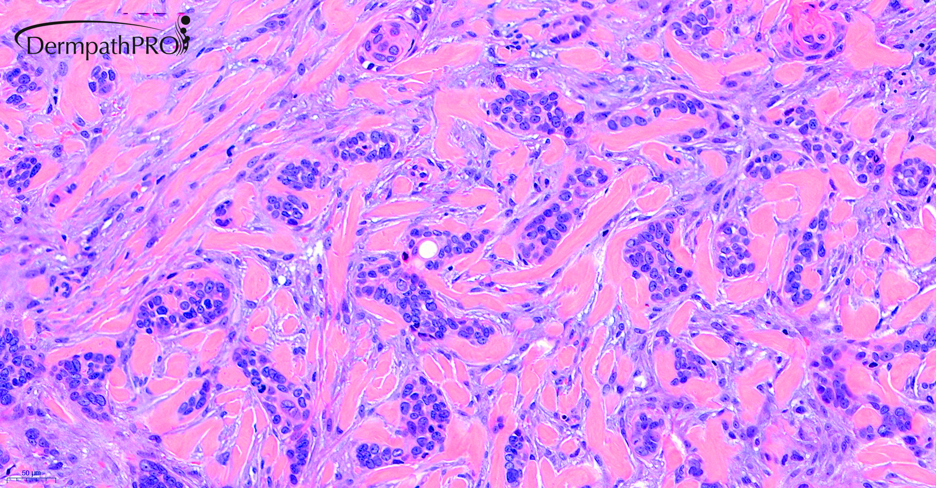
some mitotic activity seen favor MAC

Share this comment


Link to comment
share_externally

Cem Leblebici

Posted

For me, MAC is not a suitable diagnosis for this superficial tumor. DTE with ductal differantiation is a more appropriate diagnosis for this superficial tumor. 


Share this comment


Link to comment
share_externally

Saleem Taibjee

Posted

Thanks for the interesting comments.

I attach the immunohistochemistry below.

As you can see, BerEP4 was clearly positive, EMA negative, and CEA highlights the ducts evident on H&E.

This immunoprofile is a good fit for a rare example of infiltrative BCC showing ductal differentiation.

I have not come across Desmoplastic trichoepithelioma showing ductal differentiation, but can see that would be a reasonable differential diagnosis. That said, the tumour showed mitoses, as shown in the images below, and apoptotic bodies were also relatively easy to find. There is also an impression of interstitial mucin in addition to the sclerotic stroma. The tumour extends relatively deeply to the interface of dermis and subcutis. One could also request a CK20 to assess for Merkel cells, but these were not readily seen on the EMA in any case. I think overall this constellation tends to favour BCC rather than DTE. I don’t think the case is such a good fit for MAC.

46322_10.0x BerEP4 labelled.jpg

46322_10.0x EMA labelled.jpg

46322_20.0x CEA labelled.jpg


Share this comment


Link to comment
share_externally

Thanks, Saleem. I didn’t see apoptotic bodies, because many times we( I ) don’t see what we( I ) don’t want to see!

Share this comment


Link to comment
share_externally


Join the conversation

You can post now and register later. If you have an account, sign in now to post with your account.

Guest
Add a comment...

×   Pasted as rich text.   Paste as plain text instead

  Only 75 emoji are allowed.

×   Your link has been automatically embedded.   Display as a link instead

×   Your previous content has been restored.   Clear editor

×   You cannot paste images directly. Upload or insert images from URL.

Loading...

×
×
  • Create New...