Jump to content
In this section we have spot diagnoses posted on a daily basis since June 2010, now over 4000! You can review the archived cases and read the suggested diagnoses by users and the final comment by the contributors.
Case are uploaded each week day by 10 am UK time with the correct diagnosis will generally be posted at 8 pm UK time. Why not view the most recent spot diagnosis and proffer a diagnosis?

Case Number : Case 2596 - 18 June 2020 Posted By: Saleem Taibjee

Please read the clinical history and view the images by clicking on them before you proffer your diagnosis.
Submitted Date :
   (0 reviews)

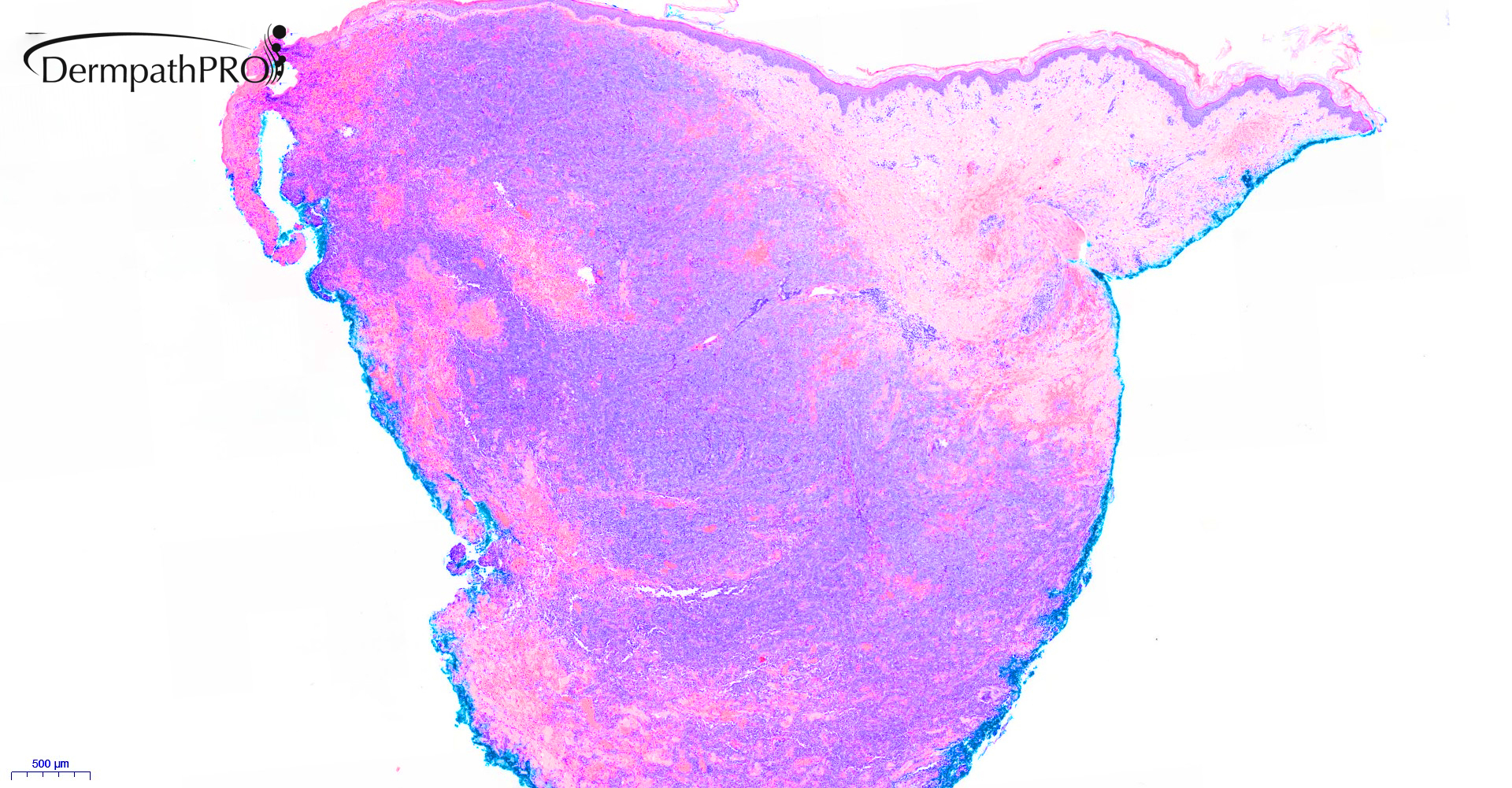
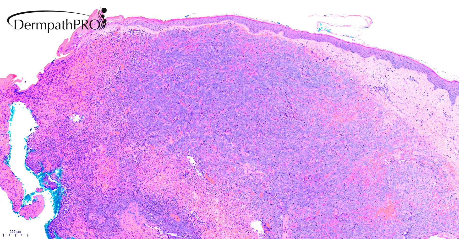
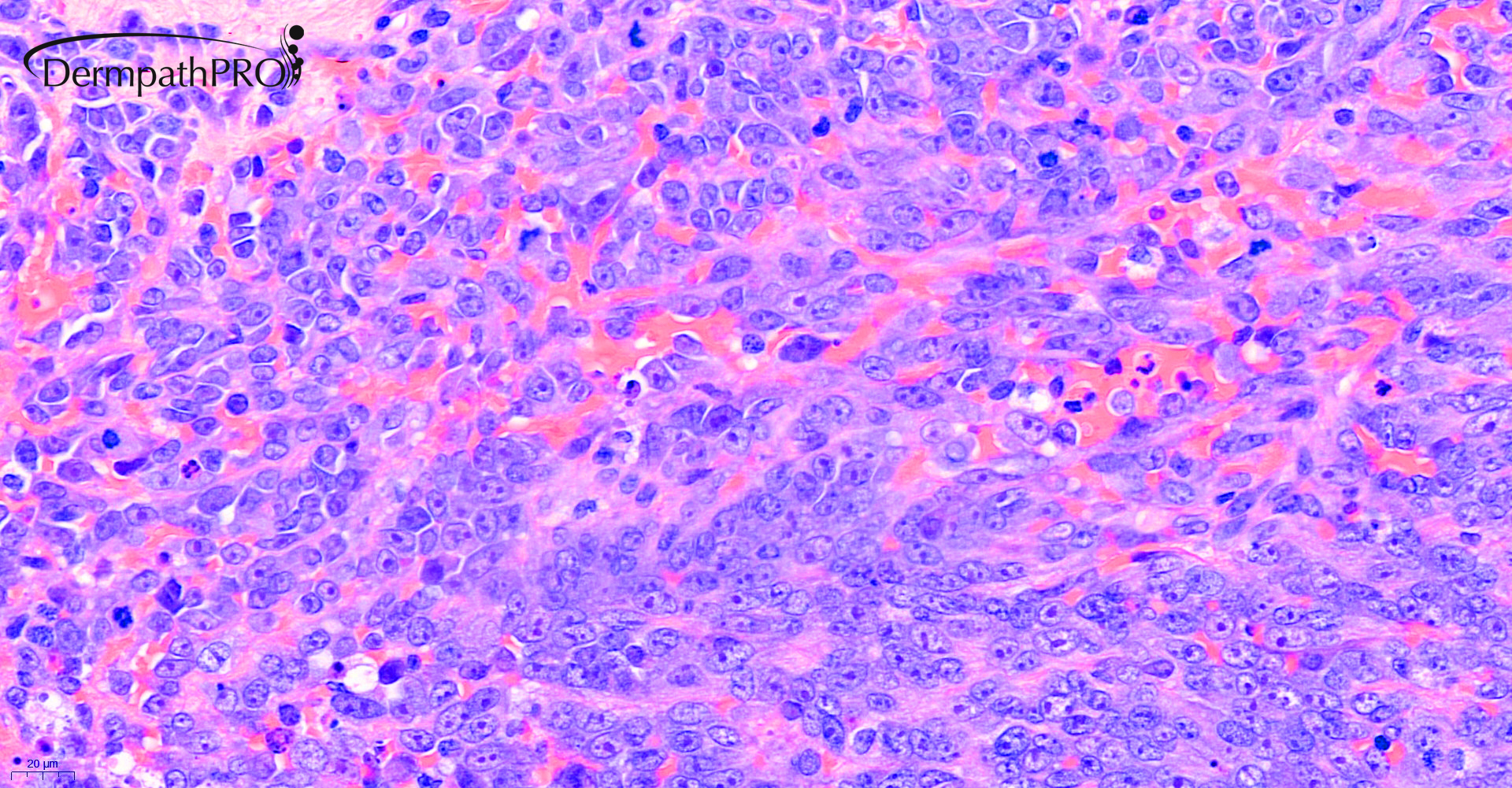
93F, Haemorrhagic blistering eruption left breast.


  • Like 1
  Report Record

User Feedback


Krishnakumar subramanian

Posted

agree with epithelioid AS

Share this comment


Link to comment
share_externally

Meenakshi Batrani

Posted

Agree with epithelioid angiosarcoma

Share this comment


Link to comment
share_externally

Dr. Mona Abdel-Halim

Posted

First thought epithelioid angiosarc

Share this comment


Link to comment
share_externally

Richard Logan

Posted

Any previous radiotherapy Saleem?

Share this comment


Link to comment
share_externally

Saleem Taibjee

Posted

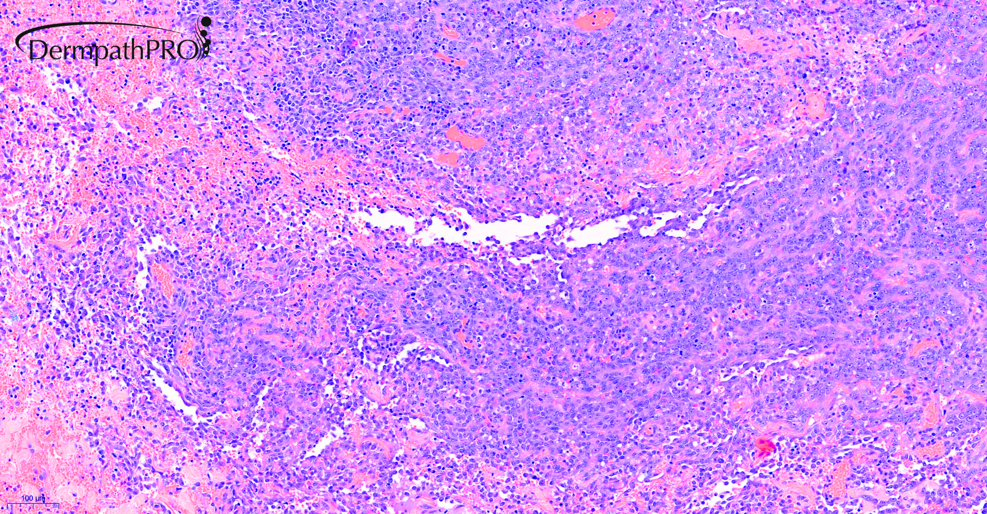
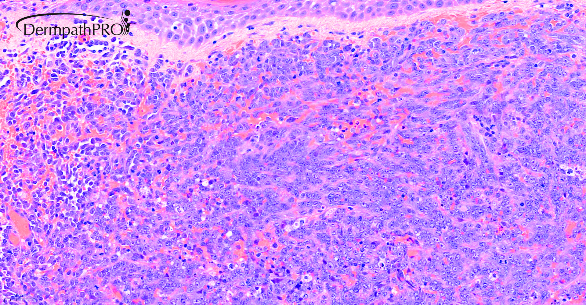
This is a recent case. Yes, correct, the patient did indeed receive prior radiotherapy for breast cancer, and this is an epithelioid variant of angiosarcoma.

Interestingly, the patient had a previous biopsy which I also reported, which was non-diagnostic (even after review). It's not the first time I have come across this scenario of a non-diagnostic first biopsy in angiosarcoma, so sampling/biopsy site does seem to be a pitfall, and it may best to advise a repeat biopsy in such non-diagnostic cases when the clinician queries angiosarcoma.

Also be aware of cases in which iron/haemosiderin can be a subtle clue.

In the present case in the 3rd image above there is a further clue with vasoformative areas (see close up below). Sometimes these may be at the periphery of the biopsy only.

BW

Saleem
02890_40.0x 2Ab.jpg


Share this comment


Link to comment
share_externally

Richard Logan

Posted

Was the earlier non-diagnostic biopsy too superficial?  Perhaps the epithelioid element of the tumour tends to be deeper in the dermis, with sometimes only subtle, easily missed vascular alterations superficially.


Inadequate superficial "shave" biopsies is one of my bugbears!

Share this comment


Link to comment
share_externally

Saleem Taibjee

Posted

Yes, precisely, Richard. The biopsy was performed by a breast surgeon. The paradox is that the general surgeon is often more sparing with a skin biopsy compared to the dermatologist.

Share this comment


Link to comment
share_externally


Join the conversation

You can post now and register later. If you have an account, sign in now to post with your account.

Guest
Add a comment...

×   Pasted as rich text.   Paste as plain text instead

  Only 75 emoji are allowed.

×   Your link has been automatically embedded.   Display as a link instead

×   Your previous content has been restored.   Clear editor

×   You cannot paste images directly. Upload or insert images from URL.

Loading...

×
×
  • Create New...