Jump to content
In this section we have spot diagnoses posted on a daily basis since June 2010, now over 4000! You can review the archived cases and read the suggested diagnoses by users and the final comment by the contributors.
Case are uploaded each week day by 10 am UK time with the correct diagnosis will generally be posted at 8 pm UK time. Why not view the most recent spot diagnosis and proffer a diagnosis?

Case Number : Case 2597 - 19 June 2020 Posted By: Dr. Richard Carr

Please read the clinical history and view the images by clicking on them before you proffer your diagnosis.
Submitted Date :
   (0 reviews)

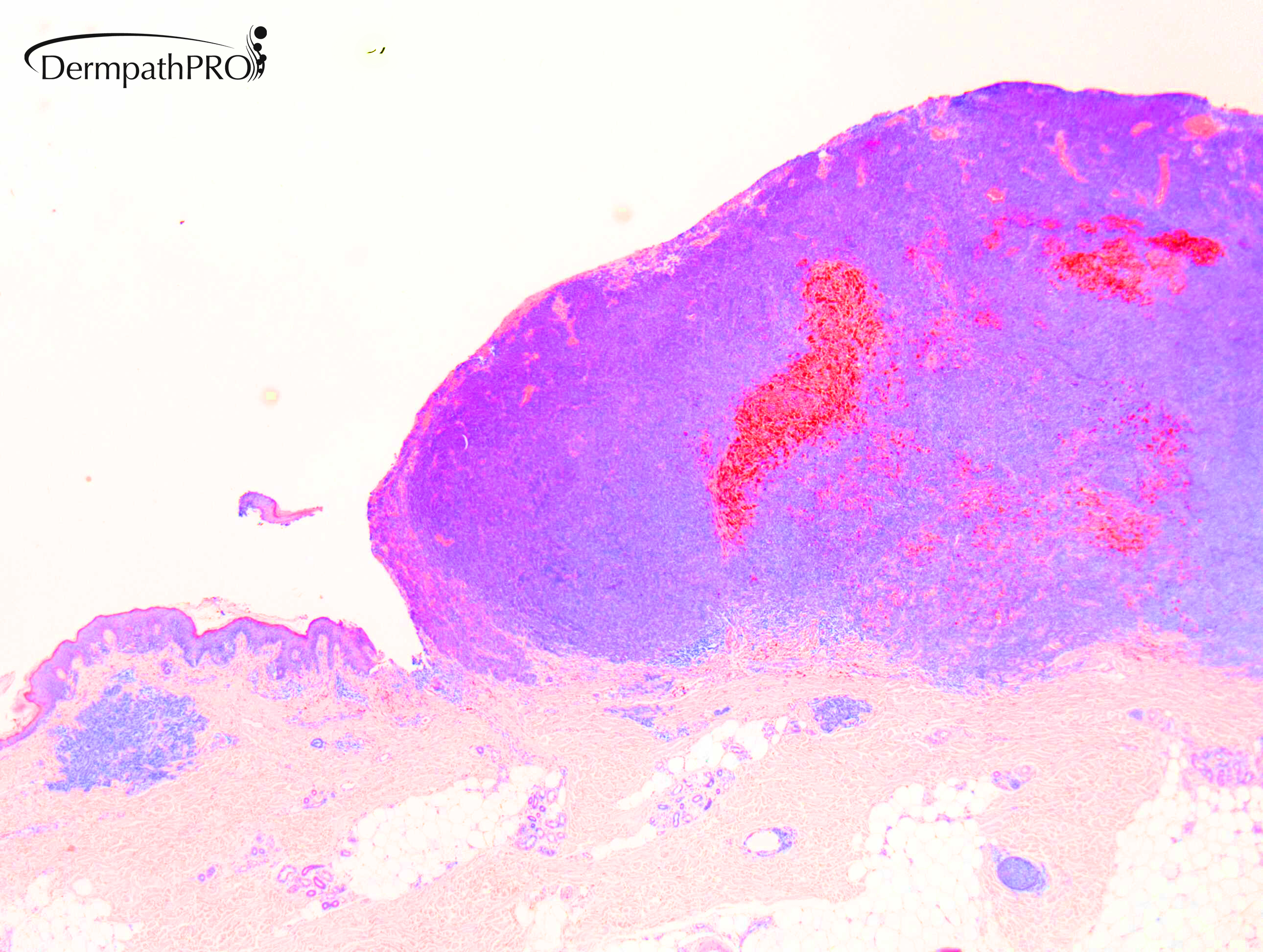
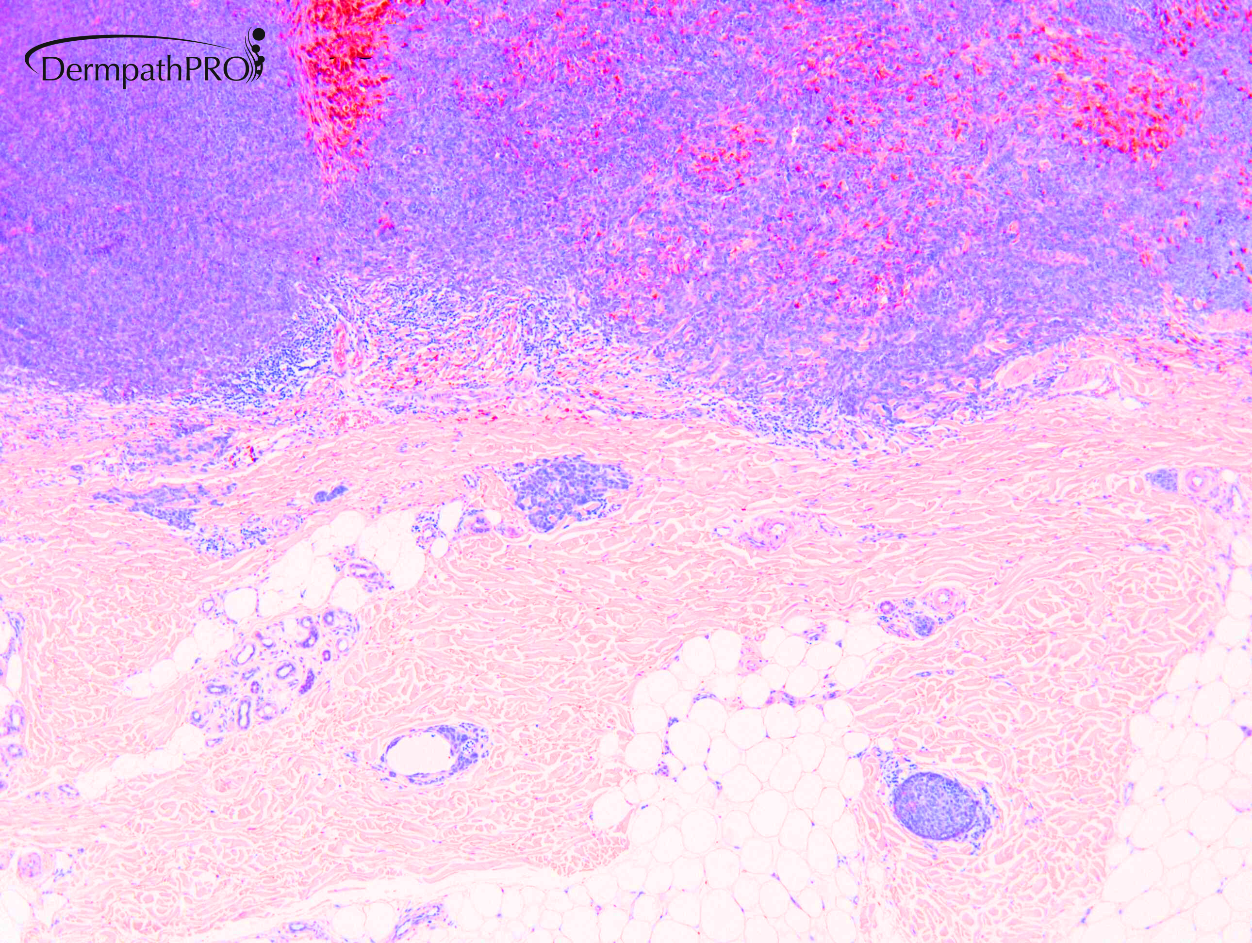
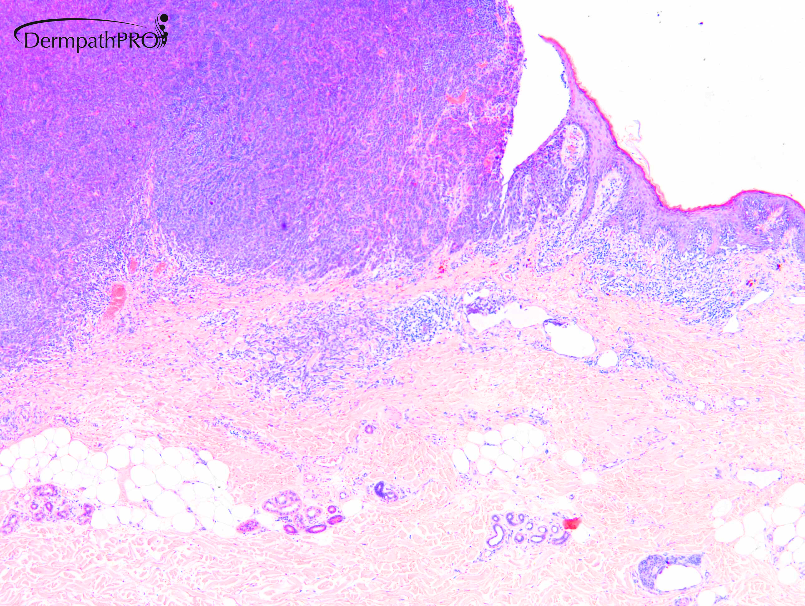
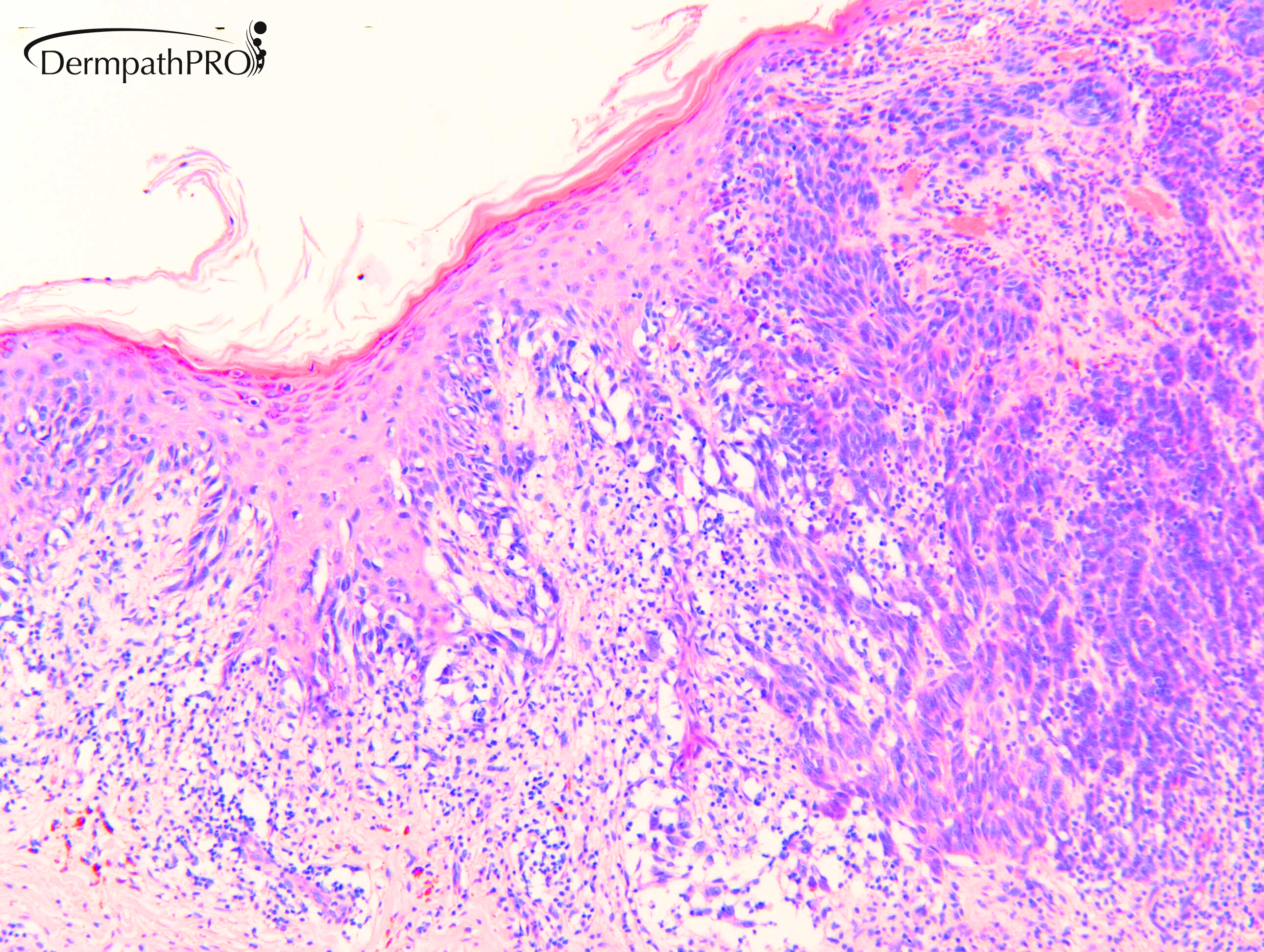
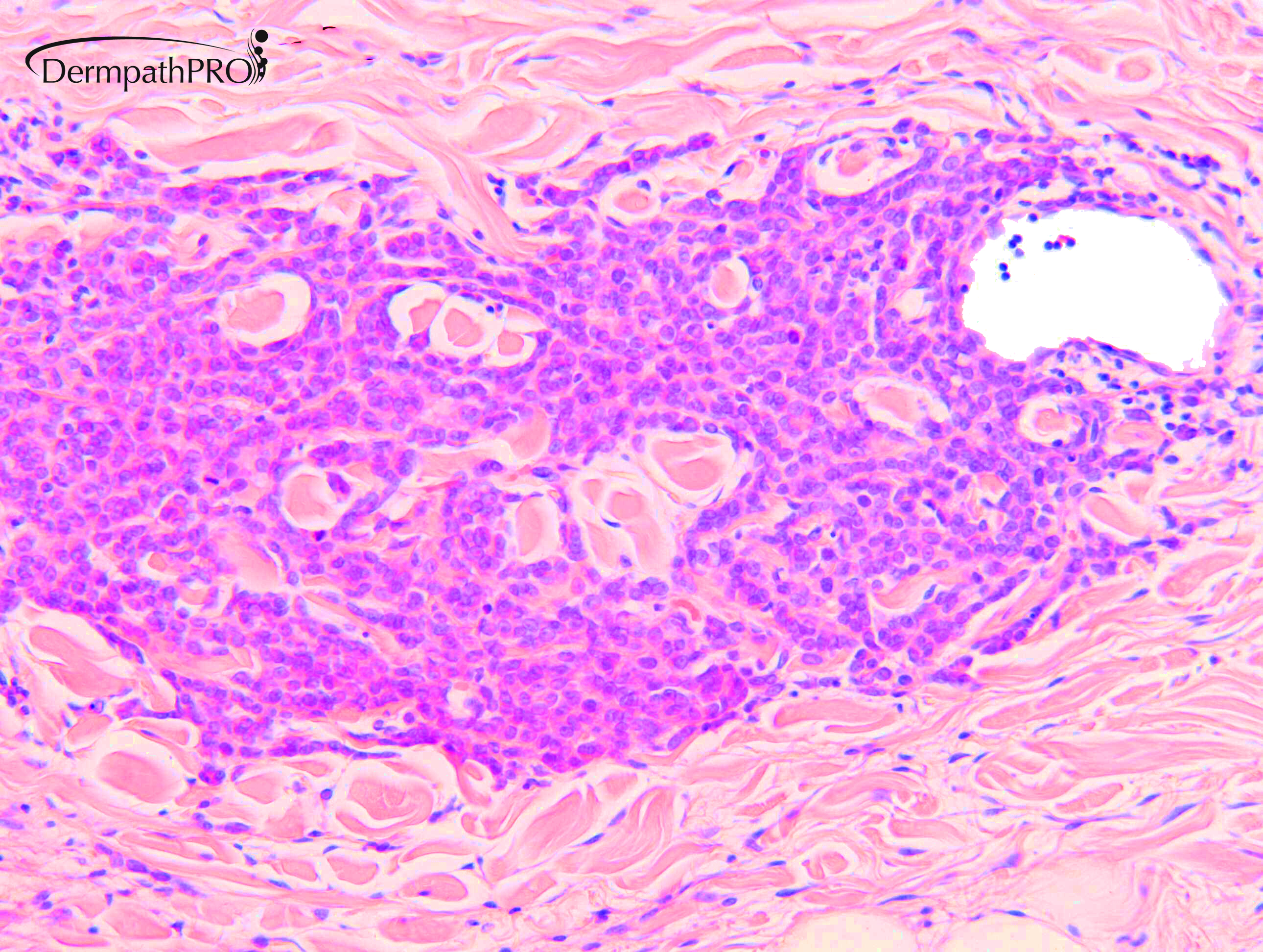
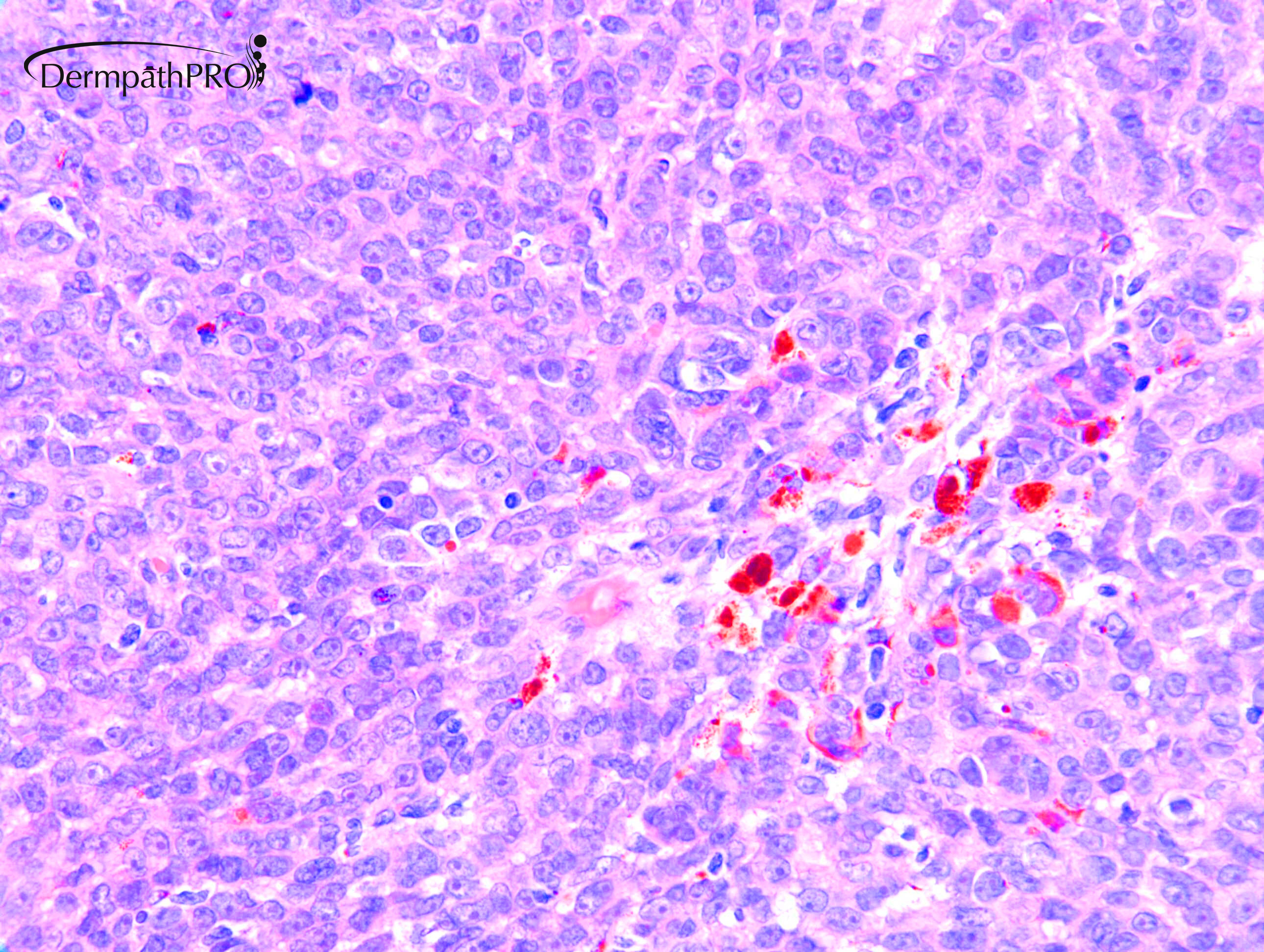
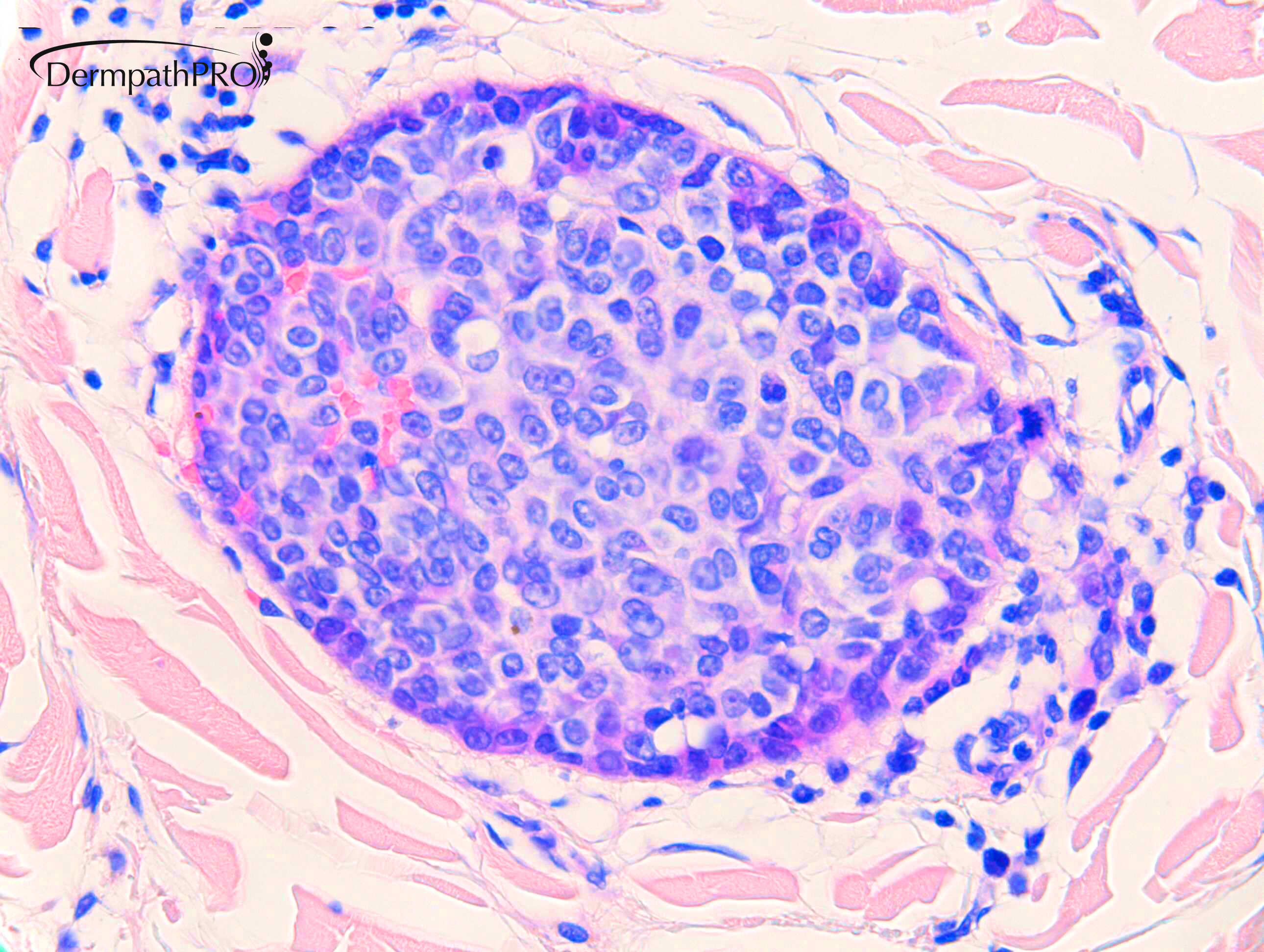
M70. Changed pigmented lesion upper arm.


  • Like 1
  Report Record

User Feedback


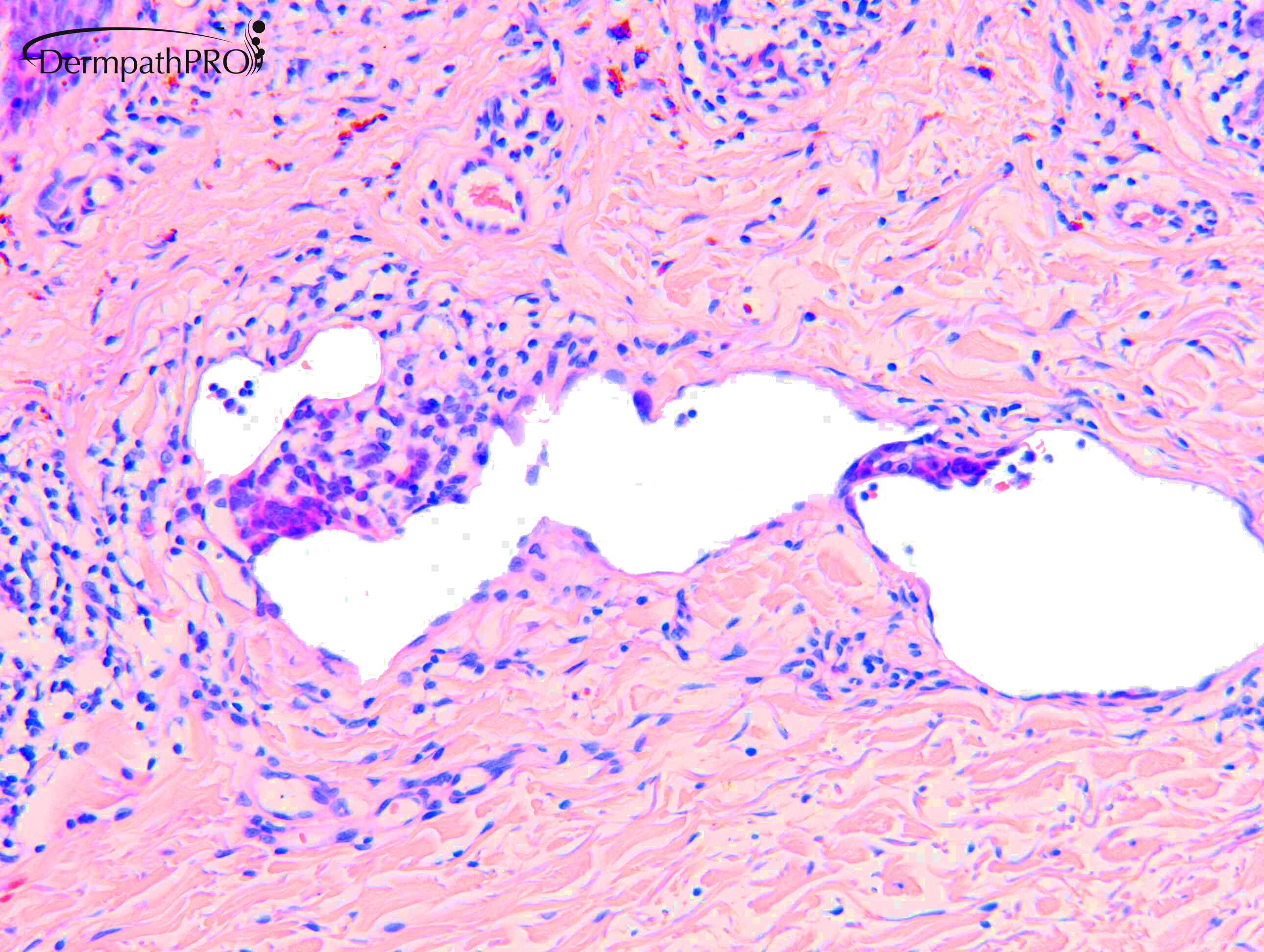
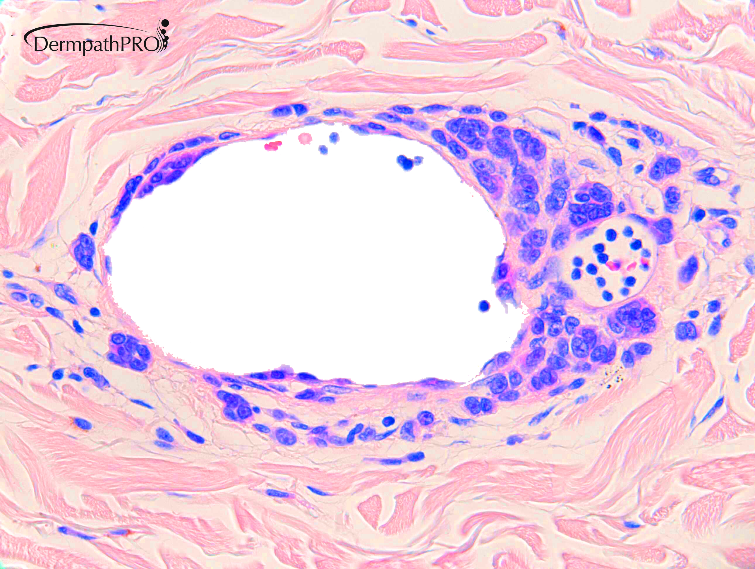
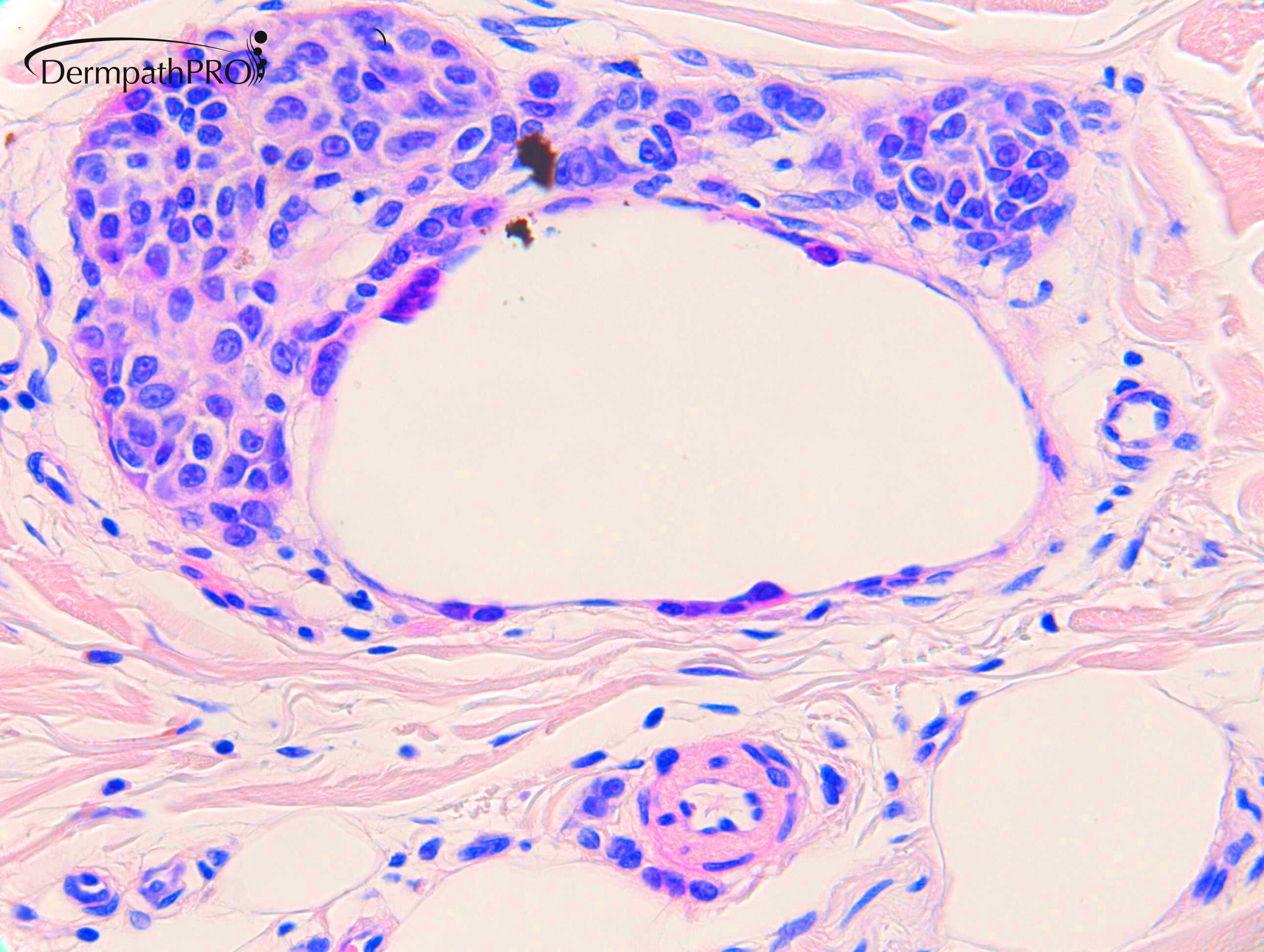
Nodular melanoma, with satellitosis and angiolymphatic peritumoral infiltration. 

Share this comment


Link to comment
share_externally

Dr. Richard Carr

Posted

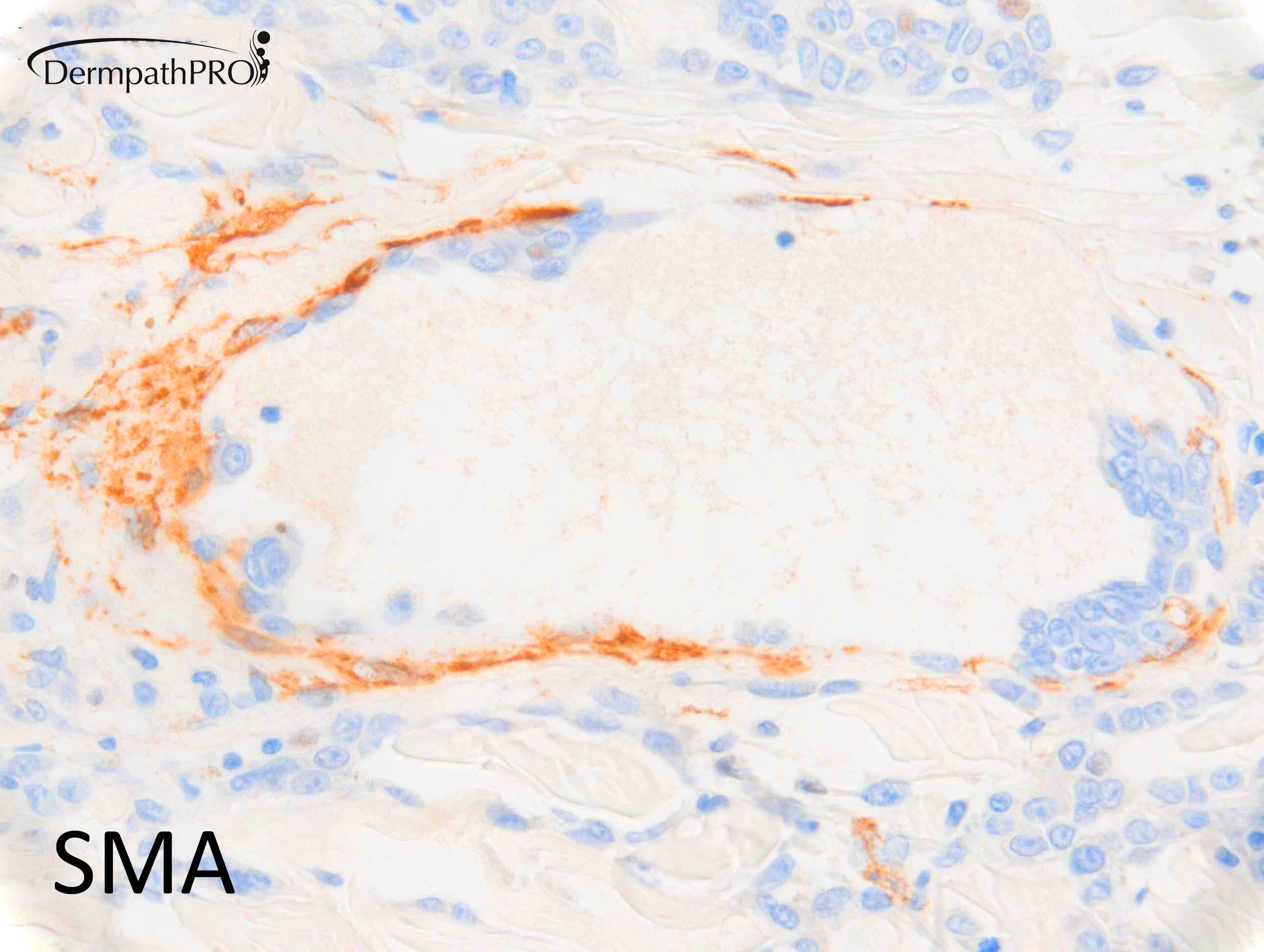
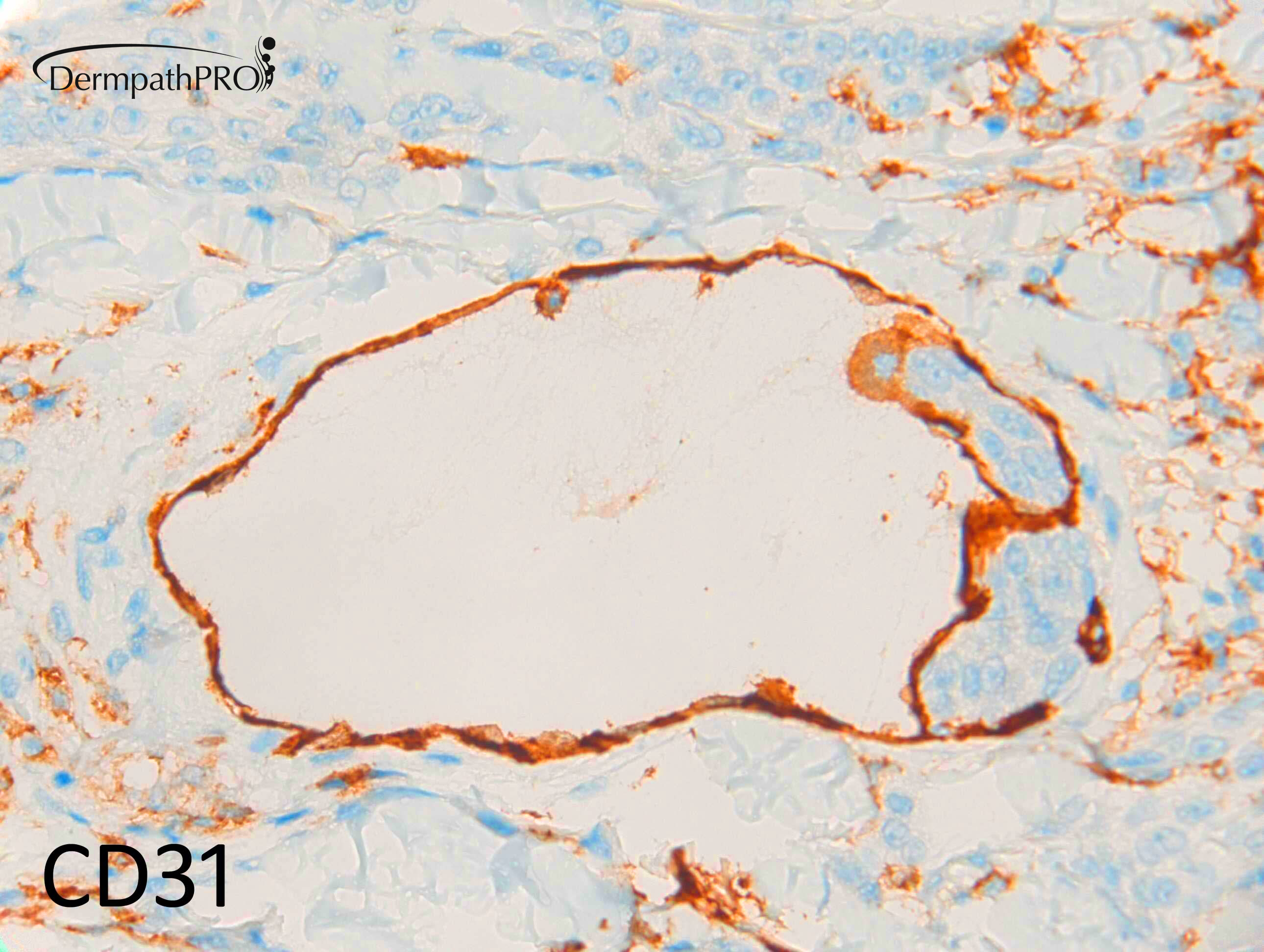
Okay this is an example of as superficial spreading (nodular overgrowth) melanoma with "extravascular migratory metastasis" which Prof. Raymond Barnhill and colleagues are actively studying. I have to admit it is the first time I've seen a case like this where the melanoma cells appear to be tropic for the vessels but are clearly located outside the endothelium (abluminal), possibly inside the muscle / pericytic cells. It has also been referred to as pericytic mimicry. Prof. Barnhill and colleagues have done some interesting cell culture studies showing melanoma cells moving along the outer aspect of small vessels. This melanoma is of a poor prognosis based on the depth, mitotic rate etc and I suppose the EVMM pattern might also confer a greater risk for loco-regional and distant metastasis.

Angiotropism, Pericytic Mimicry and Extravascular Migratory Metastasis in Melanoma: An Alternative to Intravascular Cancer Dissemination
Claire Lugassy, Sohila Zadran, Laurent A. Bentolila, Madhuri Wadehra, Roshini Prakash, S. Thomas Carmichael, Hynda K. Kleinman, Bruno Péault, Lionel Larue & Raymond L. Barnhill 
Cancer Microenvironment volume 7, pages139–152(2014)

For more than 15 years, angiotropism in melanoma has been emphasized as a marker of extravascular migration of tumor cells along the abluminal vascular surface, unveiling an alternative mechanism of tumor spread distinct from intravascular dissemination. This mechanism has been termed extravascular migratory metastasis (EVMM). During EVMM, angiotropic tumor cells migrate in a ‘pericytic-like’ manner (pericytic mimicry) along the external surfaces of vascular channels, without intravasation. Through this pathway, melanoma cells may spread to nearby or more distant sites. Angiotropism is a prognostic factor predicting risk for metastasis in human melanoma, and a marker of EVMM in several experimental models. Importantly, analogies of EVMM and pericytic mimicry include neural crest cell migration, vasculogenesis and angiogenesis, and recent studies have suggested that the interaction between melanoma cells and the abluminal vascular surface induce differential expression of genes reminiscent of cancer migration and embryonic/stem cell state transitions. A recent work revealed that repetitive UV exposure of primary cutaneous melanomas in a genetically engineered mouse model promotes metastatic progression via angiotropism and migration along the abluminal vascular surface. Finally, recent data using imaging of melanoma cells in a murine model have shown the progression of tumor cells along the vascular surfaces. Taken together, these data provide support for the biological phenomenon of angiotropism and EVMM, which may open promising new strategies for reducing or preventing melanoma metastasis.

Share this comment


Link to comment
share_externally

Dr. Richard Carr

Posted

For you interest....

Dear Raymond,
We saw a nice case of an SSMM (M70 upper arm) with your extravascular migratory deposits. It’s an NRAS mutant case.
We were not sure whether this should be called “microsatellites” with the implication for LN disease.
Looks like the cells have insinuated between the muscle layer of the thin lymphatics and the endothelium.
Warm Regards
Richard

Dear Richard,
This is a fantastic case Richard!  Angiotropism and microscopic satellites due to EV migration.  I wonder if this is linked to the NRAS mutation?   
I wonder if you could kindly send the tissue block as we are developing special IHC markers for this phenomenon.  Could you please send this H&E slide as well?  We can scan and return it to you.  
Many thanks for sharing this Richard!!!
Kindest regards,
Ray

Share this comment


Link to comment
share_externally

Richard Logan

Posted

Dear Richard,

Can I ask you to clarify your use of the description "Superficial spreading .... with nodular overgrowth" in this case?  Presumably this is because there is evidence of SSMM at the left edge of the specimen.  Of course clinically, nodular MM completely trumps SSMM.  As a clinician I was somewhat perplexed when SSMM was used in a histological report of a case like this,

Share this comment


Link to comment
share_externally

Dr. Richard Carr

Posted

Thanks Richard L,

Nodular melanoma strictly speaking don't have an SSMM like component (as in this case). It's the individual prognostic features that are important (mitotic rate, thickness etc) that matter in practice.  However....

For interest I've put the prognostic indices into the Melanoma Institute of Australia's sentinel LN predictor algorhym (it's wonderful and I'd suggest trying it).

Assuming: M70, mitotic rate = 3, Breslow: 3.8mm

for SSMM (without LVI): 34% risk of +ve SLNBx

for SSMM (with LVI): 69% risk of +ve SLNBx

for Nodular MM (without LVI): 24% risk of +ve SLNBx

for Nodular MM (with LVI): 58% risk of +ve SLNBx 

Perhaps this is NOT the result you were expecting?

I'd suggest using this indicator when you get melanoma reports, it's very easy to use and may help you decide on management.

https://www.melanomarisk.org.au/

Share this comment


Link to comment
share_externally

Richard Logan

Posted

Thank you Richard for your detailed response.  You're dead right.  It is a surprising statistic!

Share this comment


Link to comment
share_externally

Krishnakumar subramanian

Posted

Thanks a lot sir for these great points

Share this comment


Link to comment
share_externally

Meenakshi Batrani

Posted

19 hours ago, Dr. Richard Carr said:

Okay this is an example of as superficial spreading (nodular overgrowth) melanoma with "extravascular migratory metastasis" which Prof. Raymond Barnhill and colleagues are actively studying. I have to admit it is the first time I've seen a case like this where the melanoma cells appear to be tropic for the vessels but are clearly located outside the endothelium (abluminal), possibly inside the muscle / pericytic cells. It has also been referred to as pericytic mimicry. Prof. Barnhill and colleagues have done some interesting cell culture studies showing melanoma cells moving along the outer aspect of small vessels. This melanoma is of a poor prognosis based on the depth, mitotic rate etc and I suppose the EVMM pattern might also confer a greater risk for loco-regional and distant metastasis.

Angiotropism, Pericytic Mimicry and Extravascular Migratory Metastasis in Melanoma: An Alternative to Intravascular Cancer Dissemination
Claire Lugassy, Sohila Zadran, Laurent A. Bentolila, Madhuri Wadehra, Roshini Prakash, S. Thomas Carmichael, Hynda K. Kleinman, Bruno Péault, Lionel Larue & Raymond L. Barnhill 
Cancer Microenvironment volume 7, pages139–152(2014)

For more than 15 years, angiotropism in melanoma has been emphasized as a marker of extravascular migration of tumor cells along the abluminal vascular surface, unveiling an alternative mechanism of tumor spread distinct from intravascular dissemination. This mechanism has been termed extravascular migratory metastasis (EVMM). During EVMM, angiotropic tumor cells migrate in a ‘pericytic-like’ manner (pericytic mimicry) along the external surfaces of vascular channels, without intravasation. Through this pathway, melanoma cells may spread to nearby or more distant sites. Angiotropism is a prognostic factor predicting risk for metastasis in human melanoma, and a marker of EVMM in several experimental models. Importantly, analogies of EVMM and pericytic mimicry include neural crest cell migration, vasculogenesis and angiogenesis, and recent studies have suggested that the interaction between melanoma cells and the abluminal vascular surface induce differential expression of genes reminiscent of cancer migration and embryonic/stem cell state transitions. A recent work revealed that repetitive UV exposure of primary cutaneous melanomas in a genetically engineered mouse model promotes metastatic progression via angiotropism and migration along the abluminal vascular surface. Finally, recent data using imaging of melanoma cells in a murine model have shown the progression of tumor cells along the vascular surfaces. Taken together, these data provide support for the biological phenomenon of angiotropism and EVMM, which may open promising new strategies for reducing or preventing melanoma metastasis.

Thank you Dr. Carr for expounding on this relatively new concept. 

Share this comment


Link to comment
share_externally


Join the conversation

You can post now and register later. If you have an account, sign in now to post with your account.

Guest
Add a comment...

×   Pasted as rich text.   Paste as plain text instead

  Only 75 emoji are allowed.

×   Your link has been automatically embedded.   Display as a link instead

×   Your previous content has been restored.   Clear editor

×   You cannot paste images directly. Upload or insert images from URL.

Loading...

×
×
  • Create New...