Jump to content
In this section we have spot diagnoses posted on a daily basis since June 2010, now over 4000! You can review the archived cases and read the suggested diagnoses by users and the final comment by the contributors.
Case are uploaded each week day by 10 am UK time with the correct diagnosis will generally be posted at 8 pm UK time. Why not view the most recent spot diagnosis and proffer a diagnosis?

Case Number : Case 2519 - 03 March 2020 Posted By: Uma Sundram

Please read the clinical history and view the images by clicking on them before you proffer your diagnosis.
Submitted Date :
   (0 reviews)

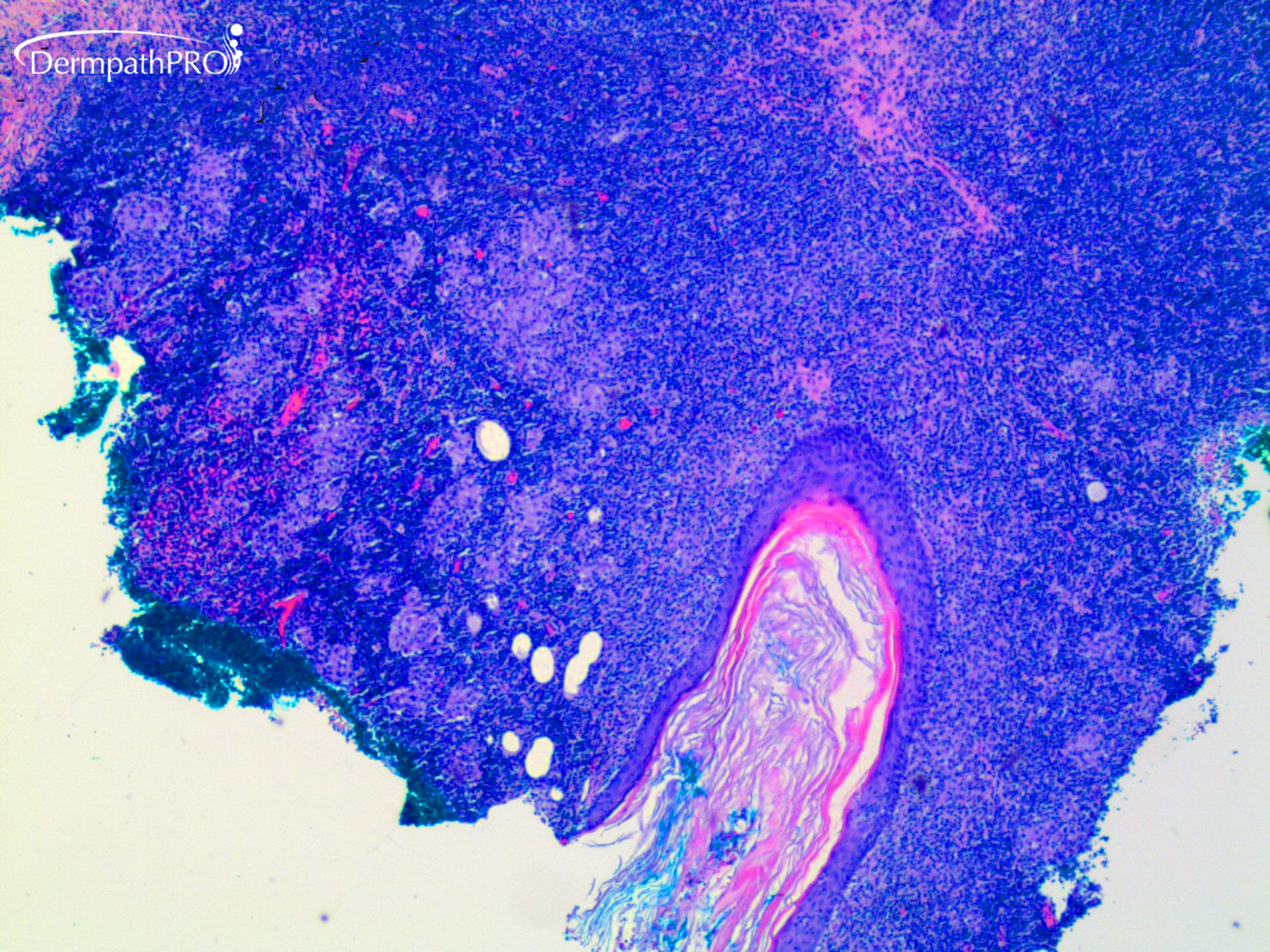
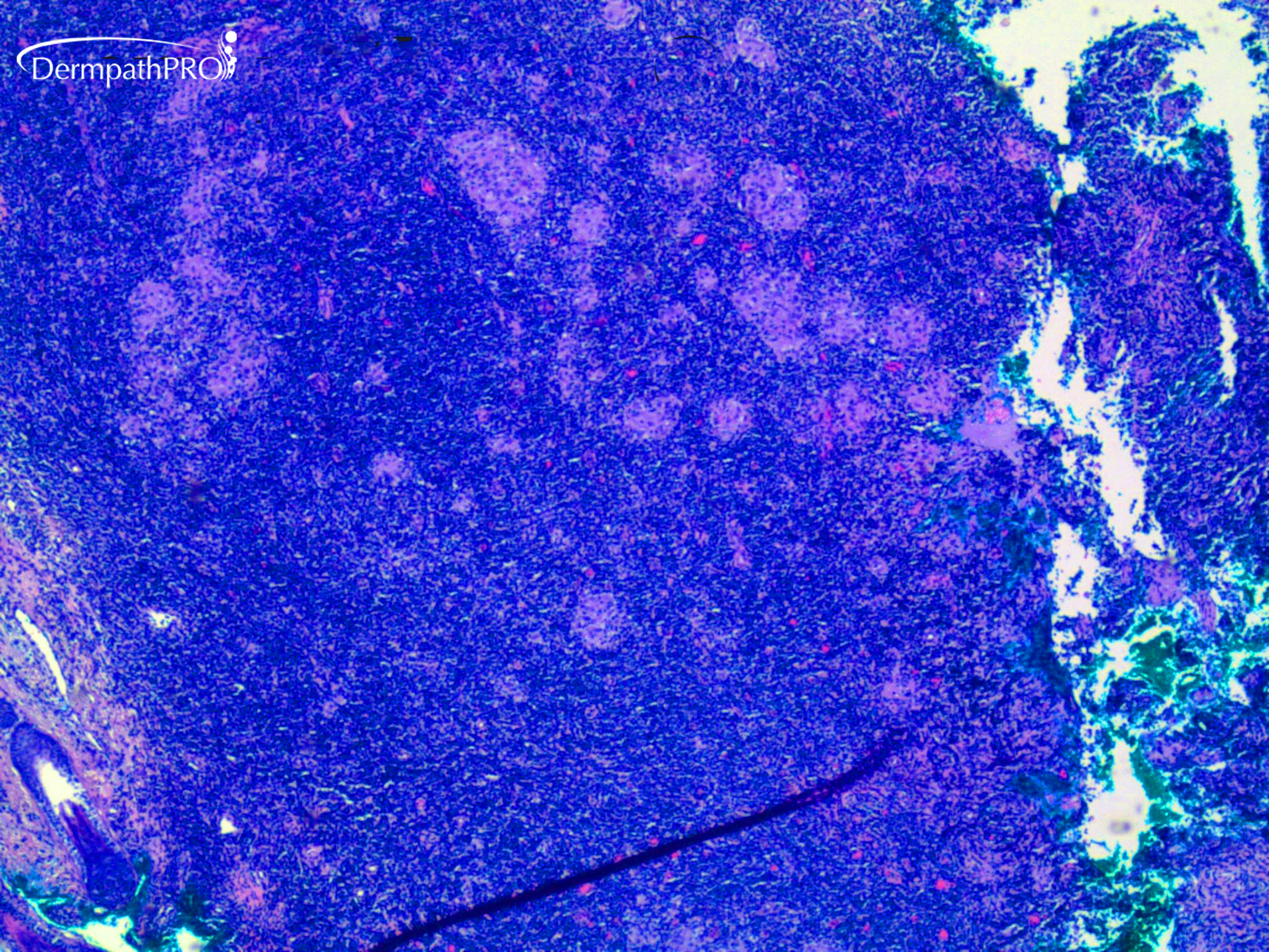
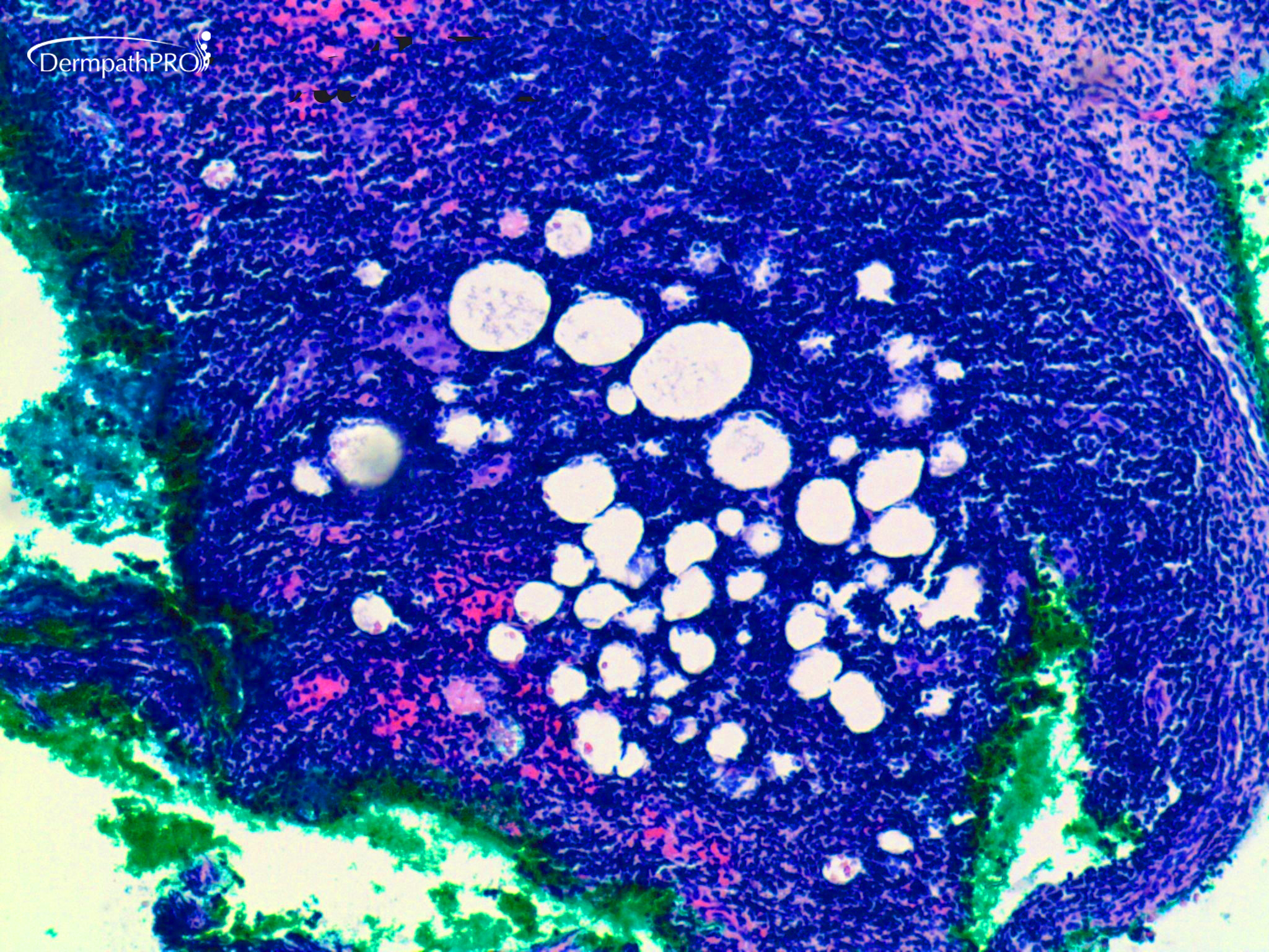
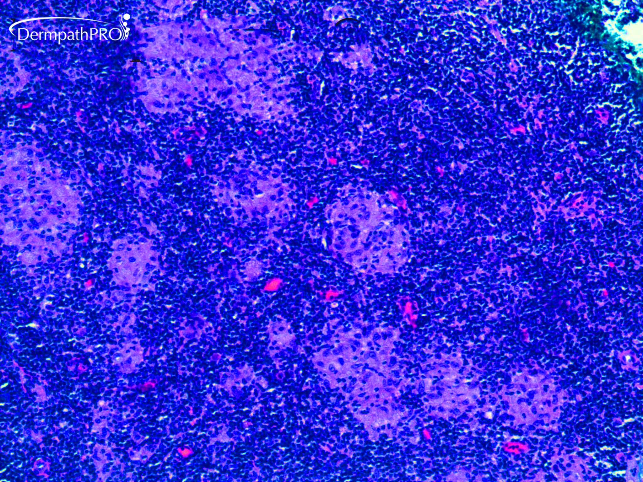
20 year old woman with enlarging lesion behind left ear.


  Report Record

User Feedback


Krishnakumar subramanian

Posted

granulomatous inflammation - no necrosis with dense collections of lymphocytes

Share this comment


Link to comment
share_externally

Krishnakumar subramanian

Posted

I was wondering whether we need to consider  lymphoepithelioid variant of peripheral T-cell lymphoma old term lenient lymphoma

I need PCR for MTB or IHC to know the clonality of the cells. please suggest

Share this comment


Link to comment
share_externally

Difficult. Granulomatous or lipogranulomatous lymphadenitis, with some toxoplasmosis-like finding ( but there aren’t hyper plastic lymphoid follicles). It needs CPC. 

Share this comment


Link to comment
share_externally

Dr. Mona Abdel-Halim

Posted

The lymphoid component is so dense and made me think like Krishnakumar in Lennert's lymphoma. Needs work up both for infectious causes as well as lymphoproliferative processes associated with granulomatous reaction. 

Share this comment


Link to comment
share_externally

Uma Sundram

Posted

I really liked all of your suggestions! It's very subtle, but the first picture has an epidermal inclusion cyst/dilated hair follicle in it. This is an exuberant cutaneous lymphoid hyperplasia in response to rupture. Although the lymphocytes are dense, they have many architectural features of a reactive process. Special stains are negative for organisms. Cultures are negative and T cell PCR is polyclonal.

Share this comment


Link to comment
share_externally


Join the conversation

You can post now and register later. If you have an account, sign in now to post with your account.

Guest
Add a comment...

×   Pasted as rich text.   Paste as plain text instead

  Only 75 emoji are allowed.

×   Your link has been automatically embedded.   Display as a link instead

×   Your previous content has been restored.   Clear editor

×   You cannot paste images directly. Upload or insert images from URL.

Loading...

×
×
  • Create New...