Jump to content
In this section we have spot diagnoses posted on a daily basis since June 2010, now over 4000! You can review the archived cases and read the suggested diagnoses by users and the final comment by the contributors.
Case are uploaded each week day by 10 am UK time with the correct diagnosis will generally be posted at 8 pm UK time. Why not view the most recent spot diagnosis and proffer a diagnosis?

Case Number : Case 2521 - 05 March 2020 Posted By: Saleem Taibjee

Please read the clinical history and view the images by clicking on them before you proffer your diagnosis.
Submitted Date :
   (0 reviews)

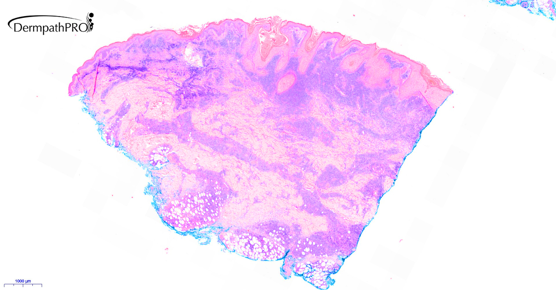
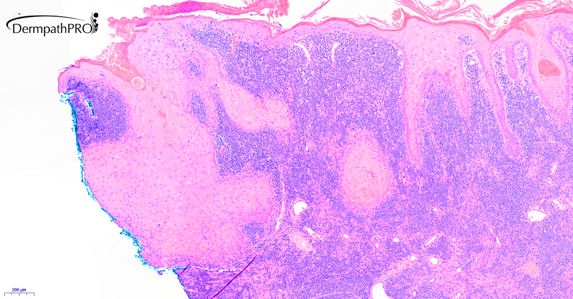
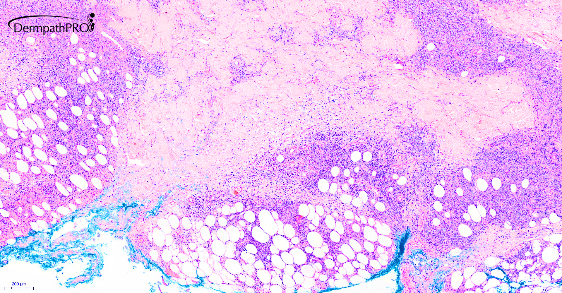
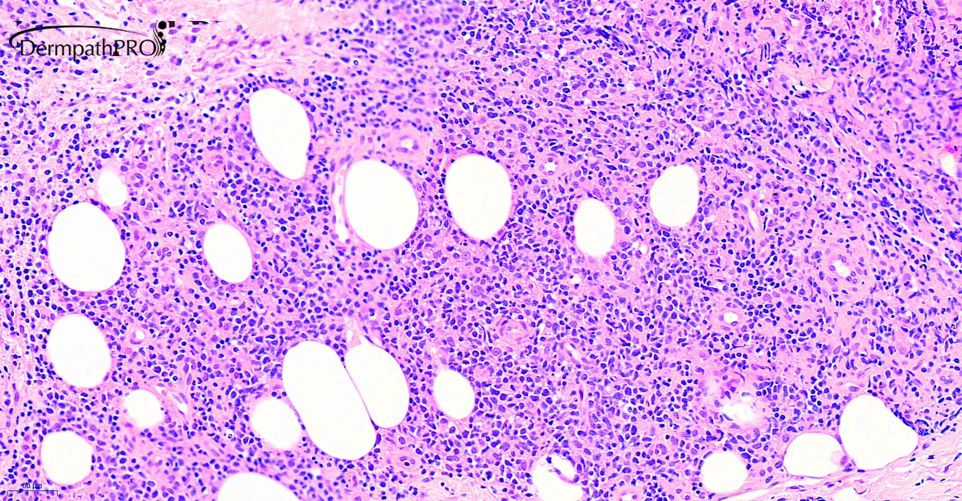
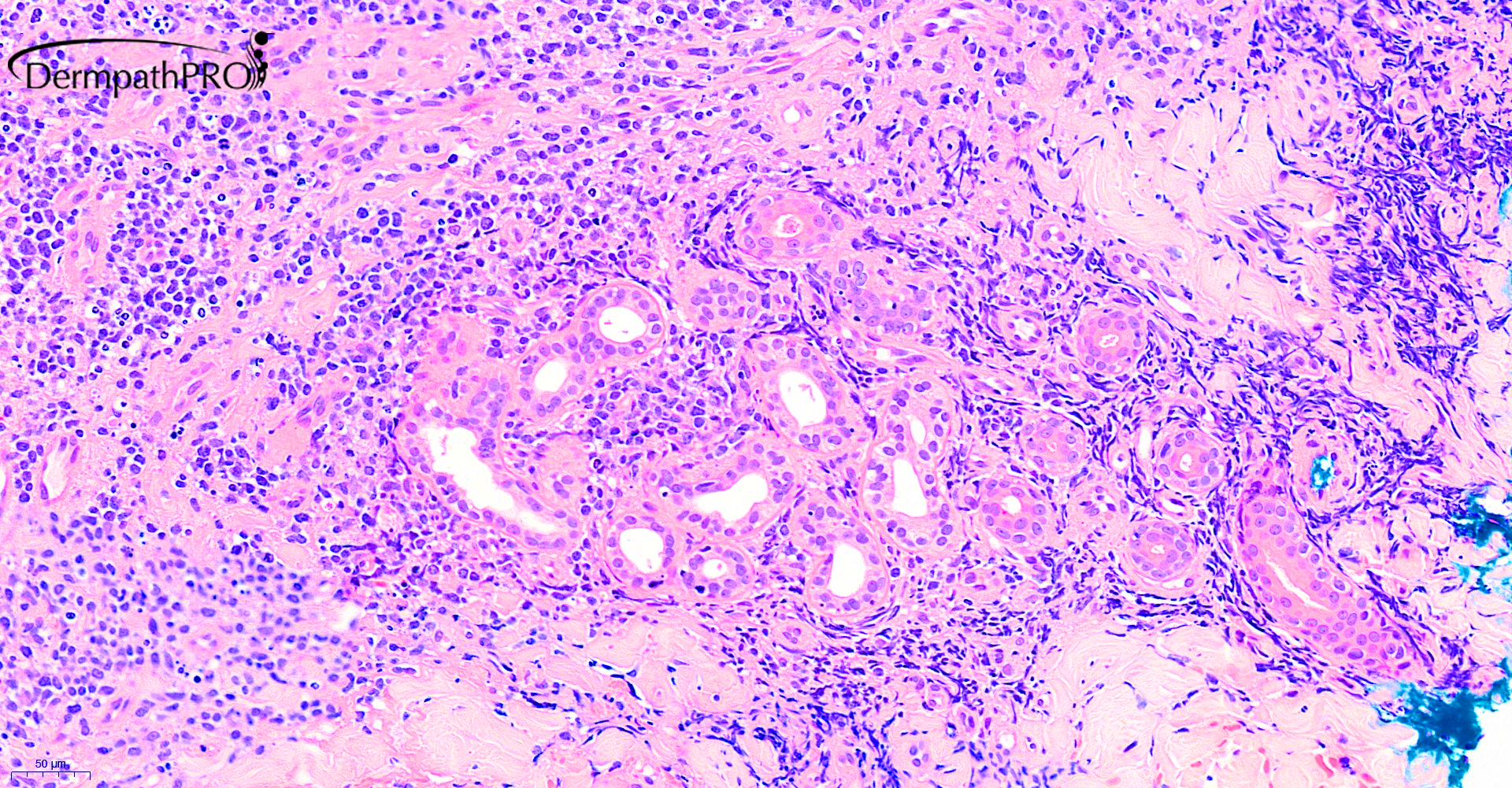
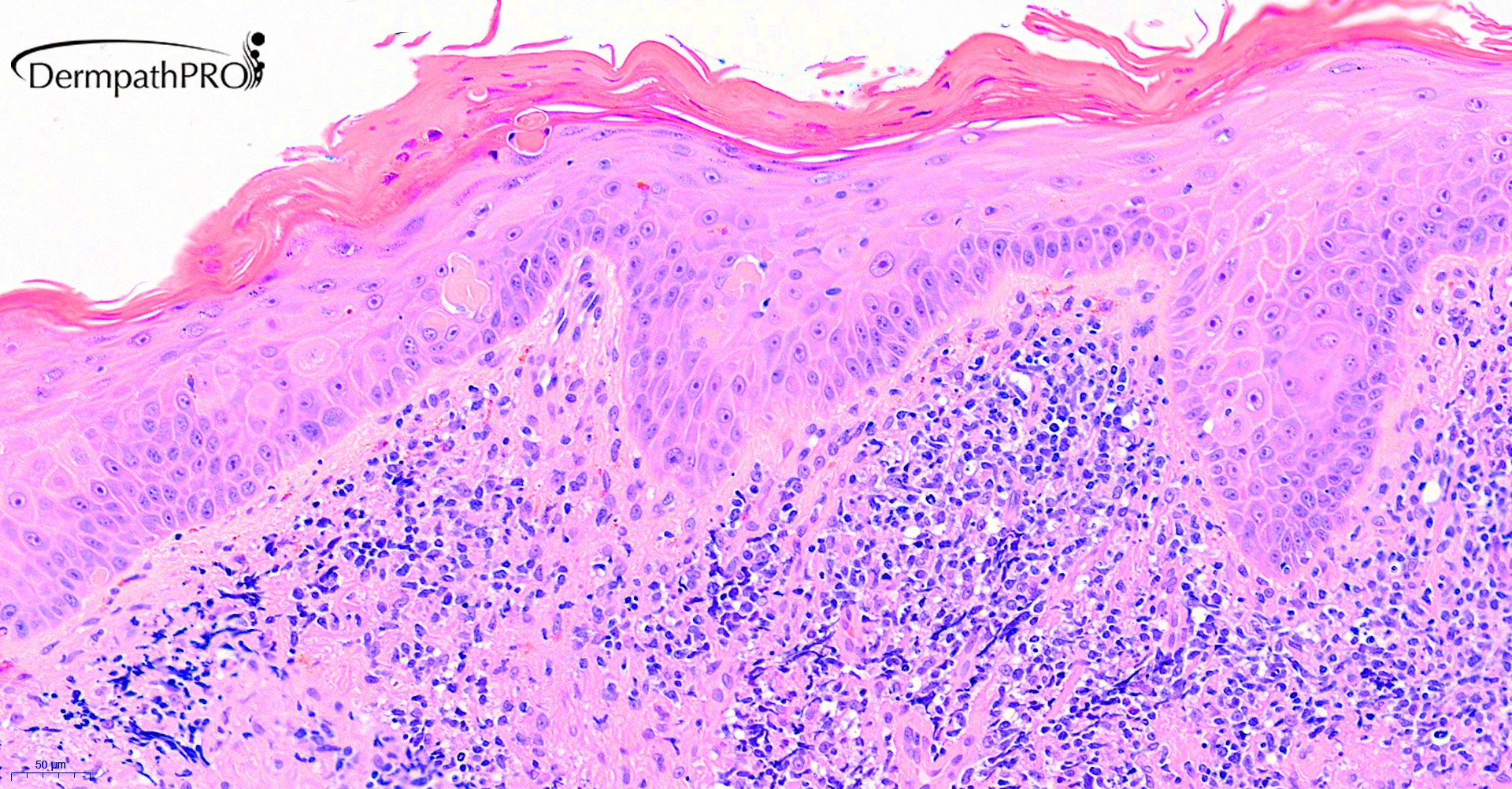
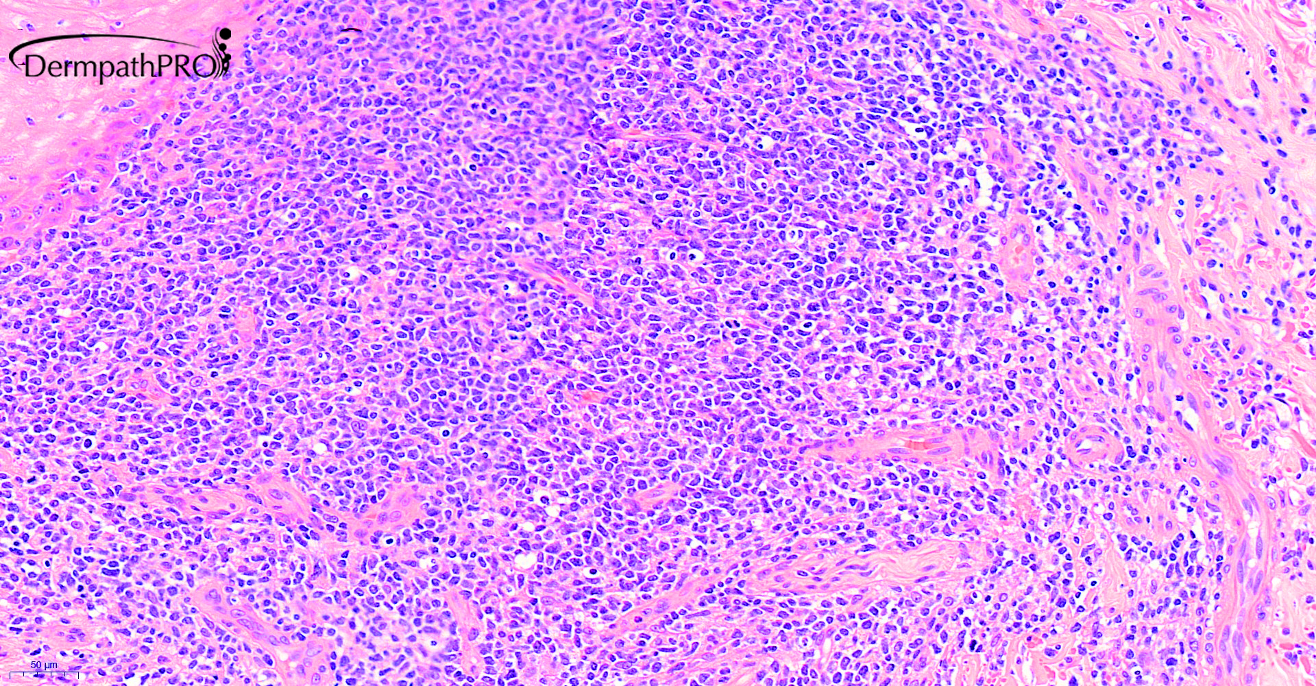
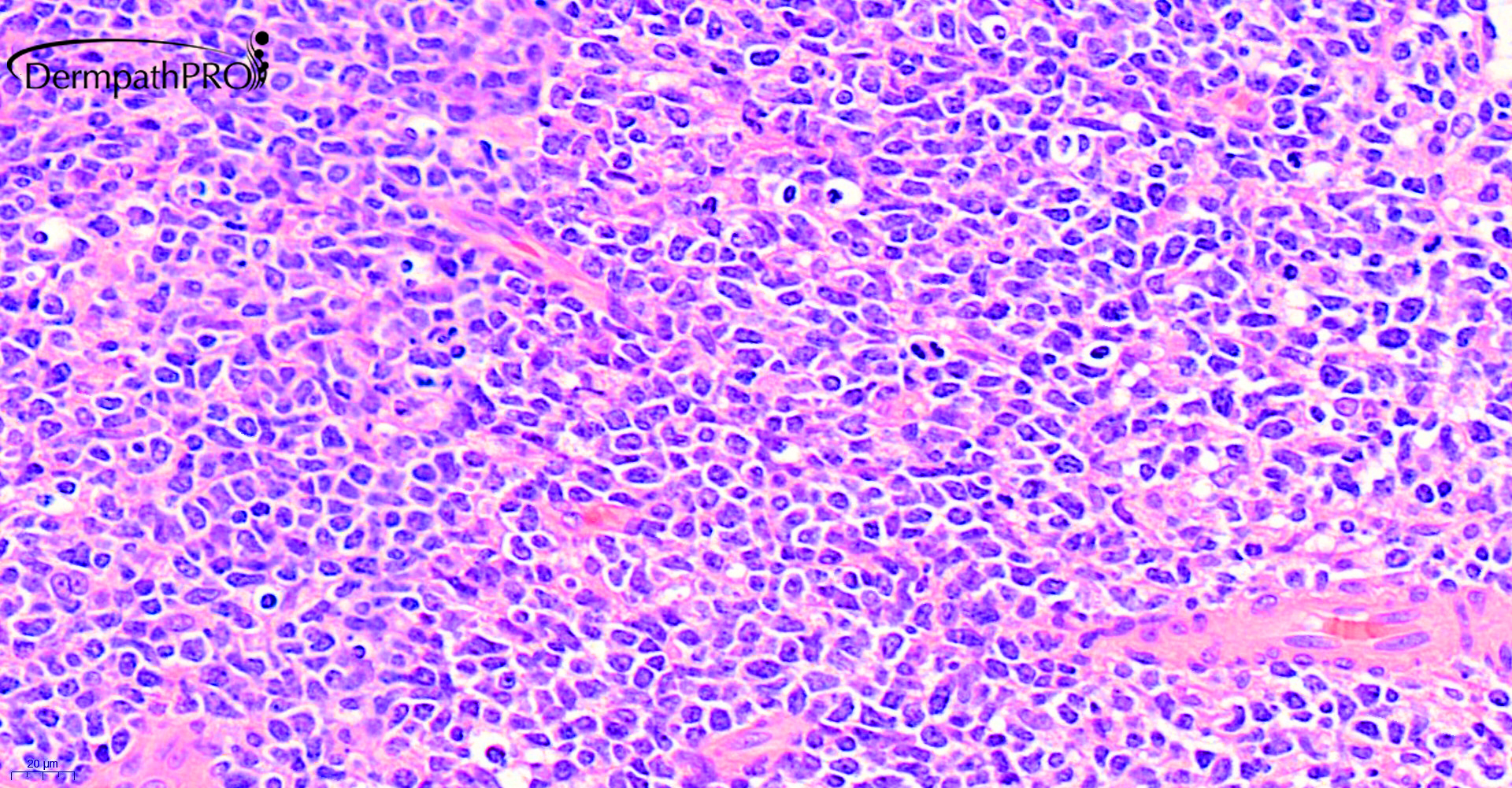
61M incisional biopsy left arm, unusual nodules and ulcers which may have followed on from injection of recreational drugs. Additional proptosis ?atypical infection ?other


  • Like 1
  Report Record

User Feedback


Last two pictures make me thjnk of Blastic Plasmocytoid Dendritic Cell Lymphoma. CD56? CD4? CD 123?

Share this comment


Link to comment
share_externally

Krishnakumar subramanian

Posted

ulcers, proptosis and dense infiltrate of atypical blast  like cells, some what a grenz zone

peripheral smear is needed urgently and IHC on the skin nodules to rule out leukemia/lymphoma/ BPDCN

Share this comment


Link to comment
share_externally

1 hour ago, Krishnakumar subramanian said:

ulcers, proptosis and dense infiltrate of atypical blast  like cells, some what a grenz zone

peripheral smear is needed urgently and IHC on the skin nodules to rule out leukemia/lymphoma/ BPDCN

Agree!

Share this comment


Link to comment
share_externally

Dr. Mona Abdel-Halim

Posted

Yes, complete IHC work up to verify leukemic/lymphoproliferative disorders

Share this comment


Link to comment
share_externally

Eman El-Nabarawy

Posted

On 05/03/2020 at 14:41, Krishnakumar subramanian said:

ulcers, proptosis and dense infiltrate of atypical blast  like cells, some what a grenz zone

peripheral smear is needed urgently and IHC on the skin nodules to rule out leukemia/lymphoma/ BPDCN

Agree.

Share this comment


Link to comment
share_externally

Krishnakumar subramanian

Posted

sir what is the final diagnosis

Share this comment


Link to comment
share_externally

Victor Delgado

Posted

Lymphoproliferative disorder. IHC is needed

Share this comment


Link to comment
share_externally

Saleem Taibjee

Posted

This was a recent case of extranodal NK/T cell lymphoma, nasal type. I think this is a truly rare skin presentation, only the second case I have personally encountered. There are a few learning points:

1. Proptosis is a recognised presentation (extra-nasal site of predilection) which I discovered after reviewing the literature.

2. In the images I tried to highlight the pseudoepitheliomatous hyperplasia. I was interested to discover that in the excellent review of pseudoepitheliomatous hyperplasia by Zayour and Lazova in Am J Dermpathol 2011;33:112-126, which includes a comprehensive table of possible causes, this particular lymphoma is listed as the first under lymphoproliferative causes (even above anaplastic large cell lymphoma). I had seemed to have previously missed this feature as a good diagnostic clue to this particular lymphoma when presenting in the skin.

3. Angiocentricity/angioinvasion is not present in all cases, and was not a conspicuous feature in this case.

4. The case also highlights the need for a good/efficient (cost-effective) screening immunohistochemistry panel for patients presenting with ulcerative nodules and a short history, when of course mycosis fungoides becomes less likely. An excellent review article is Gru AA, et al. A systematic approach to the cutaneous lymphoid infiltrates. Archives of Pathology & Laboratory Medicine 2019; 143:958-979 in which the authors present a nice algorithm. I might suggest the following: CD56 (which really assisted me in this case), cytotoxic markers (e.g. TIA-1, Granzyme or Perforin), CD2, EBER, CD30, CD123, beta-F1 as a good starting point. This will avoid breaking the bank, but provide a good starting point for working through the differential diagnosis.

In this case CD56 and EBER are shown below, also positive was granzyme, TIA-1. CD3 was also positive (membranous), reflecting that some cases also express T-cell markers, although PCR was not performed in our case.

BW

Saleem

20190514_3058.jpg

04417_20.0x CD56 labelled.jpg

04417_20.0x EBER labelled.jpg


Share this comment


Link to comment
share_externally


Join the conversation

You can post now and register later. If you have an account, sign in now to post with your account.

Guest
Add a comment...

×   Pasted as rich text.   Paste as plain text instead

  Only 75 emoji are allowed.

×   Your link has been automatically embedded.   Display as a link instead

×   Your previous content has been restored.   Clear editor

×   You cannot paste images directly. Upload or insert images from URL.

Loading...

×
×
  • Create New...