Jump to content
In this section we have spot diagnoses posted on a daily basis since June 2010, now over 4000! You can review the archived cases and read the suggested diagnoses by users and the final comment by the contributors.
Case are uploaded each week day by 10 am UK time with the correct diagnosis will generally be posted at 8 pm UK time. Why not view the most recent spot diagnosis and proffer a diagnosis?

Case Number : Case 2531 - 19 March 2020 Posted By: Saleem Taibjee

Please read the clinical history and view the images by clicking on them before you proffer your diagnosis.
Submitted Date :
   (0 reviews)

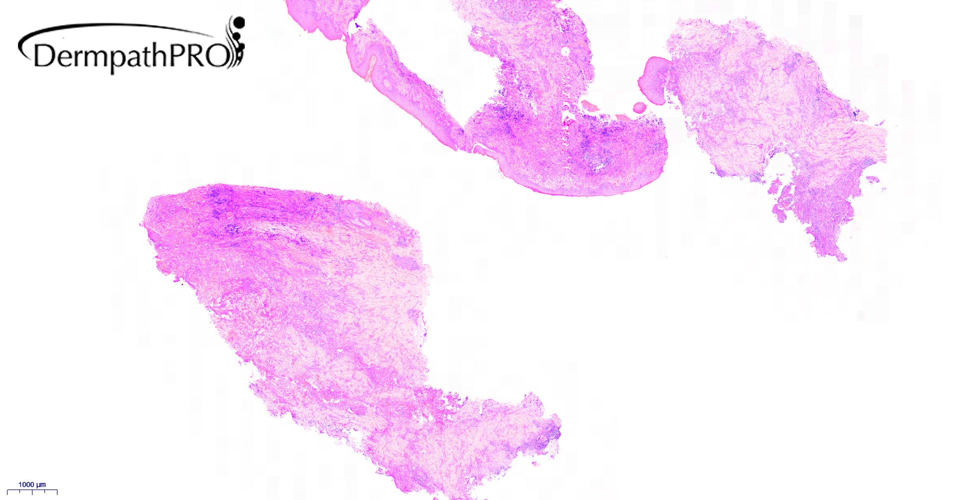
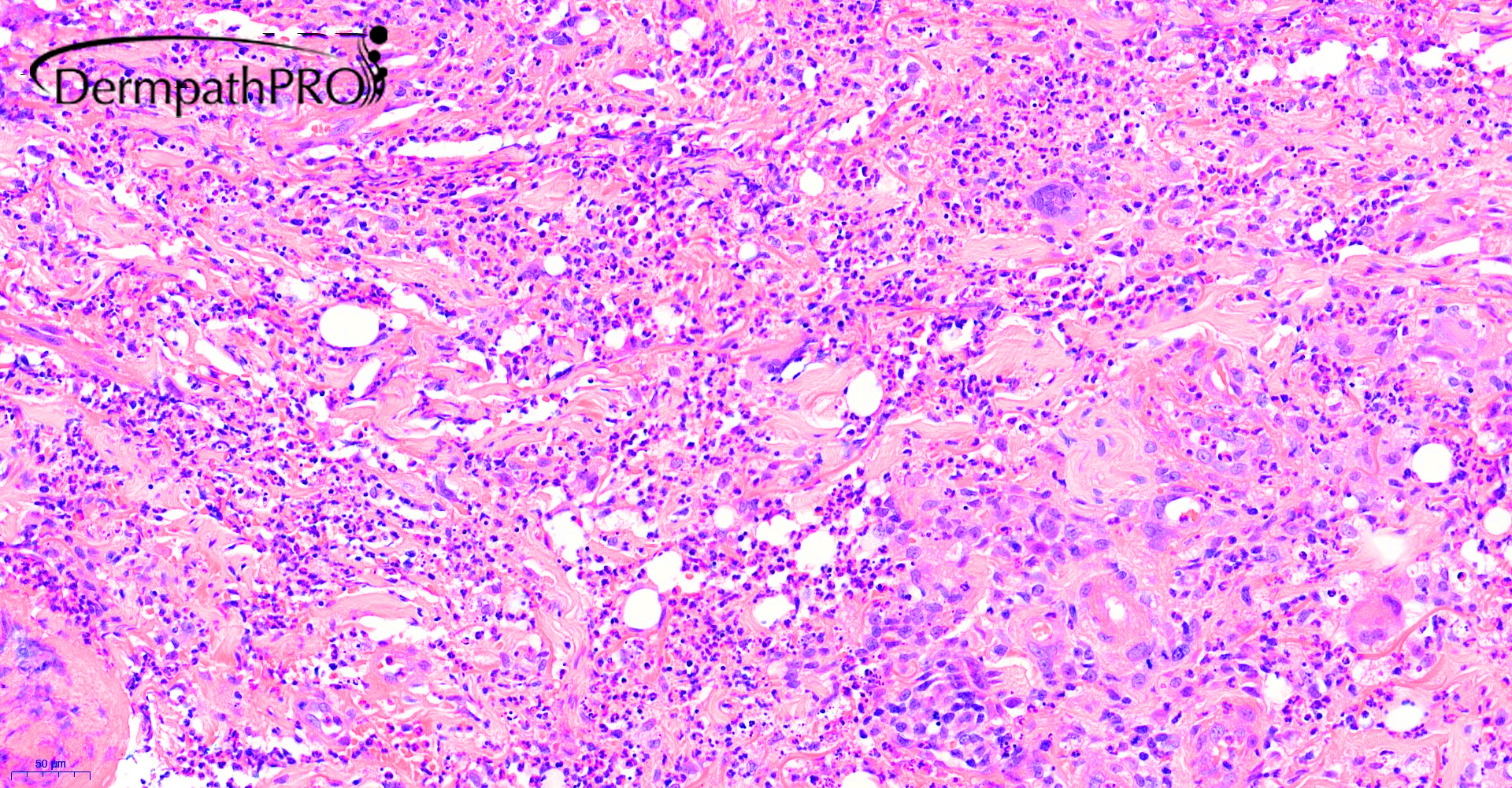
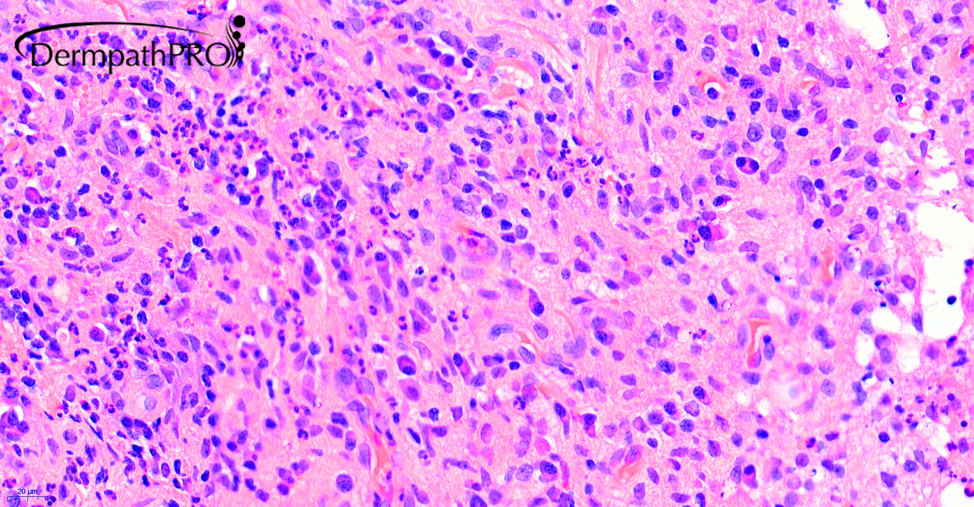
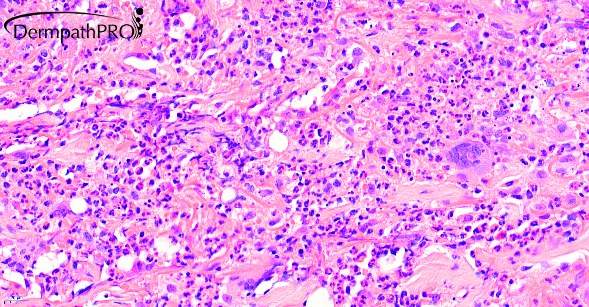
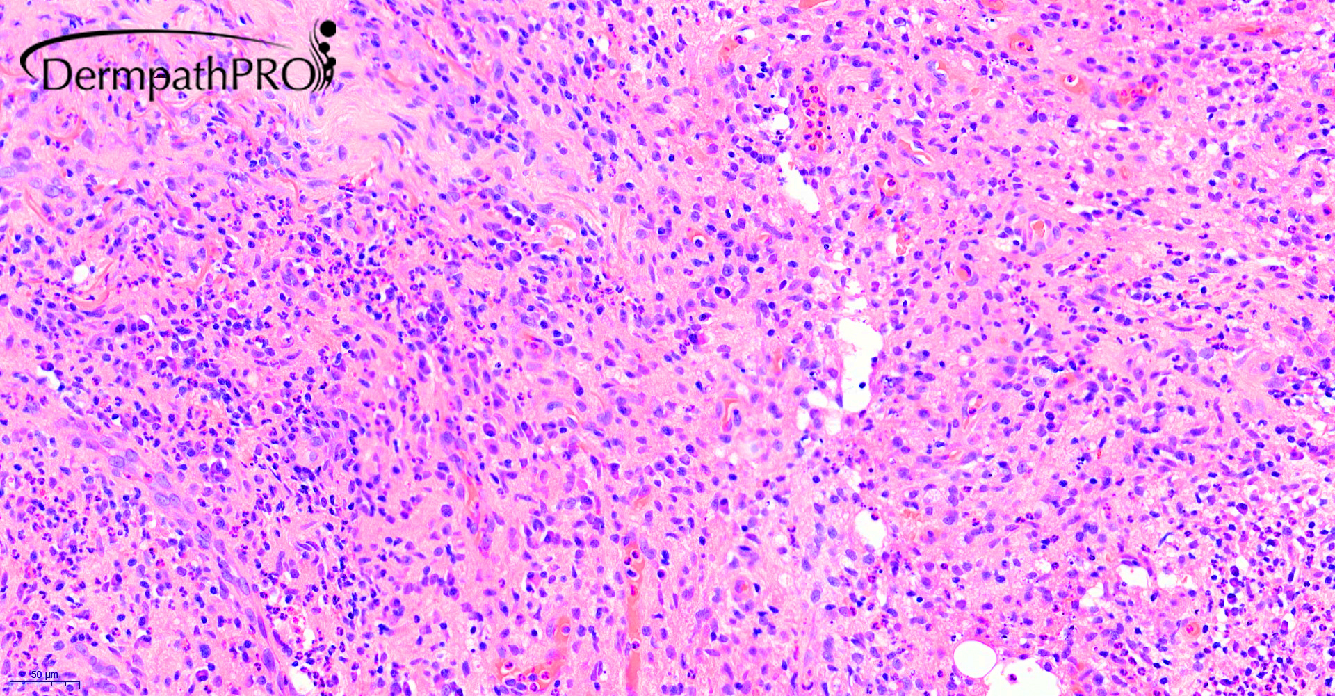
84M, Skin nodules and brain abscess.

  Report Record

User Feedback


Krishnakumar subramanian

Posted

mixed inflammatory cells with neutrophils, histiocytes, lymphocytes and plasma cells are seen. giant cells are seen

I am concerned with amebae Naegleria fowleri,  Bala muthiamandrillaris species, may be a PAS stain and GMS stain

if negative rule out vasculitis

Share this comment


Link to comment
share_externally

Thinking of EED, but there are plasma cells and there is also a brain abscess...so my spot is tertiary syphilis!

Share this comment


Link to comment
share_externally

Dr. Mona Abdel-Halim

Posted

Interesting suggestion by Vincenzo! complete infectious work up is needed.

Share this comment


Link to comment
share_externally

Meenakshi Batrani

Posted

Work up for infectious etiology. Deep fungal infections including Cryptococcus can cause brain abscess and skin lesions.

Share this comment


Link to comment
share_externally

Saleem Taibjee

Posted

Yes, you are all on the right lines. This was an unusual infection.

In addition to skin histology, fresh skin was also sent to our Microbiology reference laboratory.

The Microbiology lab report reads: "The hsp65 target is positive and sequencing has identified Nocardia cerradoensis". Our local microbiologists concluded that the clinical presentation was indeed consistent with Nocardia.

I had already scrutinised the special stains very carefully. The Wade-Fite is shown below. I found a solitary focus of staining, and prior to the Microbiology report was uncertain whether this staining represented artefactual stain deposit or mycobacteria. But in hindsight I did then wonder if perhaps this is Nocardia? Have any of you seen examples of this?

10089_40.0x Wade-Fite.jpg


Share this comment


Link to comment
share_externally

Krishnakumar subramanian

Posted

no sir, not seen a case or nocardia stained like this

do you have GMS stain

Share this comment


Link to comment
share_externally

Saleem Taibjee

Posted

Sorry, no we don't have that

Share this comment


Link to comment
share_externally

new2dp

Posted (edited)

Cool case, thank you!

Edited by new2dp
re-reading -- correction

Share this comment


Link to comment
share_externally


Join the conversation

You can post now and register later. If you have an account, sign in now to post with your account.

Guest
Add a comment...

×   Pasted as rich text.   Paste as plain text instead

  Only 75 emoji are allowed.

×   Your link has been automatically embedded.   Display as a link instead

×   Your previous content has been restored.   Clear editor

×   You cannot paste images directly. Upload or insert images from URL.

Loading...

×
×
  • Create New...