Jump to content
In this section we have spot diagnoses posted on a daily basis since June 2010, now over 4000! You can review the archived cases and read the suggested diagnoses by users and the final comment by the contributors.
Case are uploaded each week day by 10 am UK time with the correct diagnosis will generally be posted at 8 pm UK time. Why not view the most recent spot diagnosis and proffer a diagnosis?

Case Number : Case 2535 - 25 March 2020 Posted By: Dr. Hafeez Diwan

Please read the clinical history and view the images by clicking on them before you proffer your diagnosis.
Submitted Date :
   (0 reviews)

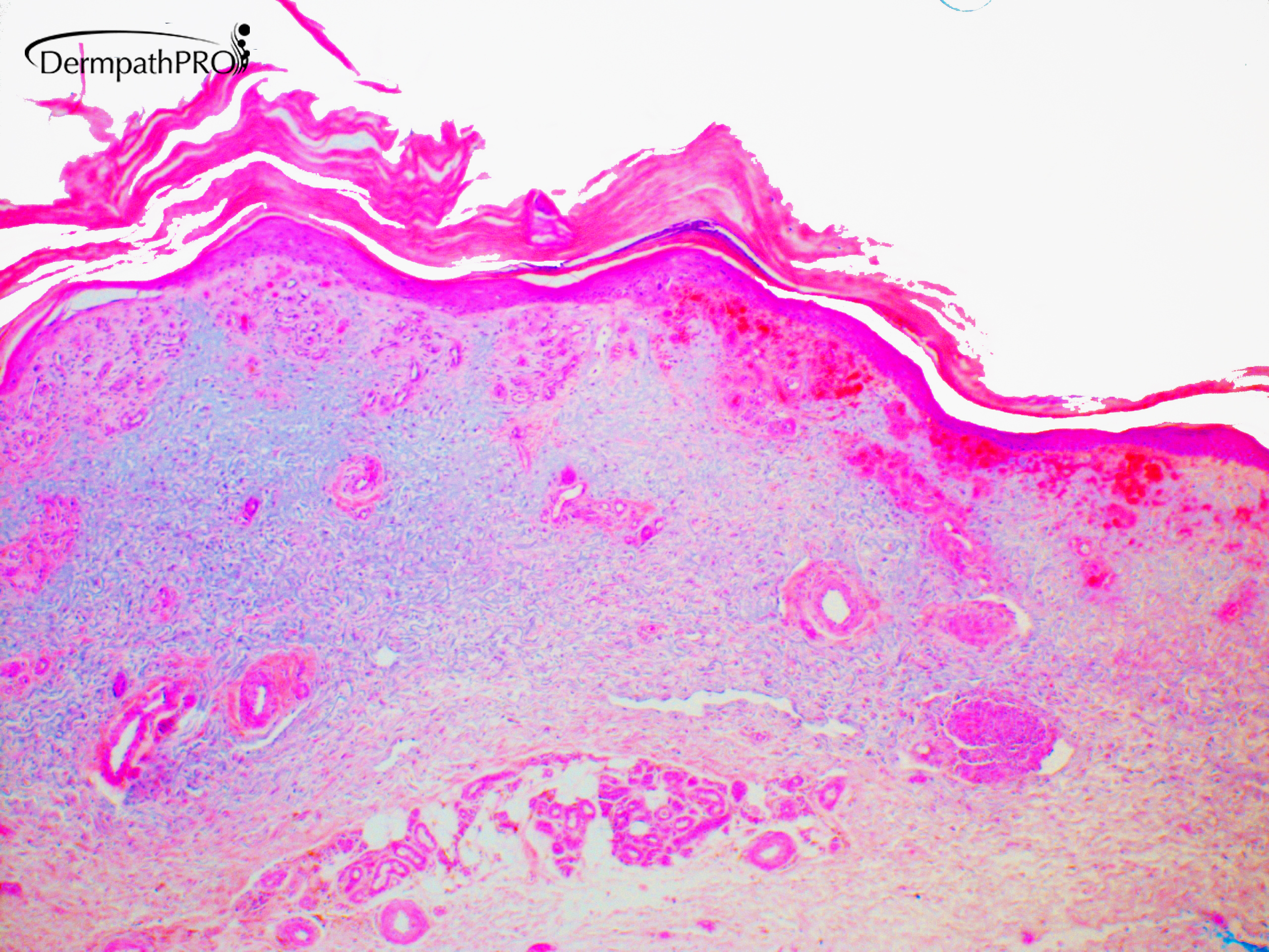
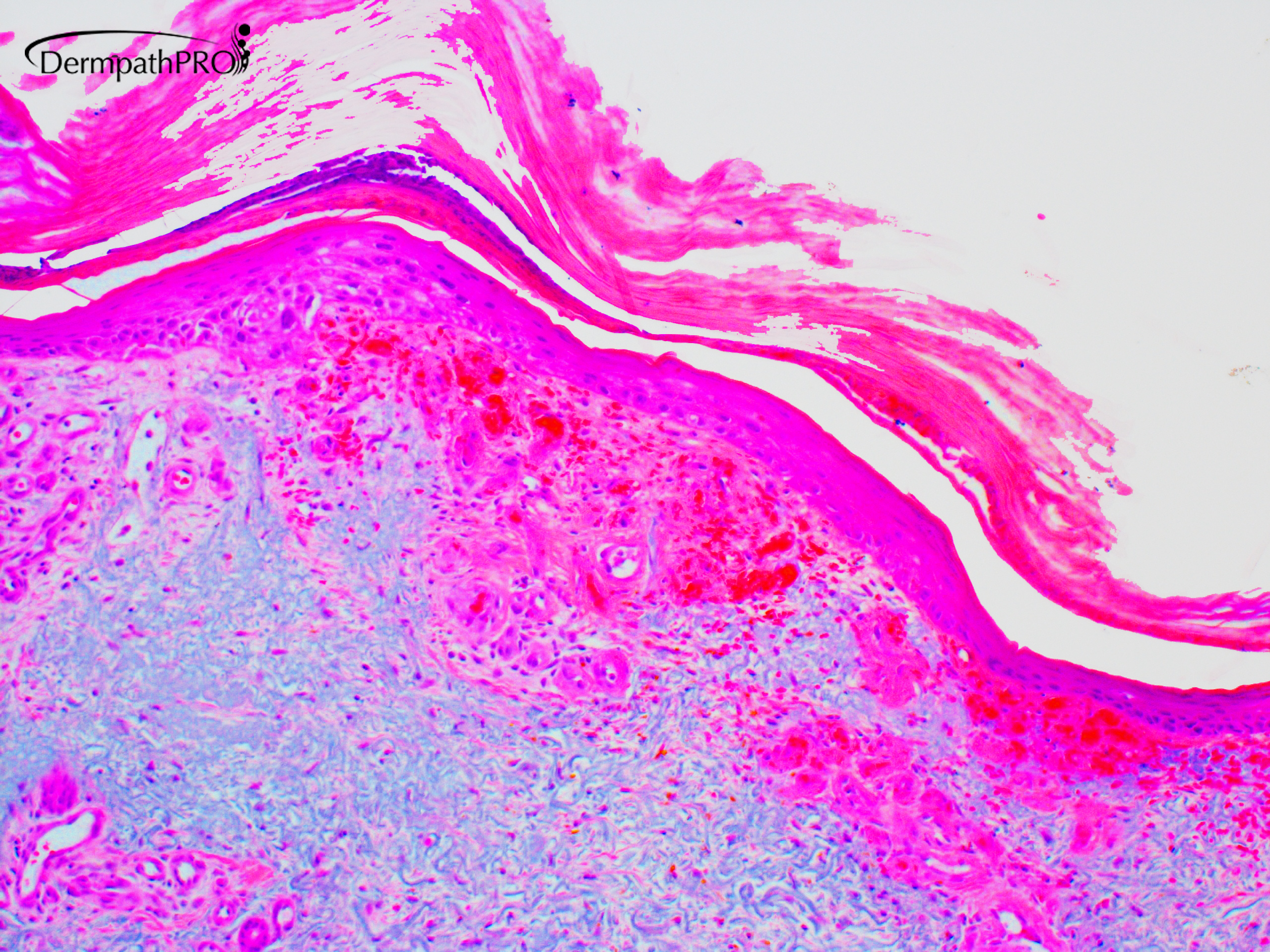
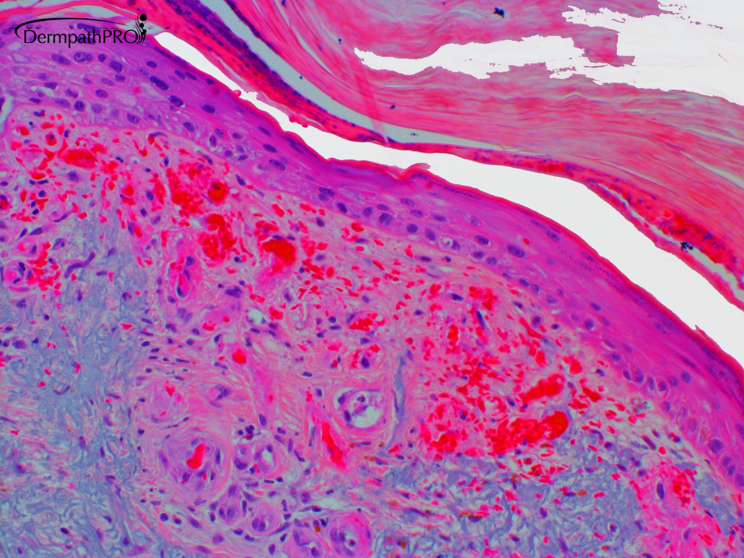
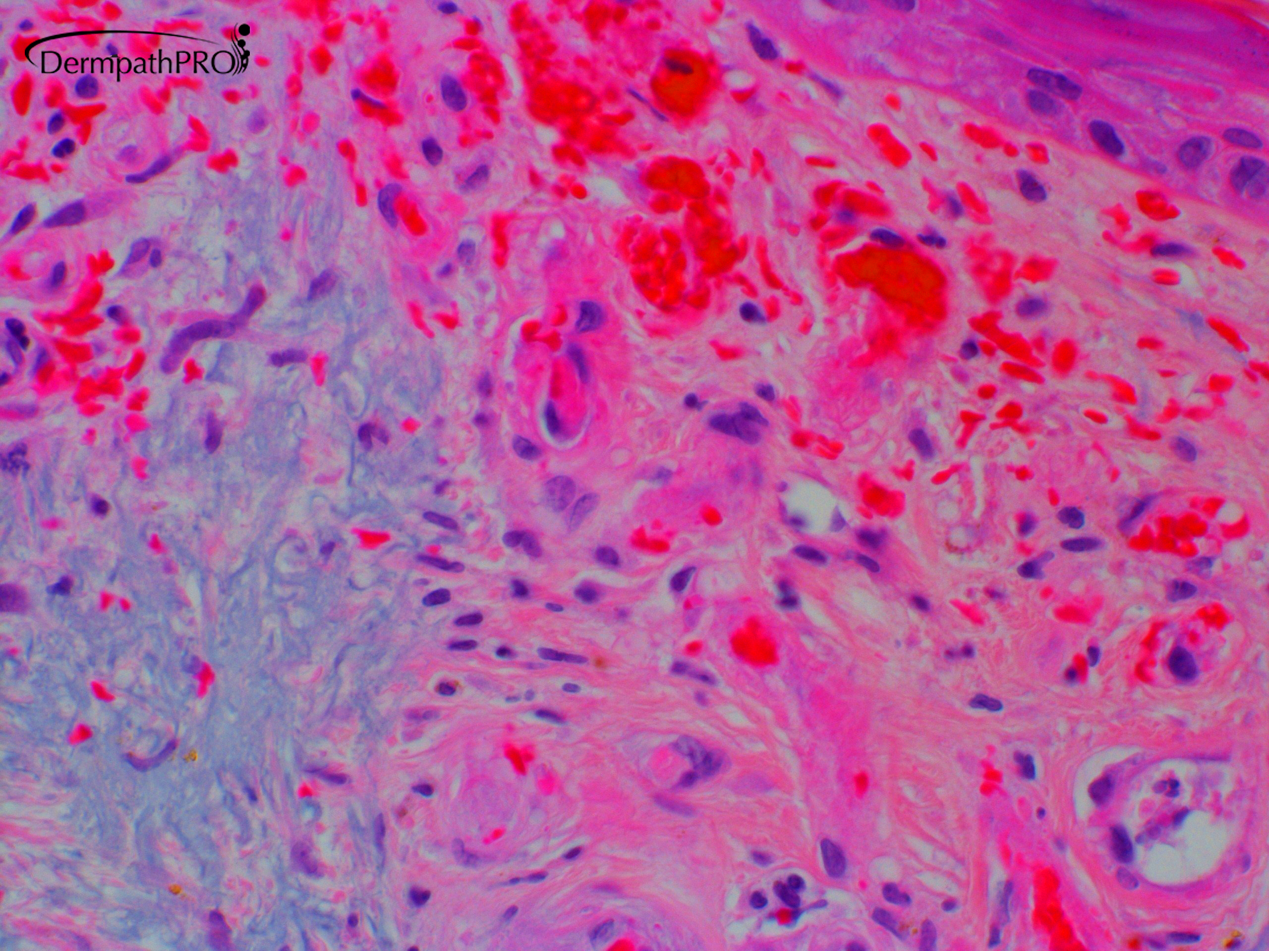
50 year-old female with biopsy from left ankle.


  Report Record

User Feedback


An angiomatous growth in a actinic skin, with elastic damage, could lead to thinking of an acquired elastic hemangioma, but this lesion show vascular channels not horizontally oriented, and with fibrinous microtrombosis. There is erythrocytic extravasation, also. So favor a stasis dermatitis, interviewing in a actinic skin.

Share this comment


Link to comment
share_externally

Dr. Mona Abdel-Halim

Posted

Or lividoid vasculopathy? 

Share this comment


Link to comment
share_externally

Victor Delgado

Posted

There is epidermal atrophy with mild atypia, probably due to dermal changes, where we can see extravasated RBCs, vascular congestion, width vascular walls, fibrinoid material on them and a few thrombus. Agree with Mona, I would prefer to call this Livedoid Vasculopathy.

Share this comment


Link to comment
share_externally

Yes, Mona. I was off road. Livedoid Vasculopathy is the right diagnosis. 

Share this comment


Link to comment
share_externally

dermpath1

Posted

There is a mucin in the dermis.Better to exclude lupus.

Share this comment


Link to comment
share_externally

I would suggest stasis, as it is ankle; epidermis is hyperkeratotic; there is lobular small vessel hyperplasia with fibrin thrombi, probably focal fibrinoid necrosis and extravasation of erytrocytes. There are neutrofiles within stratum corneum, and I would do PAS. There is severe solar damage. But I wonder, if something even more important is hiding.

Share this comment


Link to comment
share_externally

Dr. Hafeez Diwan

Posted

Atrophie blanche/livedoid vasculopathy.  There is a background of stasis.

Share this comment


Link to comment
share_externally

Dr. Richard Carr

Posted

Agree with stasis - severe. Needs CPC of course.

Share this comment


Link to comment
share_externally


Join the conversation

You can post now and register later. If you have an account, sign in now to post with your account.

Guest
Add a comment...

×   Pasted as rich text.   Paste as plain text instead

  Only 75 emoji are allowed.

×   Your link has been automatically embedded.   Display as a link instead

×   Your previous content has been restored.   Clear editor

×   You cannot paste images directly. Upload or insert images from URL.

Loading...

×
×
  • Create New...