Jump to content
In this section we have spot diagnoses posted on a daily basis since June 2010, now over 4000! You can review the archived cases and read the suggested diagnoses by users and the final comment by the contributors.
Case are uploaded each week day by 10 am UK time with the correct diagnosis will generally be posted at 8 pm UK time. Why not view the most recent spot diagnosis and proffer a diagnosis?

Case Number : Case 2536 - 26 March 2020 Posted By: Saleem Taibjee

Please read the clinical history and view the images by clicking on them before you proffer your diagnosis.
Submitted Date :
   (0 reviews)

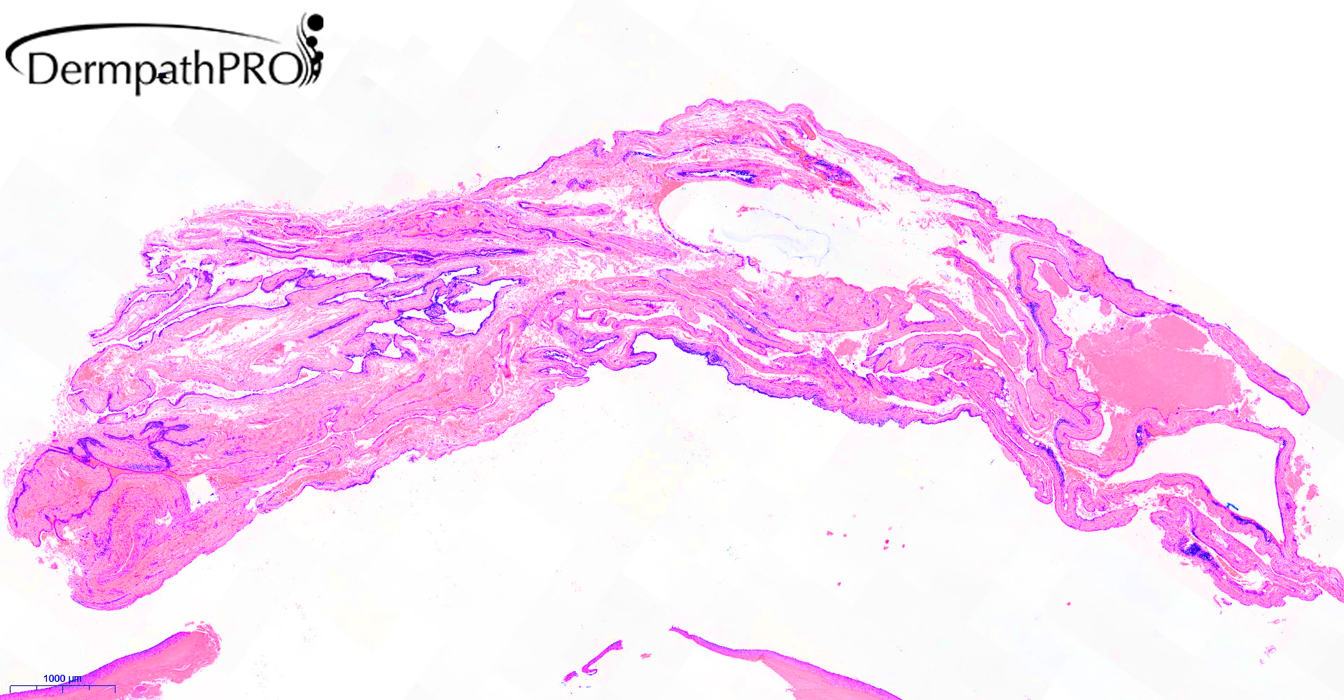
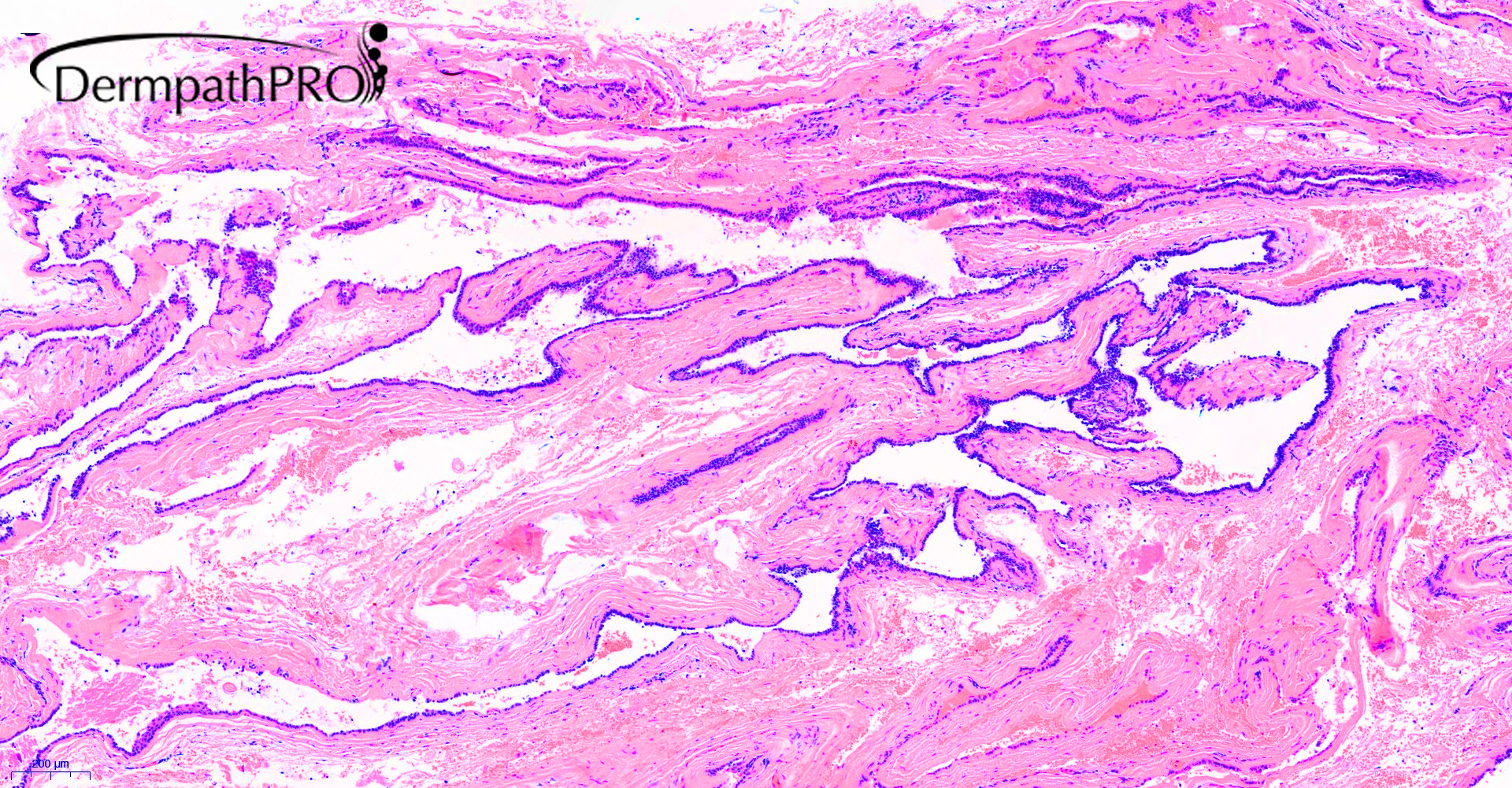
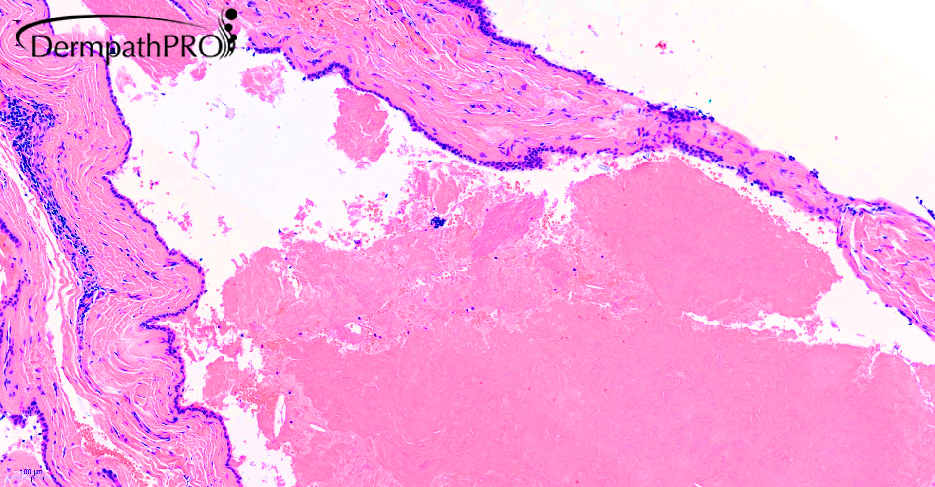
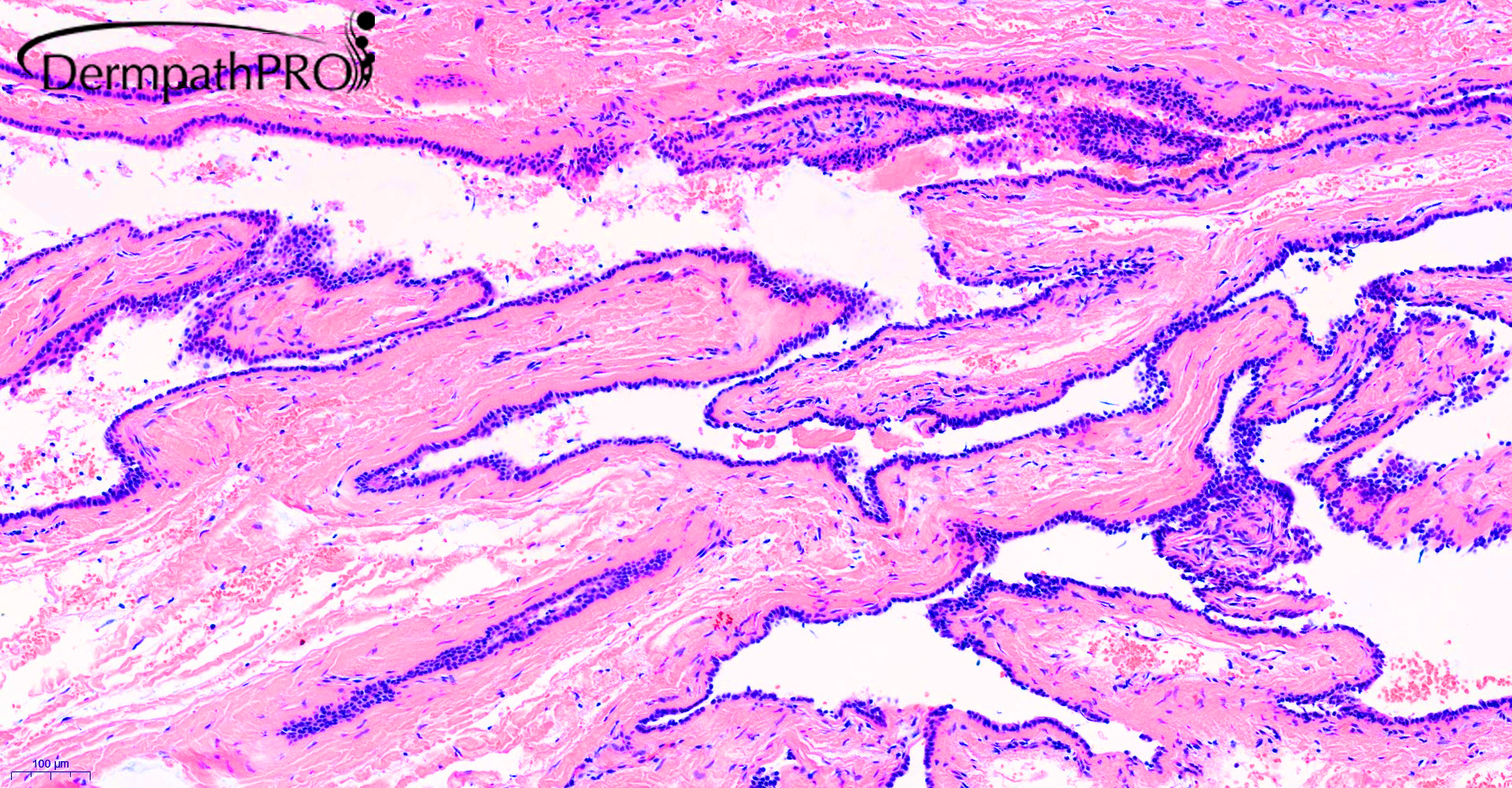
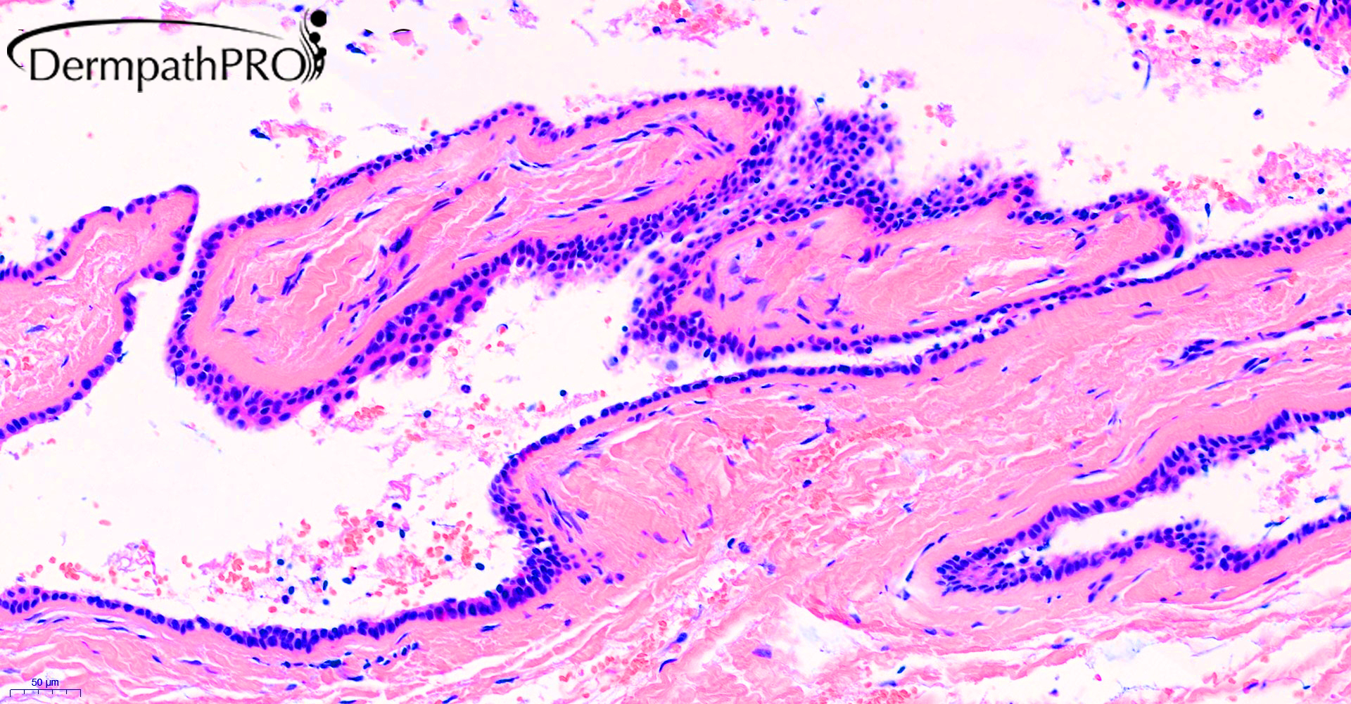
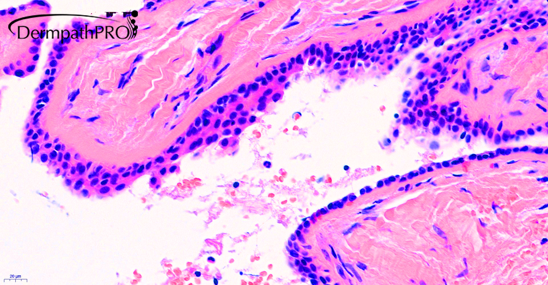
42F, suspected cyst on scalp.


  Report Record

User Feedback


Cem Leblebici

Posted

Do we only observe the basal layer of trichilemmal cyst with suprabasal seperation?

Share this comment


Link to comment
share_externally

Saleem Taibjee

Posted

Yes, Cem is spot on. This is a great and notorious pitfall, in which suprabasal separation of the pilar (tricholemmal) cyst wall (presumably at the time of surgery) leaves a remnant which can produce a mimic of hidrocystoma. This is a particularly impressive example. The clue is in the first image, where at the bottom of the field I included more typical pilar cyst at the edge. Below I include other parts of the section which I had deliberately omitted in the first set of images.

Arrowed image.jpg

16023_0.6x.jpg

16023_5.0xb.jpg

16023_20.0xb.jpg


Share this comment


Link to comment
share_externally


Join the conversation

You can post now and register later. If you have an account, sign in now to post with your account.

Guest
Add a comment...

×   Pasted as rich text.   Paste as plain text instead

  Only 75 emoji are allowed.

×   Your link has been automatically embedded.   Display as a link instead

×   Your previous content has been restored.   Clear editor

×   You cannot paste images directly. Upload or insert images from URL.

Loading...

×
×
  • Create New...