Jump to content
In this section we have spot diagnoses posted on a daily basis since June 2010, now over 4000! You can review the archived cases and read the suggested diagnoses by users and the final comment by the contributors.
Case are uploaded each week day by 10 am UK time with the correct diagnosis will generally be posted at 8 pm UK time. Why not view the most recent spot diagnosis and proffer a diagnosis?

Case Number : Case 2538 - 30 March 2020 Posted By: Iskander H. Chaudhry

Please read the clinical history and view the images by clicking on them before you proffer your diagnosis.
Submitted Date :
   (0 reviews)

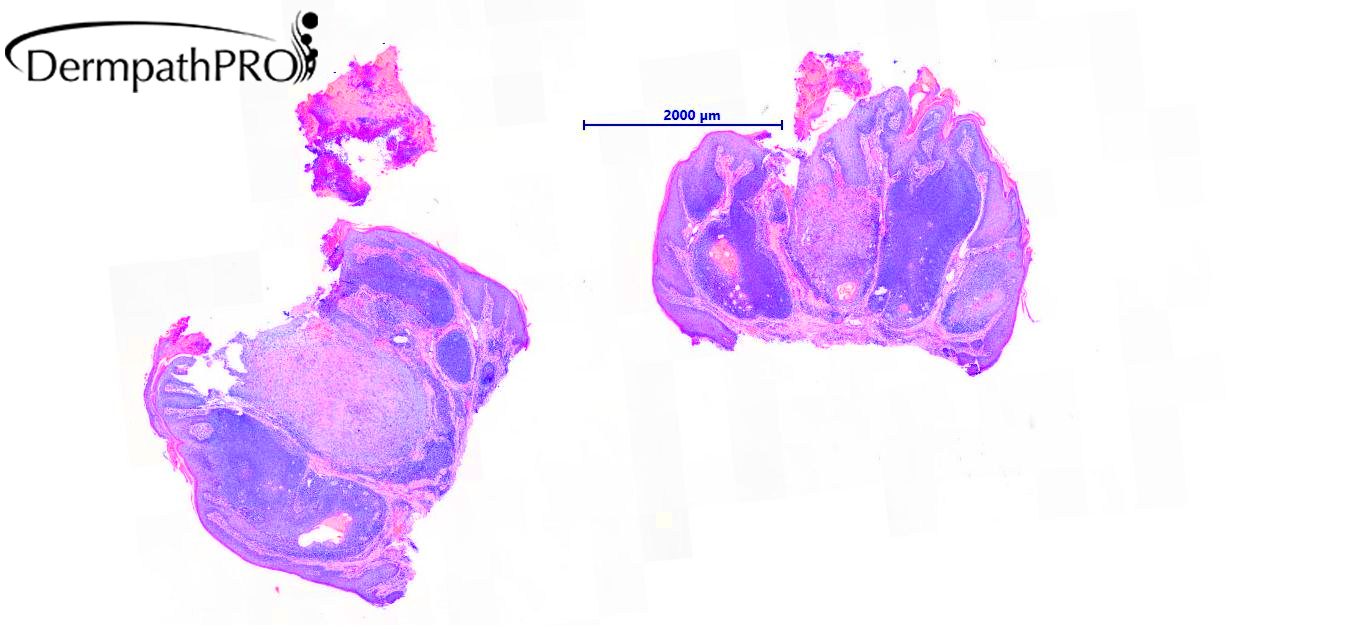
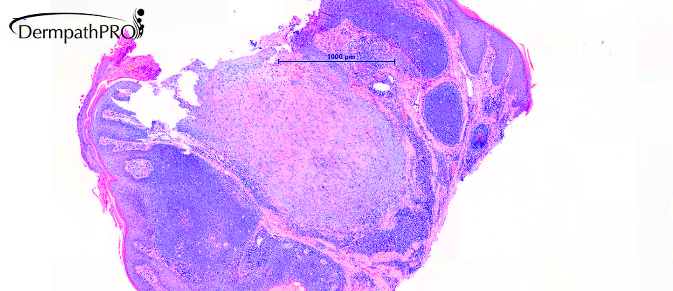
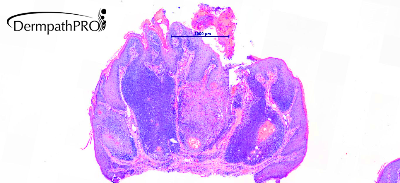
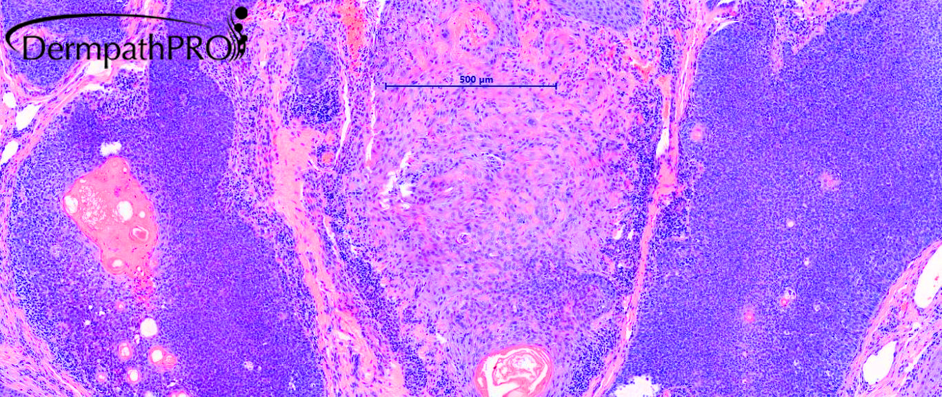
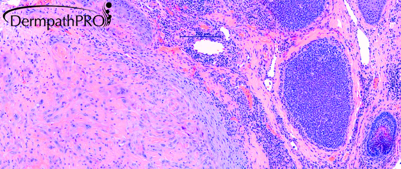
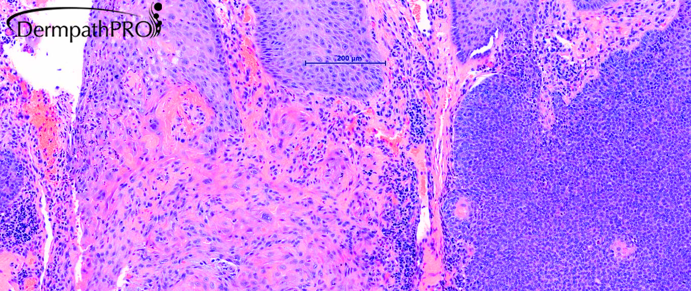
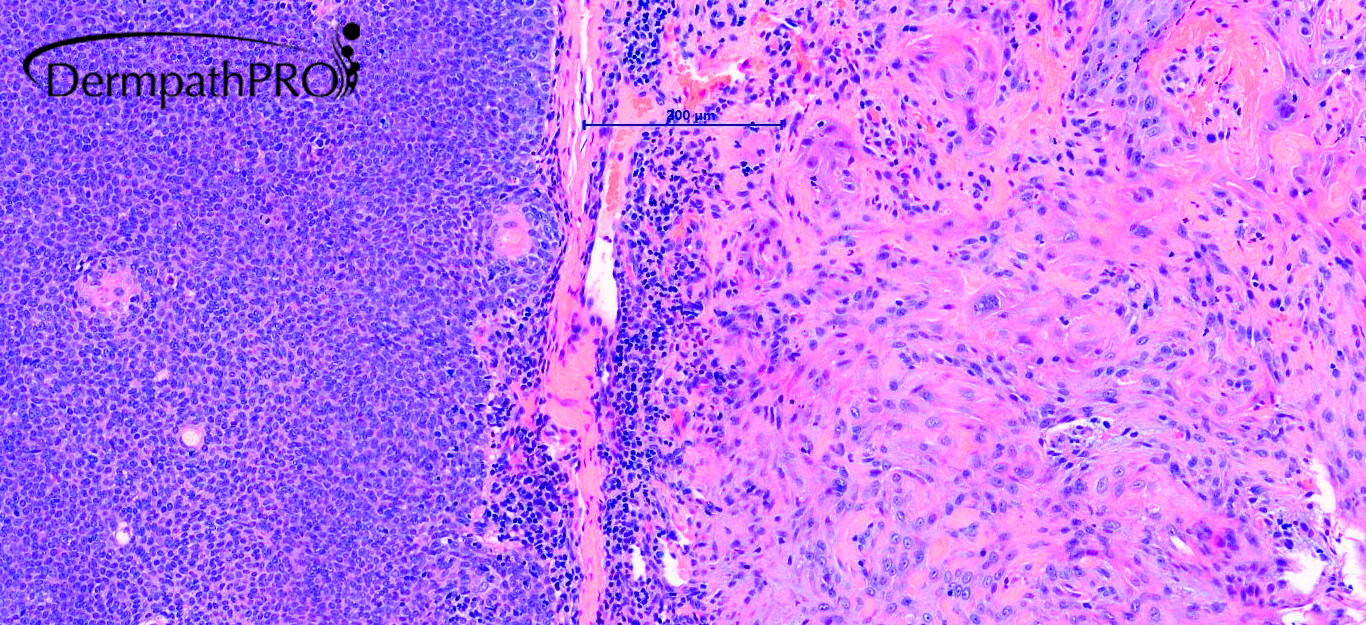
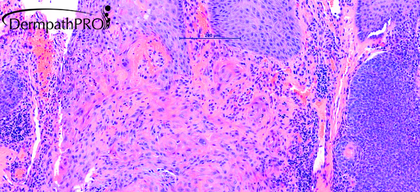
77M, Left chin C+C (double cycle)- ?Viral wart ?SCC ?KA ?AK


  Report Record

User Feedback


Cem Leblebici

Posted

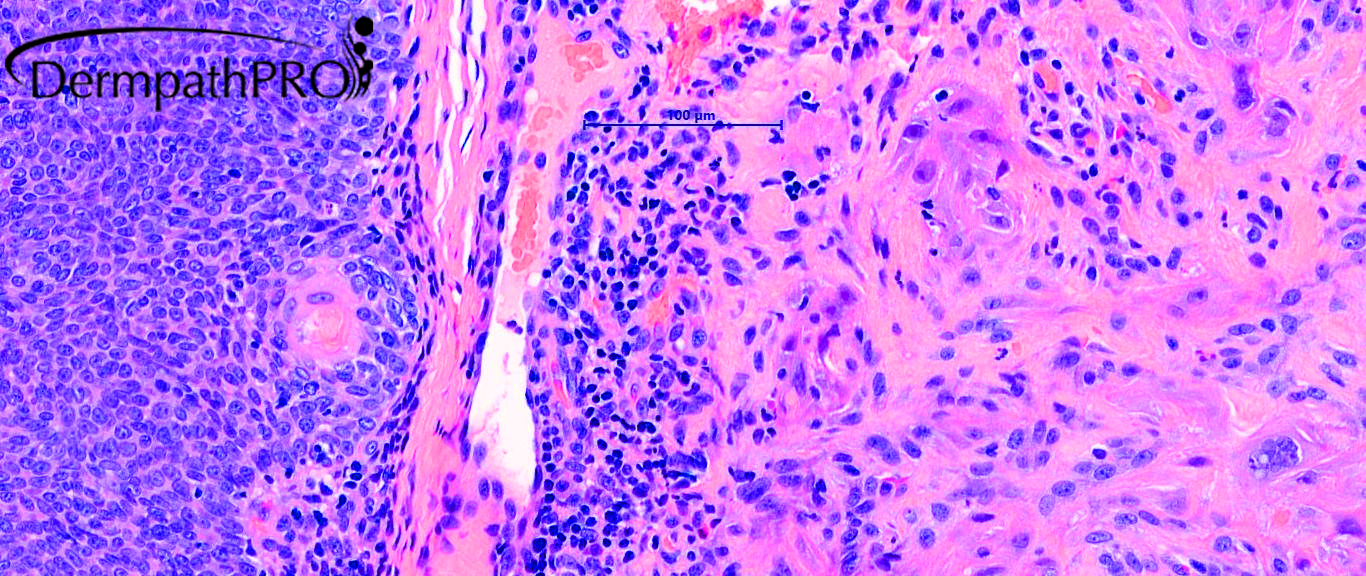
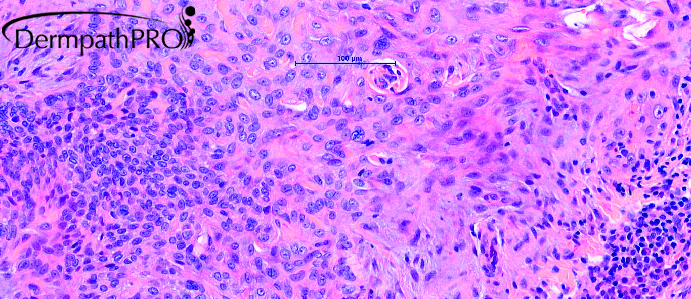
Poroid neoplasm (probably poroma) with squamous differentiation.

Share this comment


Link to comment
share_externally

I see trichilemmal and matrichal follicular differentiation. Favor a desmoplastic trichilemmoma, with basaloid follicular differentiation

Share this comment


Link to comment
share_externally

Admin_Dermpath

Posted

Dr Chaudhry - See below:


Skin with a lesion marked by the expansion of the follicular infundibulum over a number of follicles. The component cells are in part small and basaloid and in part squamoid. The distended infundibulum has a minor sebaceous component as well as a few rudimentary ducts some of which are enlarged and distorted to form squamous cysts. Nuclear palisading is not very apparent. Mitoses are not a feature and there are no apoptotic figures and no necrosis. These expanded lobules are surrounded by a plasma cell infiltrate.


The lesion is diffusely negative for CD34 that also shows no specialized stroma and highlights delicate vasculature. Immunochemistry for BerEP4 is diffusely negative. PRAME shows only staining of mature sebocytes only and weak nuclear staining in the central more squamoid lobule. EMA shows plasma cell staining and staining in the
more squamous areas. The lesion is close to the deep resection margin. This is an in situ lesion of infundibular origin.


The presence of cells with sebaceous differentiation raises the possibility of sebaceoma with a central desmoplastic trichilemmoma despite the lack of CD34. 

Share this comment


Link to comment
share_externally


Join the conversation

You can post now and register later. If you have an account, sign in now to post with your account.

Guest
Add a comment...

×   Pasted as rich text.   Paste as plain text instead

  Only 75 emoji are allowed.

×   Your link has been automatically embedded.   Display as a link instead

×   Your previous content has been restored.   Clear editor

×   You cannot paste images directly. Upload or insert images from URL.

Loading...

×
×
  • Create New...