Jump to content
In this section we have spot diagnoses posted on a daily basis since June 2010, now over 4000! You can review the archived cases and read the suggested diagnoses by users and the final comment by the contributors.
Case are uploaded each week day by 10 am UK time with the correct diagnosis will generally be posted at 8 pm UK time. Why not view the most recent spot diagnosis and proffer a diagnosis?

Case Number : Case 2562 - 01 May 2020 Posted By: Dr. Richard Carr

Please read the clinical history and view the images by clicking on them before you proffer your diagnosis.
Submitted Date :
   (0 reviews)

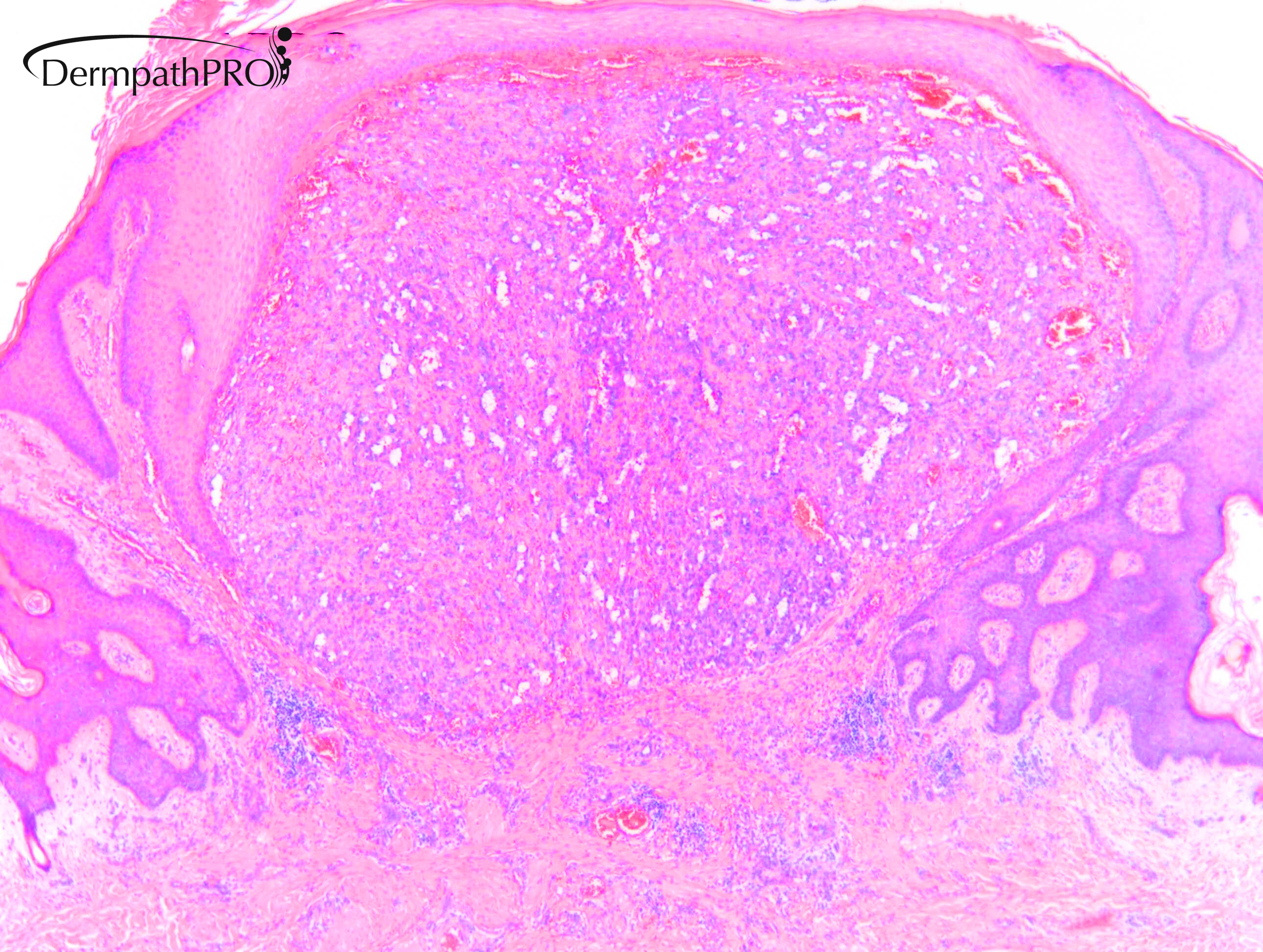
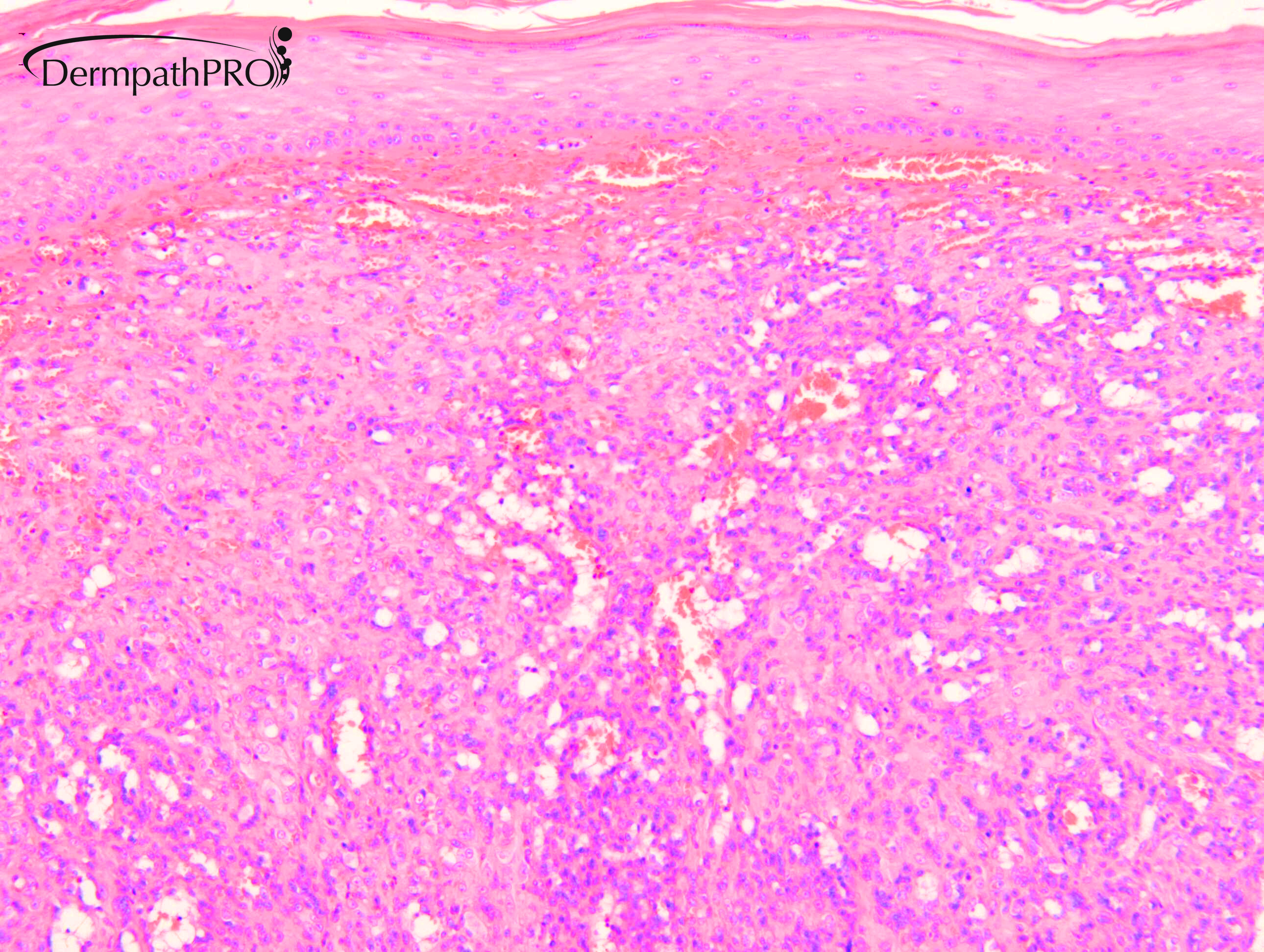
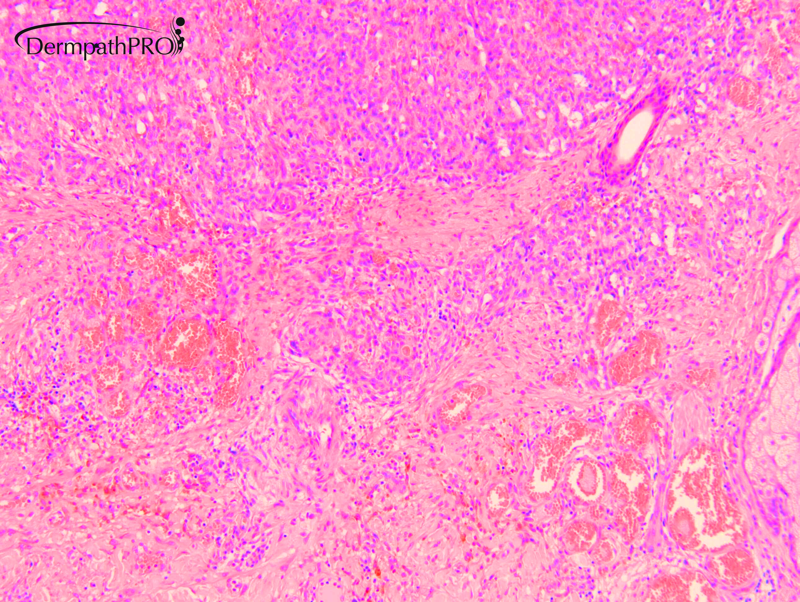
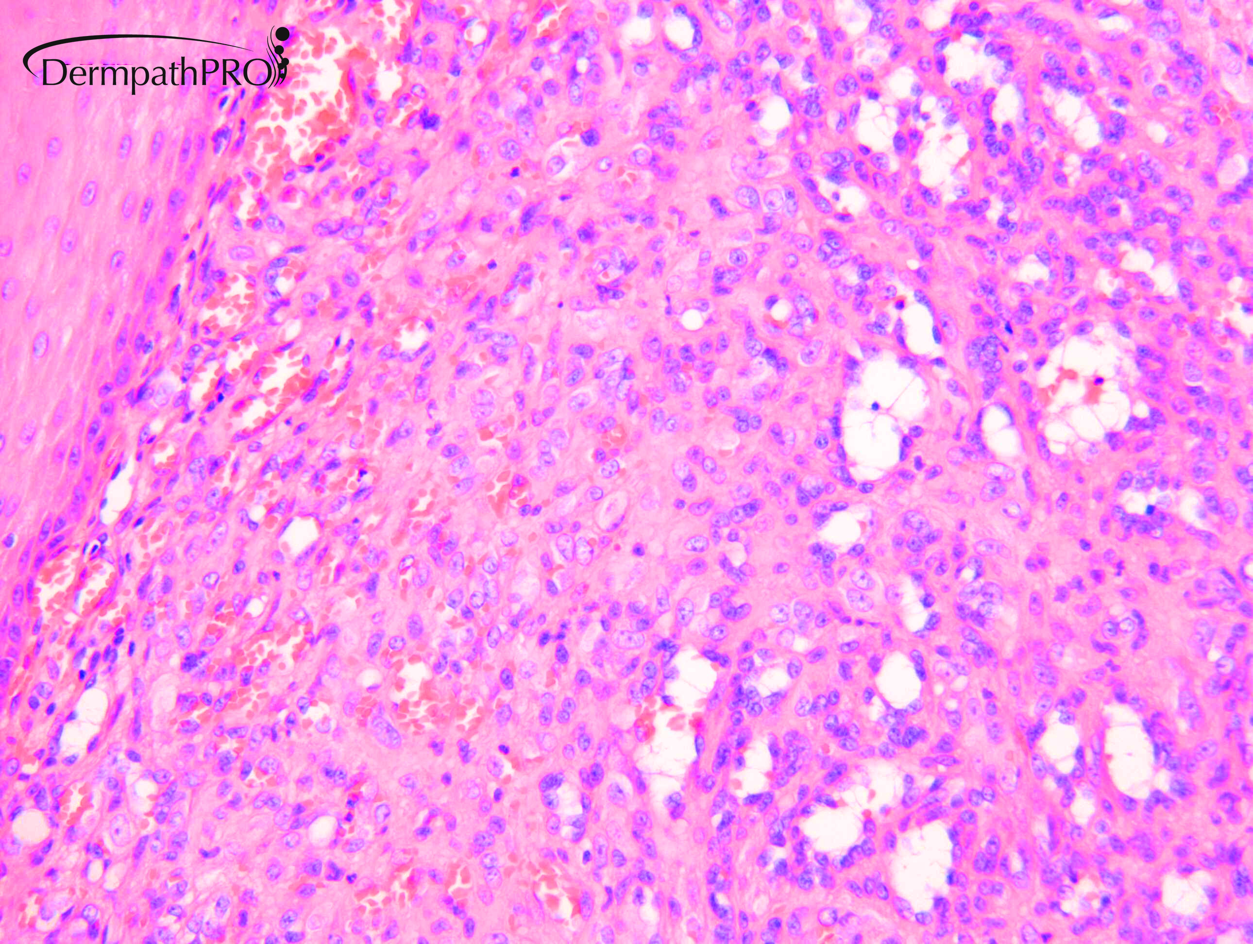
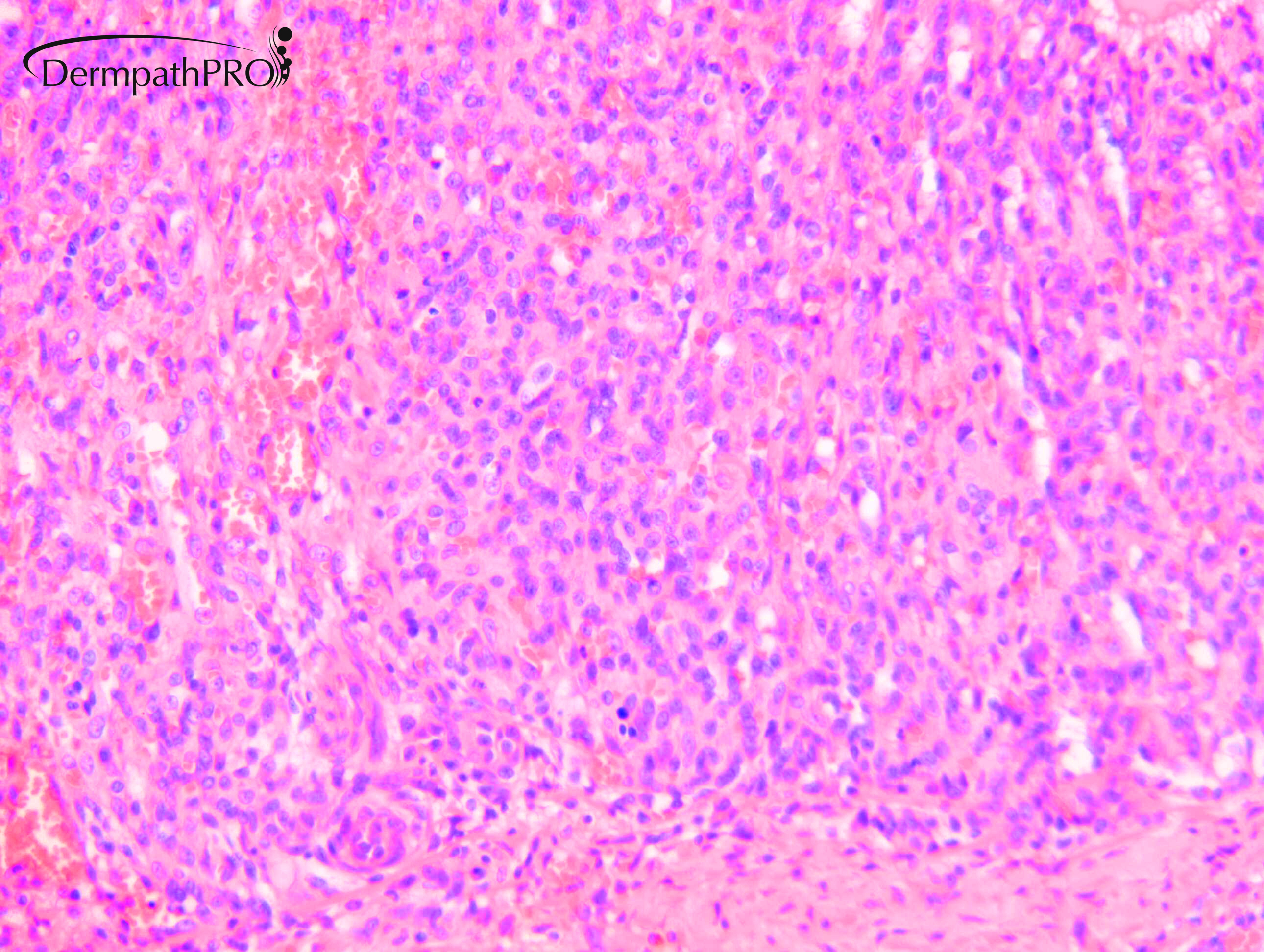
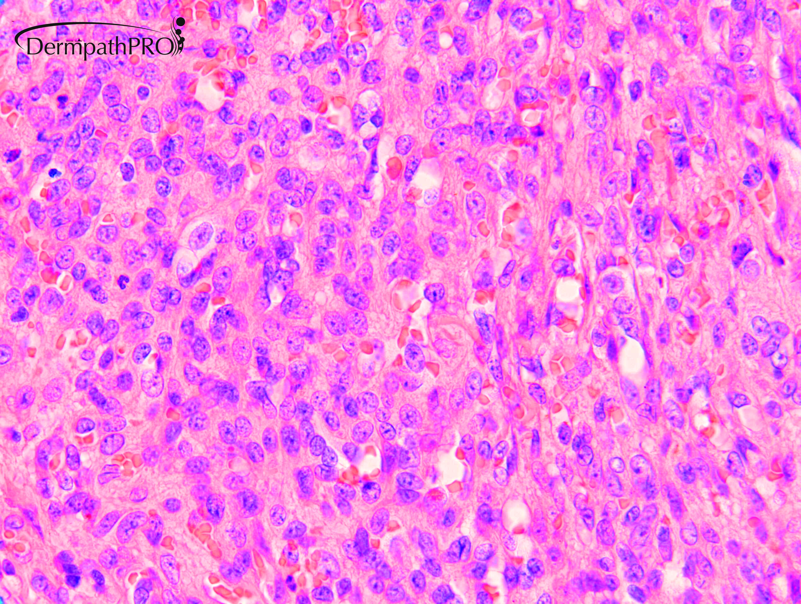
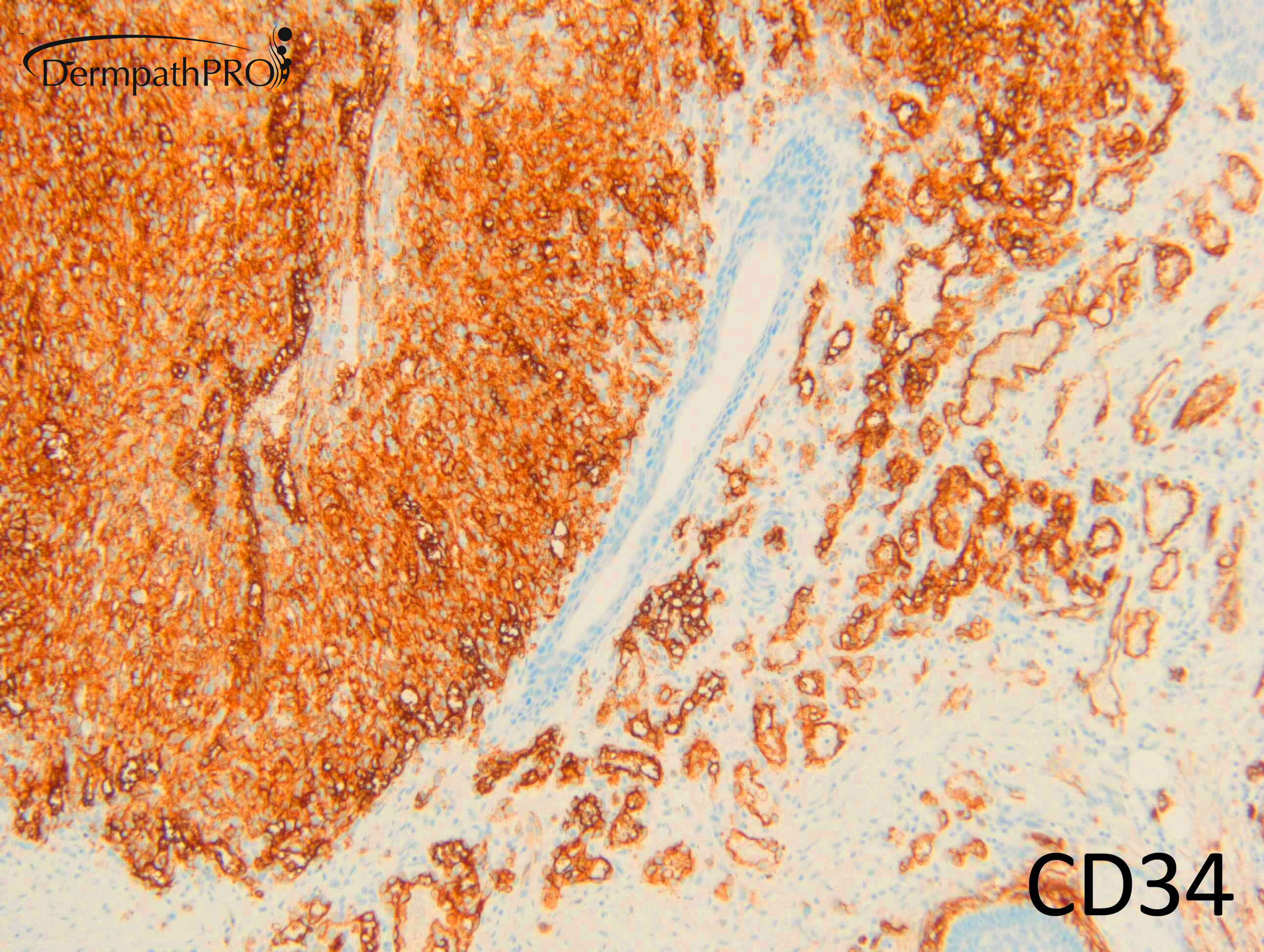
M25. Left chest. Tender nodule 6/52. Case referred c/o Dr Vivek Mudaliar


  • Like 2
  Report Record

User Feedback


Dr. Mona Abdel-Halim

Posted

Cutaneous epithelioid angiomatous nodule

Share this comment


Link to comment
share_externally

vincenzo

Posted

I was making the same diagnosis. 
So agree with Mona.  
There is only an important differential here: Bacillary Angiomatosis. But I don’t see any purplish grey bacterial colonies, and BA is a very rare condition nowadays. 
 

Share this comment


Link to comment
share_externally

30 minutes ago, Dr. Mona Abdel-Halim said:

Cutaneous epithelioid angiomatous nodule

Agree

Share this comment


Link to comment
share_externally

Krishnakumar subramanian

Posted

looks like capillary angioma


Share this comment


Link to comment
share_externally

Dr. Richard Carr

Posted

Thank you for the contibutions. The proliferaiton in cutaneous epithelioid angiomatous nodule should be as follows: "Histologically, the lesions were located superficially in dermis (or submucosa) and consisted of a circumscribed, unilobular, mainly solid proliferation of large polygonal epithelioid endothelial cells with vesicular nuclei and conspicuous nucleoli."

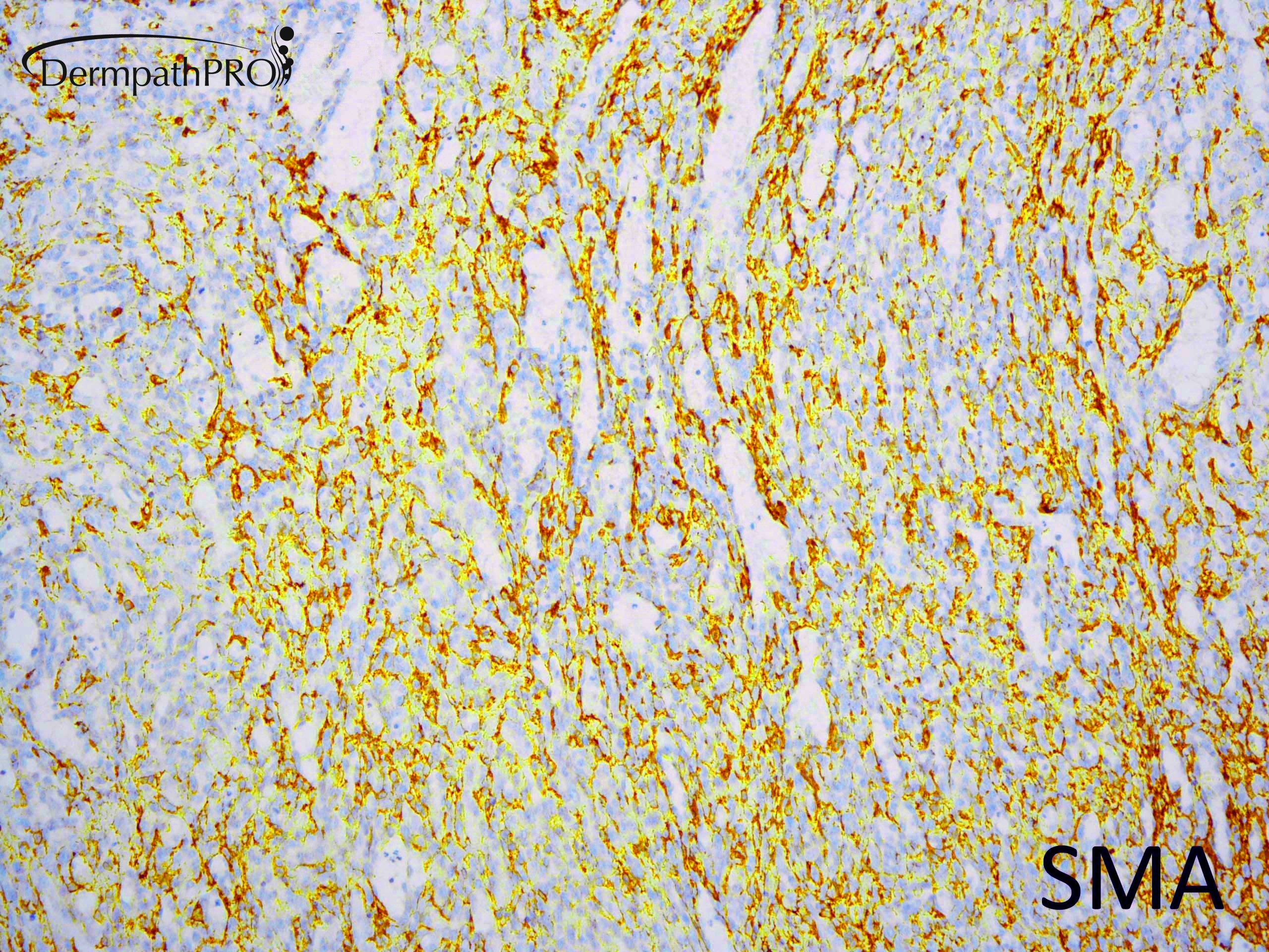
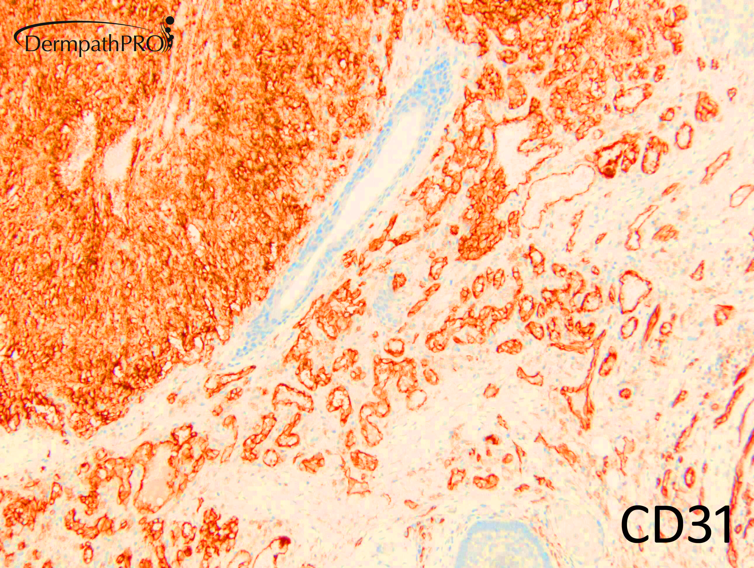
I think the lesion illustrated here has a dual population of endothelial (CD31 / CD34) and pericytes / smooth muscle cells (SMA) so I guess this lesion is just a rather solid appearing lobular capillary haemangioma (pyogenic granuloma). I've only diagnosed CEAN once and unfortunately the referring pathologist had already cut the lesion through with levels so it was not possilble to do smooth muscle actin. It's benign so I guess this is somewhat academic.

Share this comment


Link to comment
share_externally

Meenakshi Batrani

Posted

8 hours ago, Dr. Richard Carr said:

Thank you for the contibutions. The proliferaiton in cutaneous epithelioid angiomatous nodule should be as follows: "Histologically, the lesions were located superficially in dermis (or submucosa) and consisted of a circumscribed, unilobular, mainly solid proliferation of large polygonal epithelioid endothelial cells with vesicular nuclei and conspicuous nucleoli."

I think the lesion illustrated here has a dual population of endothelial (CD31 / CD34) and pericytes / smooth muscle cells (SMA) so I guess this lesion is just a rather solid appearing lobular capillary haemangioma (pyogenic granuloma). I've only diagnosed CEAN once and unfortunately the referring pathologist had already cut the lesion through with levels so it was not possilble to do smooth muscle actin. It's benign so I guess this is somewhat academic.

Thanks Dr. Carr for this explanation. I was also thinking of pyogenic granuloma in this case. I have seen an occasional case of PG exhibiting epithelioid appearing endothelial cells, but overall silhouette and features are of PG. While working on these cases, I came across an old publication which mentions "the occasional epithelioid appearance of the lining endothelial cells may cause confusion with epithelioid hemangioma" as one of the pitfalls in diagnosis of PG.

Kapadia SB, Heffner DK. Pitfalls in the histopathologic diagnosis of pyogenic granuloma. Eur Arch Otorhinolaryngol. 1992;249(4):195‐200.


Share this comment


Link to comment
share_externally

Dr. Richard Carr

Posted

Thanks very much. Certainly I believe you can see quite plump endothelial (and pericytic cells) in a variety of benign lesions including PG. Often this is not in the publications or textbook. Curiously the original papers on CEAN don't discuss the differential of the the humble PG!

Share this comment


Link to comment
share_externally

vincenzo

Posted

Thanks Richard. Nice learning point!

Share this comment


Link to comment
share_externally

Josie Bisi

Posted

On 01/05/2020 at 12:42, Dr. Mona Abdel-Halim said:

Cutaneous epithelioid angiomatous nodule

I agree, nice case.


Share this comment


Link to comment
share_externally


Join the conversation

You can post now and register later. If you have an account, sign in now to post with your account.

Guest
Add a comment...

×   Pasted as rich text.   Paste as plain text instead

  Only 75 emoji are allowed.

×   Your link has been automatically embedded.   Display as a link instead

×   Your previous content has been restored.   Clear editor

×   You cannot paste images directly. Upload or insert images from URL.

Loading...

×
×
  • Create New...