Jump to content
In this section we have spot diagnoses posted on a daily basis since June 2010, now over 4000! You can review the archived cases and read the suggested diagnoses by users and the final comment by the contributors.
Case are uploaded each week day by 10 am UK time with the correct diagnosis will generally be posted at 8 pm UK time. Why not view the most recent spot diagnosis and proffer a diagnosis?

Case Number : Case 2566 - 07 May 2020 Posted By: Saleem Taibjee

Please read the clinical history and view the images by clicking on them before you proffer your diagnosis.
Submitted Date :
   (0 reviews)

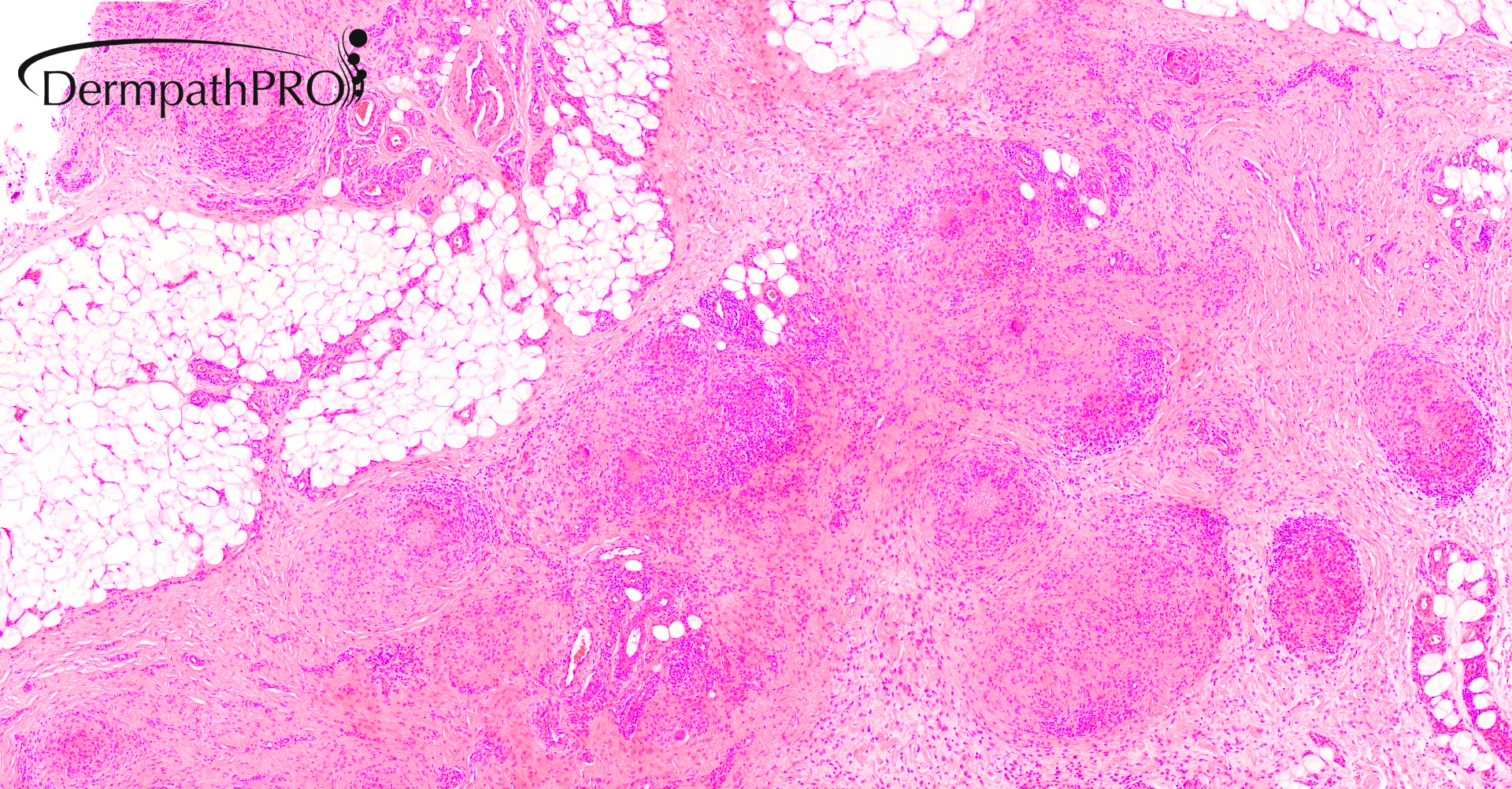
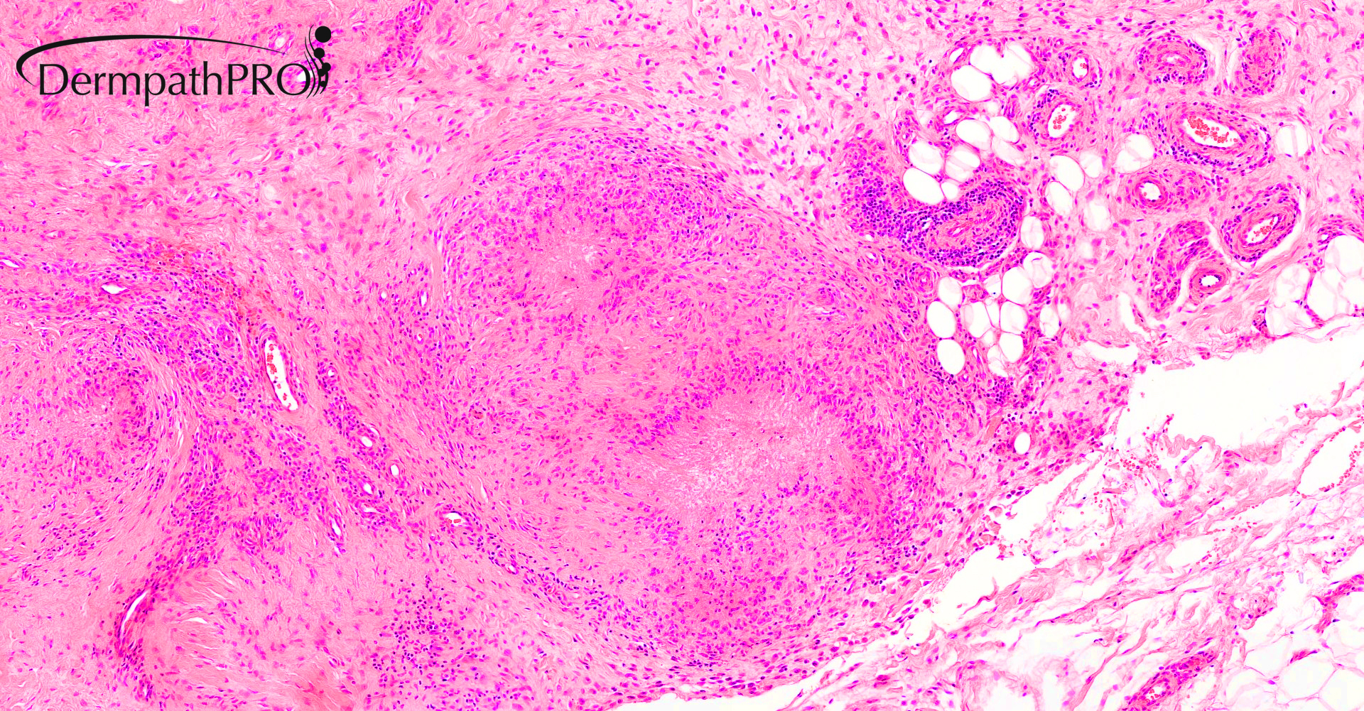
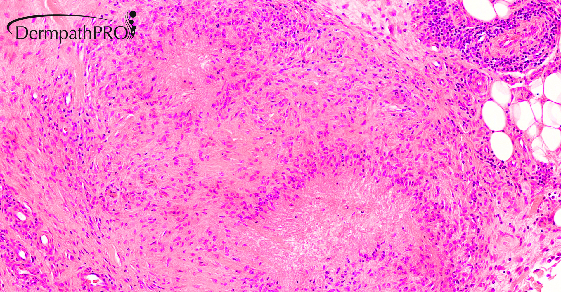
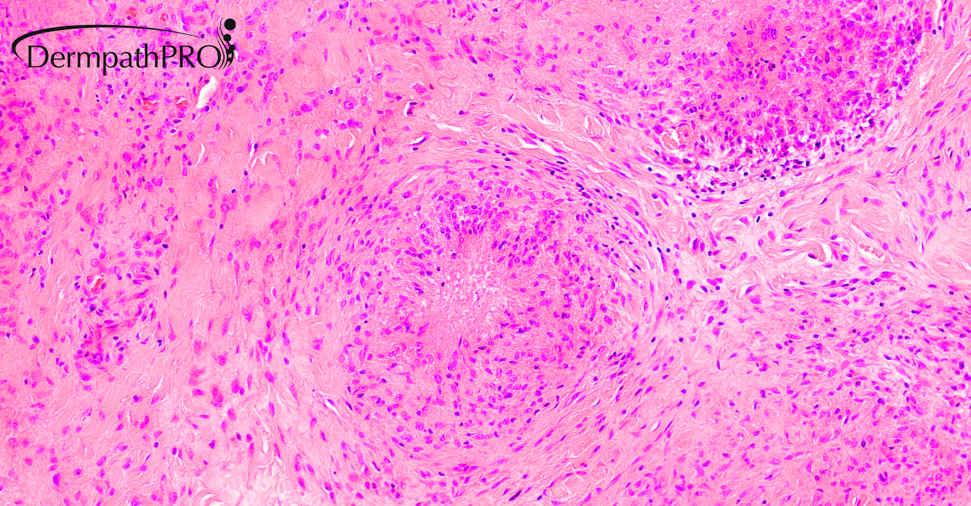
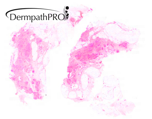
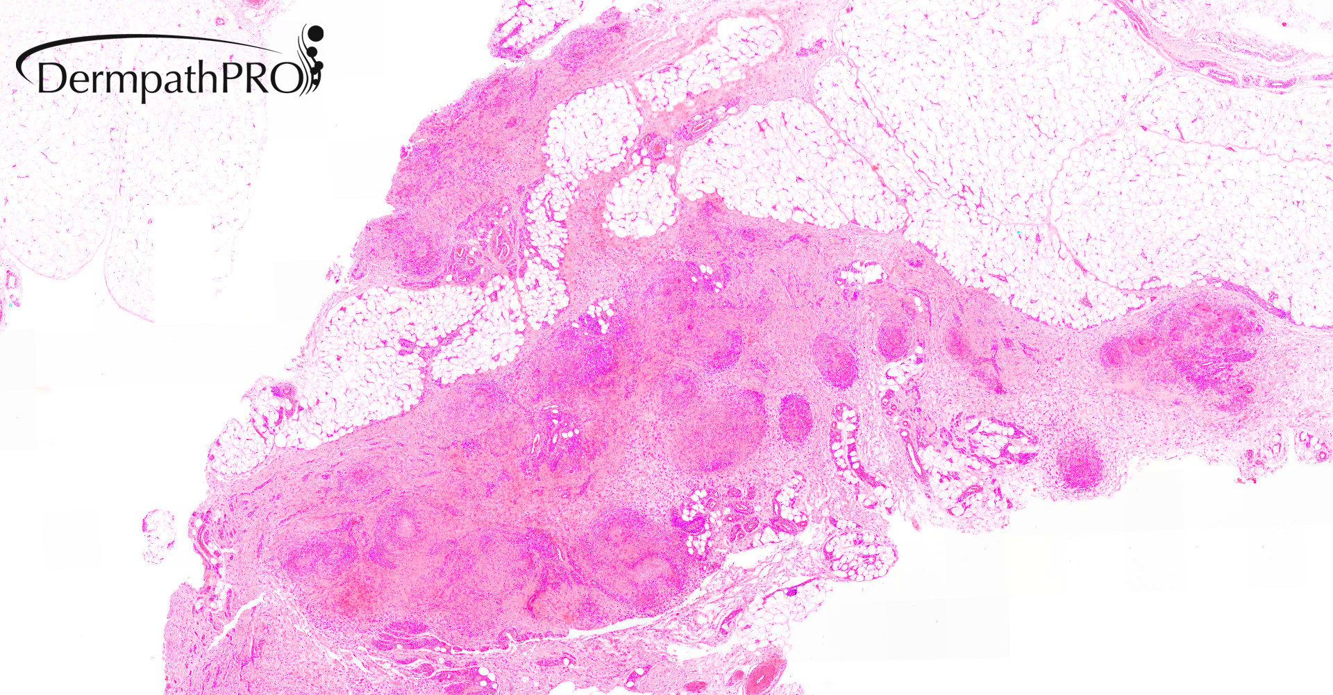
51M, fibrous fatty lesion/nodule left forearm. Multiple lesions over forearms, ex-tensor aspects.

  Report Record

User Feedback


Saleem Taibjee

Posted

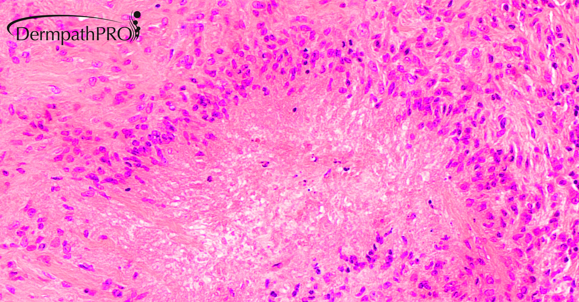
OK, Vincenzo. As requested! Alcian blue x 2 areas

01541_20.0xAB.2 labelled.jpg

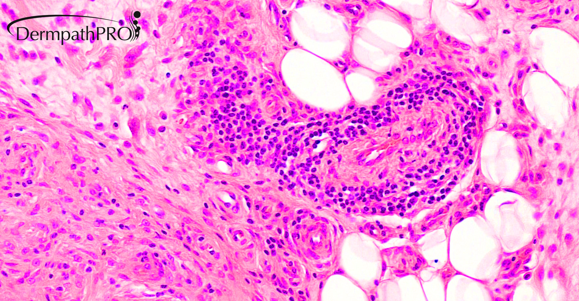
01541_20.0xAB.3 labelled.jpg


Share this comment


Link to comment
share_externally

Krishnakumar subramanian

Posted

sub cutaneous Granuloma annulare


Share this comment


Link to comment
share_externally

John Zhang

Posted

If we can rule out mycobacterial infections, this can be either rheumatoid nodule (if history of rheumatoid arthritis) or deep GA. Anatomic site is good for rheumatoid nodule.

Share this comment


Link to comment
share_externally

Meenakshi Batrani

Posted

Juxta-articular GA (Pseudorehumatoid nodule). But clinical correlation and other ancillary tests to exclude Rheumatoid nodule, and also Fite to exclude atypical mycobacterial infection. 

Share this comment


Link to comment
share_externally

Victor Delgado

Posted

Agree with all of you guys. Deep GA vs. Rehumatoid Nodule. Thanks for sharing. Beautiful!!!

Share this comment


Link to comment
share_externally

Saleem Taibjee

Posted

Thanks for the interesting comments. 

Yes, the main differential diagnosis in this case is deep granuloma annulare vs rheumatoid nodule. In fact, this case was understandably originally signed out as Rh nodule by the original pathologist.

The table from McKee is shown below showing the main discriminating features between palisading granulomatous inflammation.

Differential diagnosis.jpg

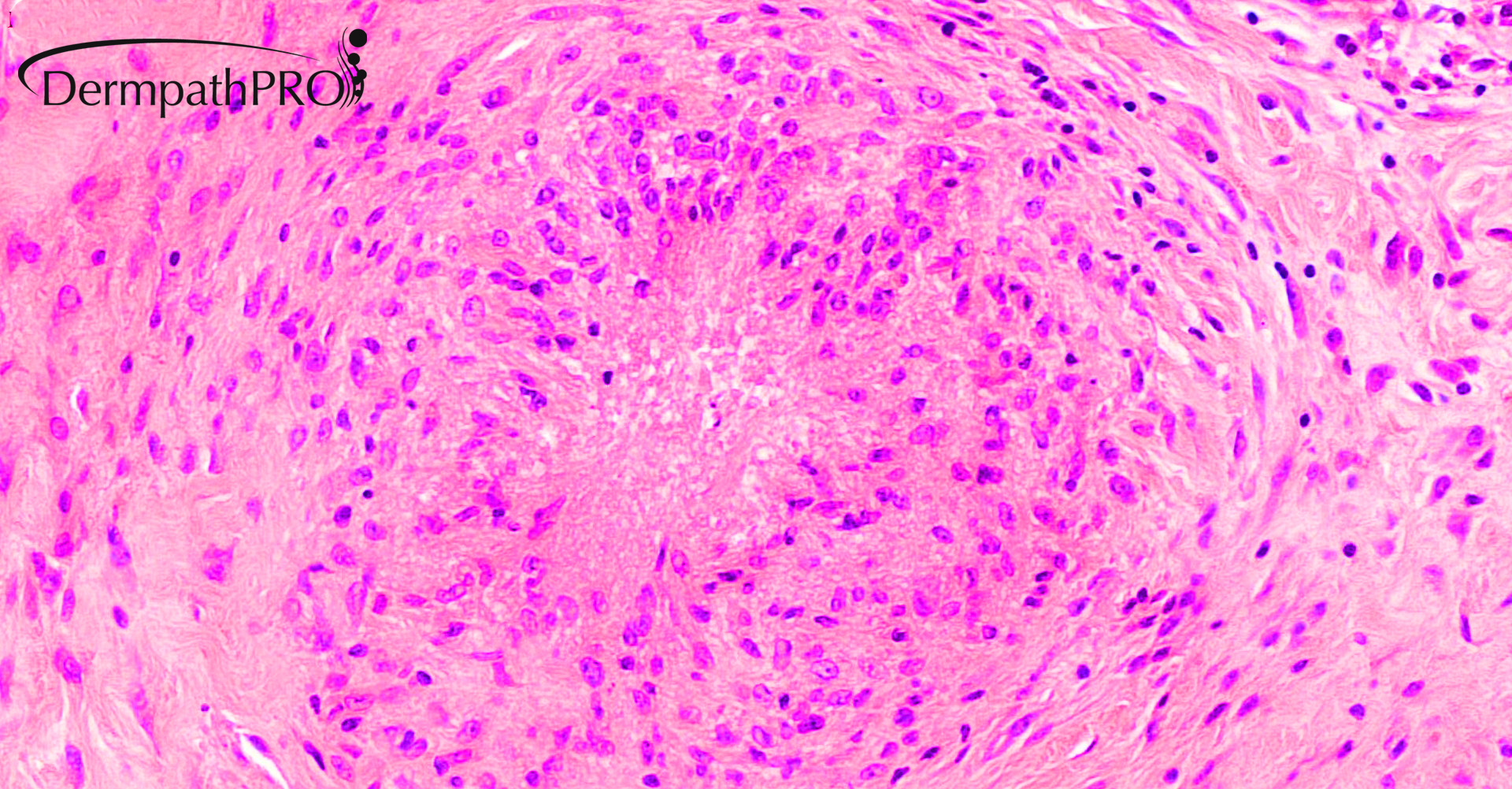
As you can see, detection of mucin (either on H&E or Alcian blue) can be very helpful in deep GA. This case also had sarcoidal features, which is a further variation of GA histologically. I also show the Martius Scarlet Blue below, which seems negative i.e. no evidence of fibrin, as would be perhaps expected if Rh nodule.

01541_10.0x MSB labelled.jpg

It later transpired, once the patient was seen in Dermatology, that the patient had a polymorphous cutaneous picture (see example photo below of annular plaques in this patient), much more in keeping with GA. Interestingly other biopsies on this patient showed more 'conventional' GA or 'interstitial' GA patterns. A good example of the need for clinical correlation and multiple biopsies.

GA clinical.jpg

Share this comment


Link to comment
share_externally

Dr. Mona Abdel-Halim

Posted

Lovely! CPC is the key in dermatopathology!

Share this comment


Link to comment
share_externally

vincenzo

Posted

Nice case and interesting learning points. 

Share this comment


Link to comment
share_externally

Krishnakumar subramanian

Posted

Thanks a lot for sharing this case Dr

Retrospective must we have also thought of epithelioid sarcoma in DD. i sew there is no atypia, inputs welcome

Share this comment


Link to comment
share_externally

John Zhang

Posted

Awesome case. Thanks Dr. Taibjee!

Share this comment


Link to comment
share_externally


Join the conversation

You can post now and register later. If you have an account, sign in now to post with your account.

Guest
Add a comment...

×   Pasted as rich text.   Paste as plain text instead

  Only 75 emoji are allowed.

×   Your link has been automatically embedded.   Display as a link instead

×   Your previous content has been restored.   Clear editor

×   You cannot paste images directly. Upload or insert images from URL.

Loading...

×
×
  • Create New...