Jump to content
In this section we have spot diagnoses posted on a daily basis since June 2010, now over 4000! You can review the archived cases and read the suggested diagnoses by users and the final comment by the contributors.
Case are uploaded each week day by 10 am UK time with the correct diagnosis will generally be posted at 8 pm UK time. Why not view the most recent spot diagnosis and proffer a diagnosis?

Case Number : Case 2571 - 14 May 2020 Posted By: Saleem Taibjee

Please read the clinical history and view the images by clicking on them before you proffer your diagnosis.
Submitted Date :
   (0 reviews)

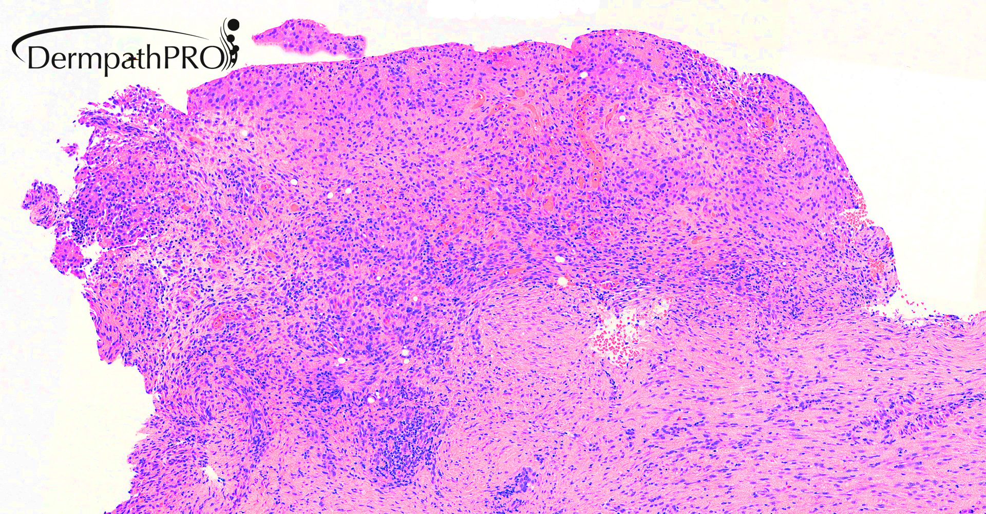
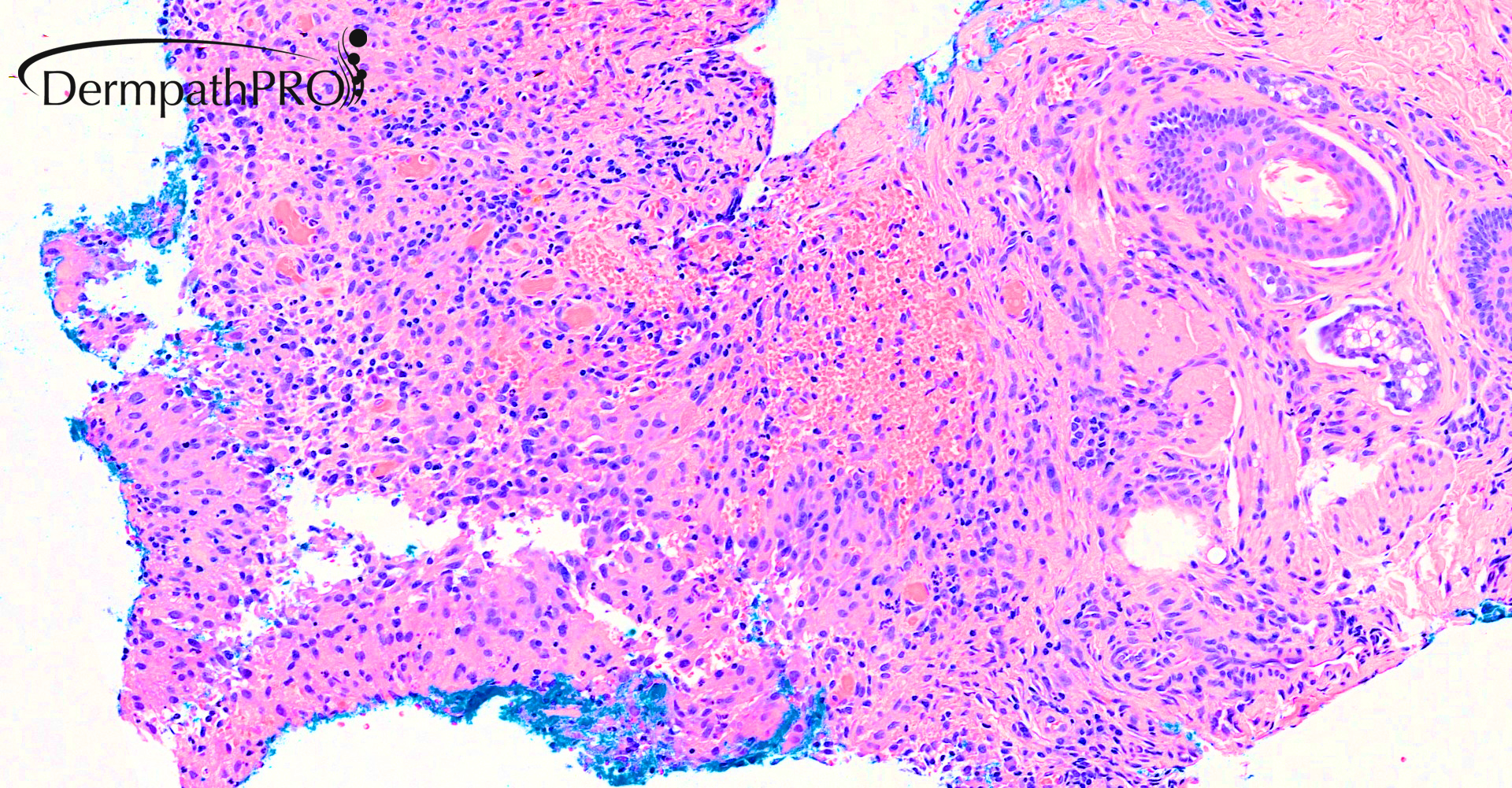
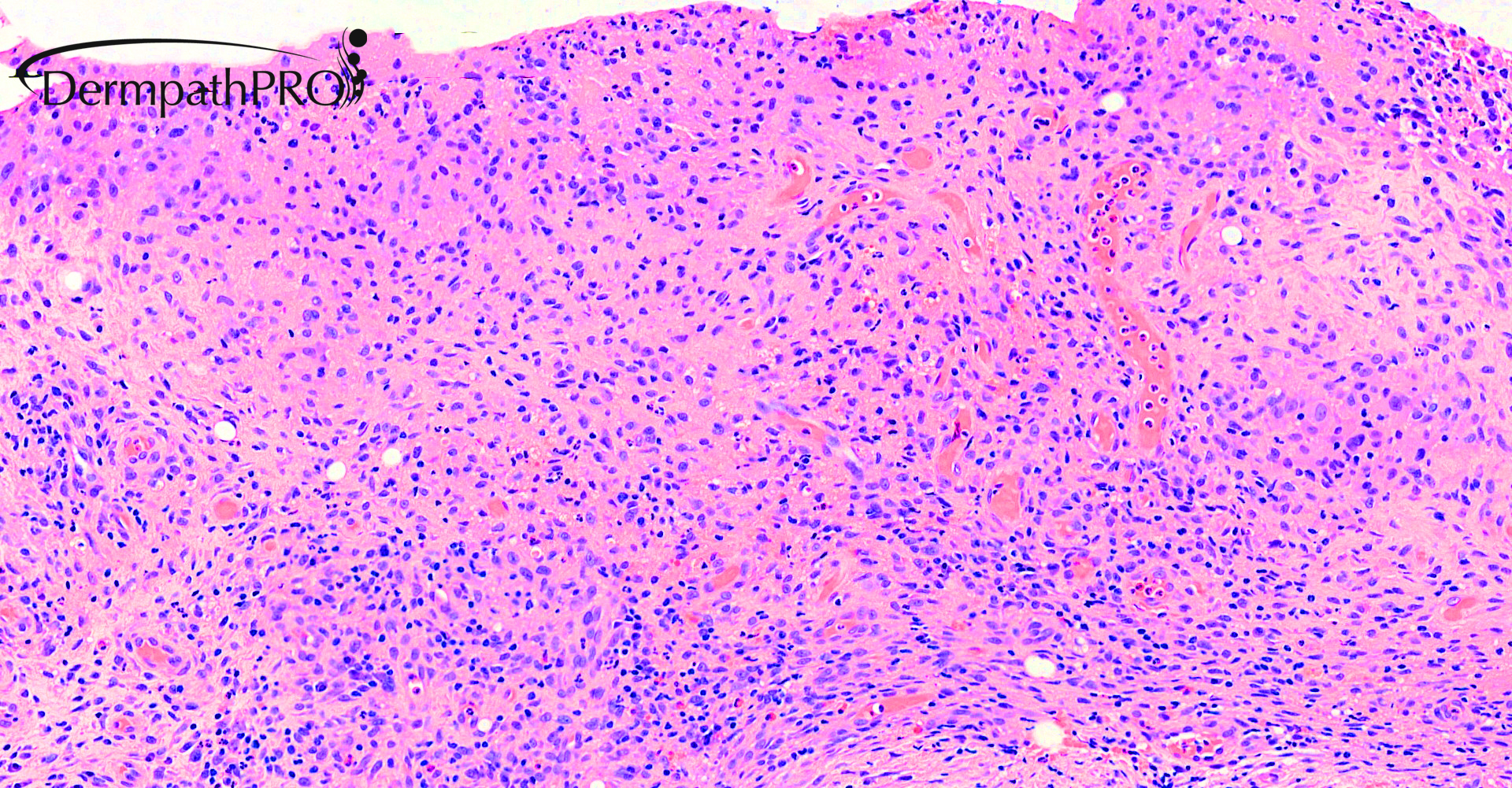
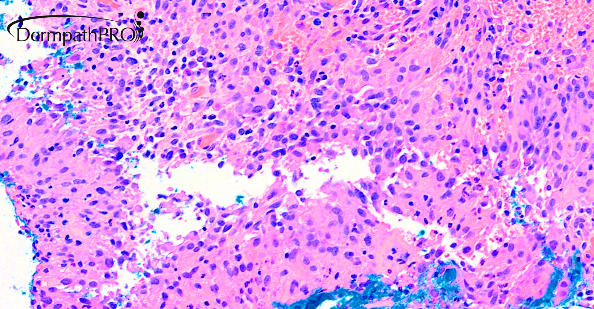
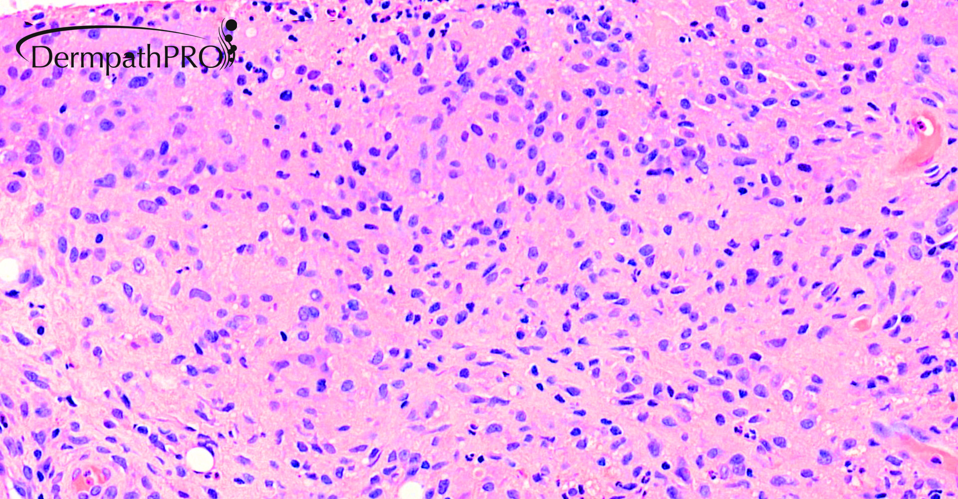
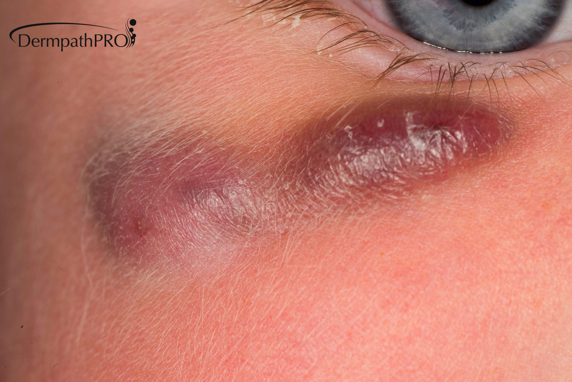
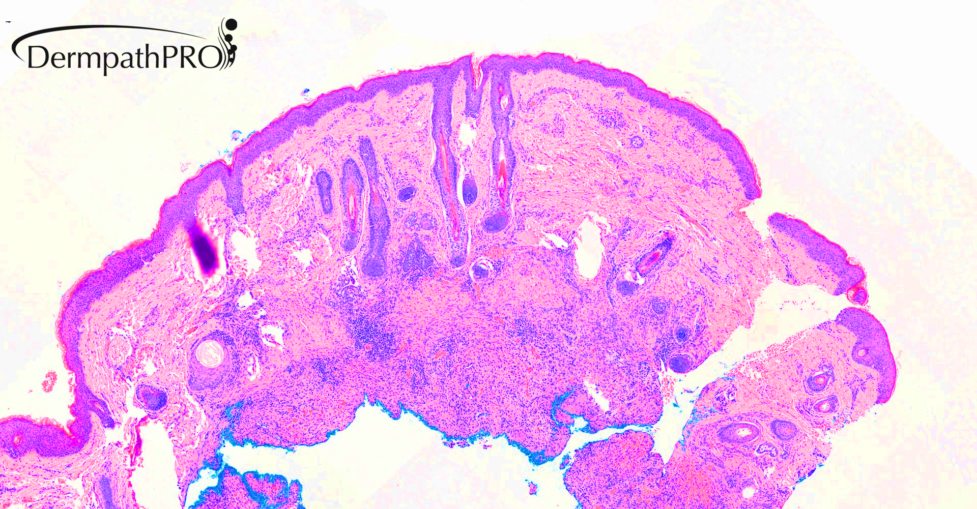
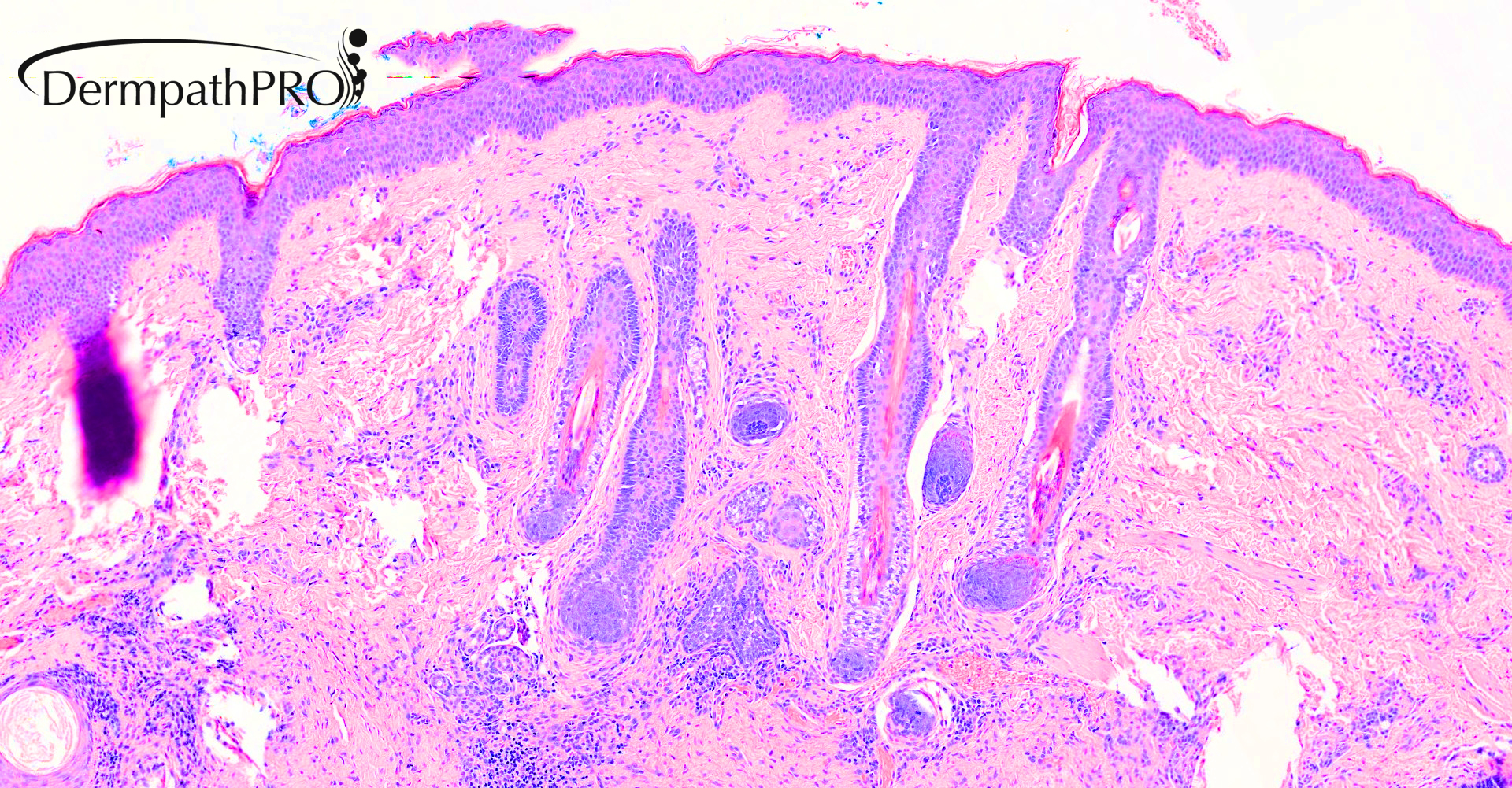
Incisional biopsy right upper cheek: 2 year old girl, 4 month history of enlarging haemorrhagic lump/swelling

  • Like 1
  • Thanks 1
  Report Record

User Feedback


vincenzo

Posted

Difficult. It's a subcutaneous, faintly granulomatous, histiocytosis, whereas LCH is usually dermal with epidermotropism. It's a single lesion, whereas cutaneous LCH is usually multifocal ( can be solitary when arises in bone )...but my spot is Langherans Cell Histiocytosis.

Share this comment


Link to comment
share_externally

Krishnakumar subramanian

Posted

it is a granulomatous inflammation exclude infections like tuberculosis. I have not seen any idiopathic facial granulomatous inflammation  in my part of the world. if tb is negative i would also do s100, Cd68 to see whether it is  Non LCH type histiocytic infiltrate then do BRAFF mutation etc

Share this comment


Link to comment
share_externally

Eman El-Nabarawy

Posted

Same thoughts here.. I like the idea of Idiopathic facial aseptic granuloma (IFAG).

Share this comment


Link to comment
share_externally

vincenzo

Posted

14 hours ago, Krishnakumar subramanian said:

it is a granulomatous inflammation exclude infections like tuberculosis. I have not seen any idiopathic facial granulomatous inflammation  in my part of the world. if tb is negative i would also do s100, Cd68 to see whether it is  Non LCH type histiocytic infiltrate then do BRAFF mutation etc

I never saw this pathology, as well. 

Share this comment


Link to comment
share_externally

Anil Patki

Posted

Epithelioid fibrous histiocytoma

Share this comment


Link to comment
share_externally

Saleem Taibjee

Posted

This was a patient I had seen in clinic, the haemorrhagic quality and initial progression of the lesion rather confused me, and this was compounded by an ultrasound scan in which the radiologist also considered a vascular lesion.

However, the histology was rather granulomatous, special stains were negative.

Hence, I too concluded that this was a case of 'idiopathic facial aseptic granuloma'. I have seen a few cases over the years.

The original description was from a group from France. Boralevi et al. Idiopathic facial aseptic granuloma: a multicentre
prospective study of 30 cases. Pediatric Dermatology 2007. The clinical images from the original publication are shown below.

Cases are exclusively in children, and generally resolve spontaneously, as in this case. The original paper shows the anatomical distribution in their cases, cheek is far and away the most common. Also see below.

BW
Saleem

collage original paper IFAG.jpg

IFAG distribution.jpg

Share this comment


Link to comment
share_externally

Dr. Mona Abdel-Halim

Posted

Nice case! Have never seen it before! Thanks for sharing!

Share this comment


Link to comment
share_externally


Join the conversation

You can post now and register later. If you have an account, sign in now to post with your account.

Guest
Add a comment...

×   Pasted as rich text.   Paste as plain text instead

  Only 75 emoji are allowed.

×   Your link has been automatically embedded.   Display as a link instead

×   Your previous content has been restored.   Clear editor

×   You cannot paste images directly. Upload or insert images from URL.

Loading...

×
×
  • Create New...