Jump to content
In this section we have spot diagnoses posted on a daily basis since June 2010, now over 4000! You can review the archived cases and read the suggested diagnoses by users and the final comment by the contributors.
Case are uploaded each week day by 10 am UK time with the correct diagnosis will generally be posted at 8 pm UK time. Why not view the most recent spot diagnosis and proffer a diagnosis?

Case Number : Case 2576 - 21 May 2020 Posted By: Saleem Taibjee

Please read the clinical history and view the images by clicking on them before you proffer your diagnosis.
Submitted Date :
   (0 reviews)

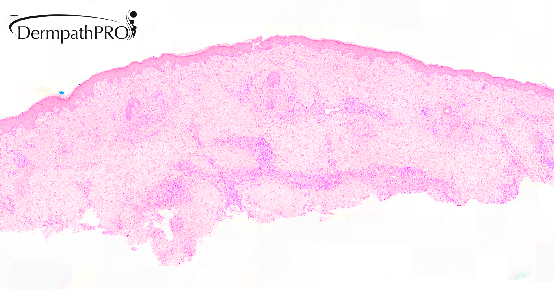
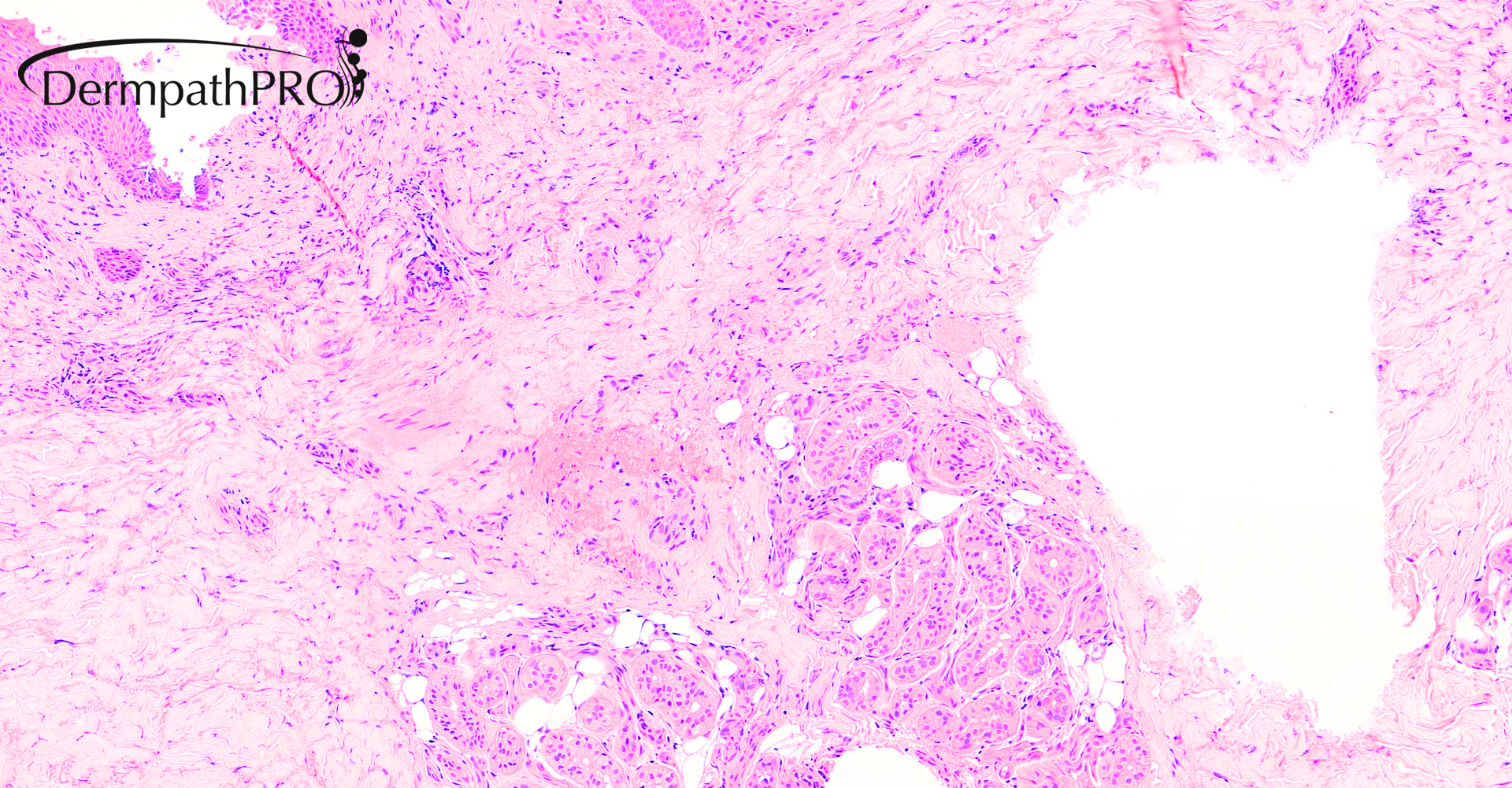
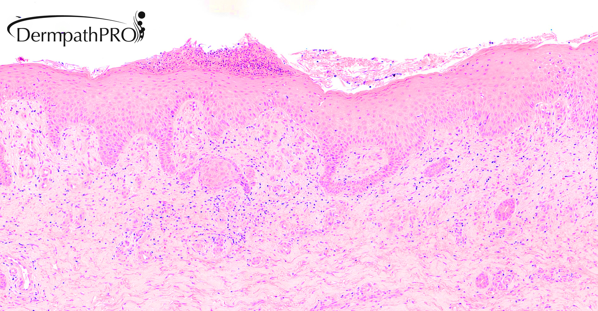
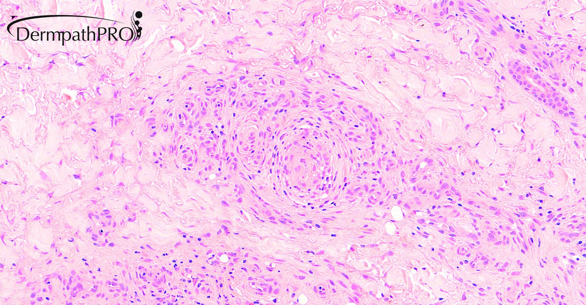
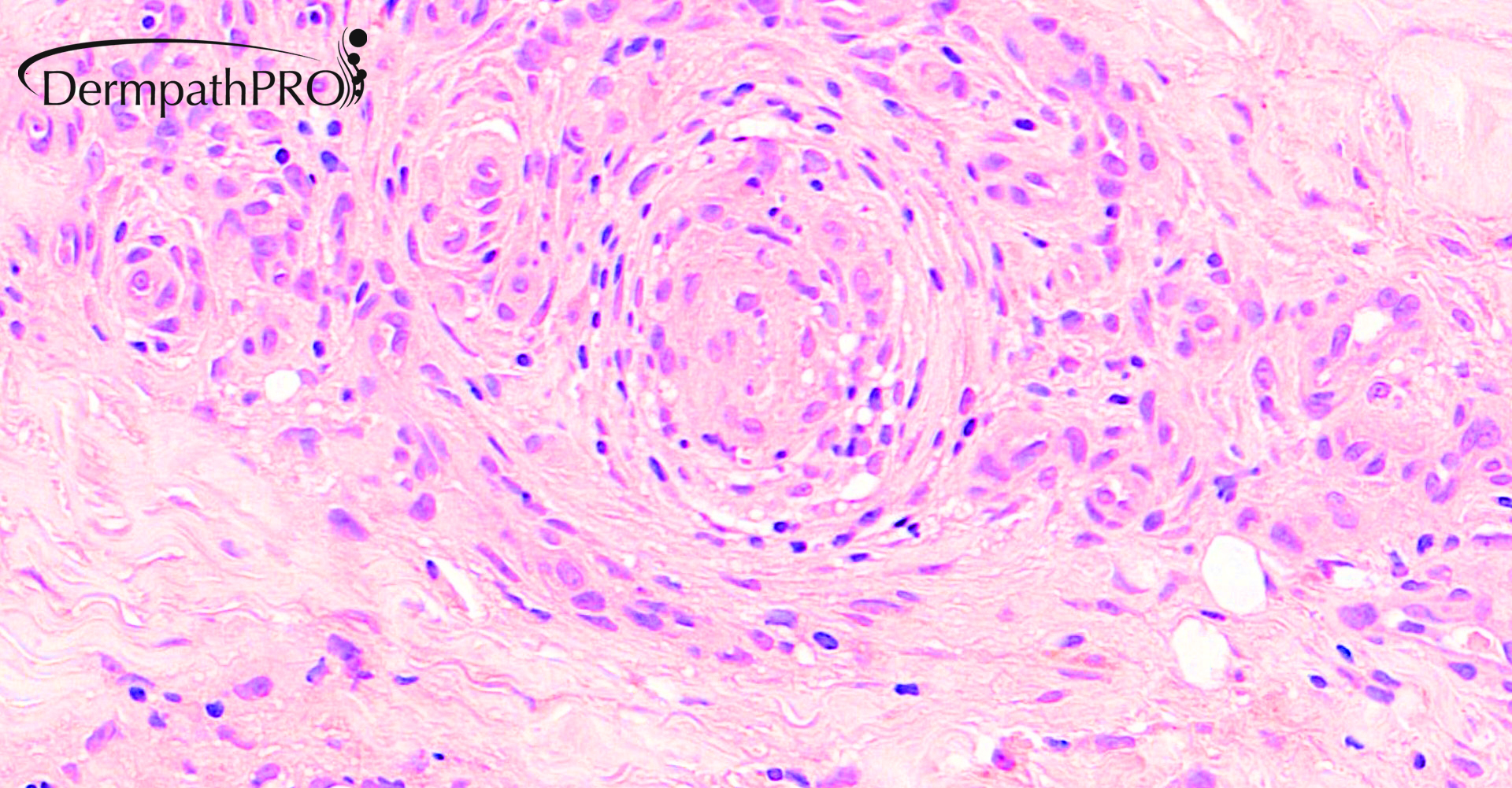
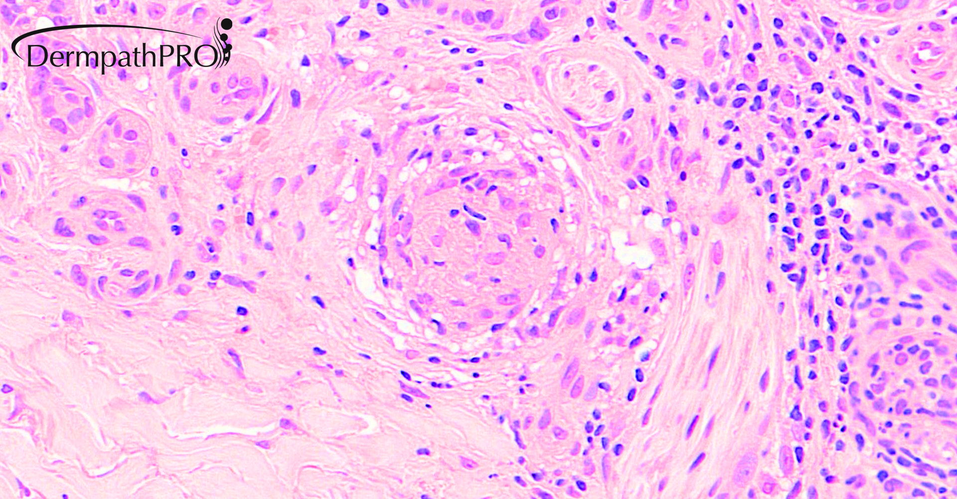
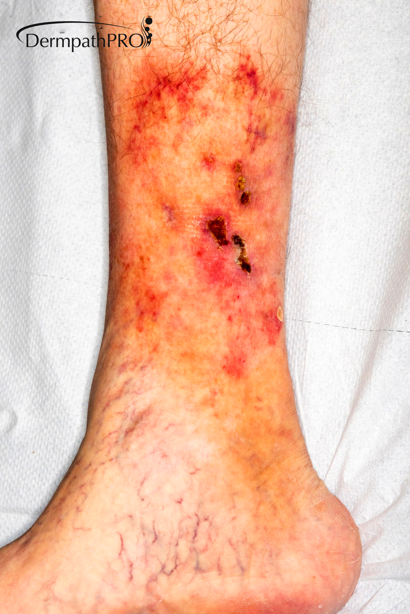
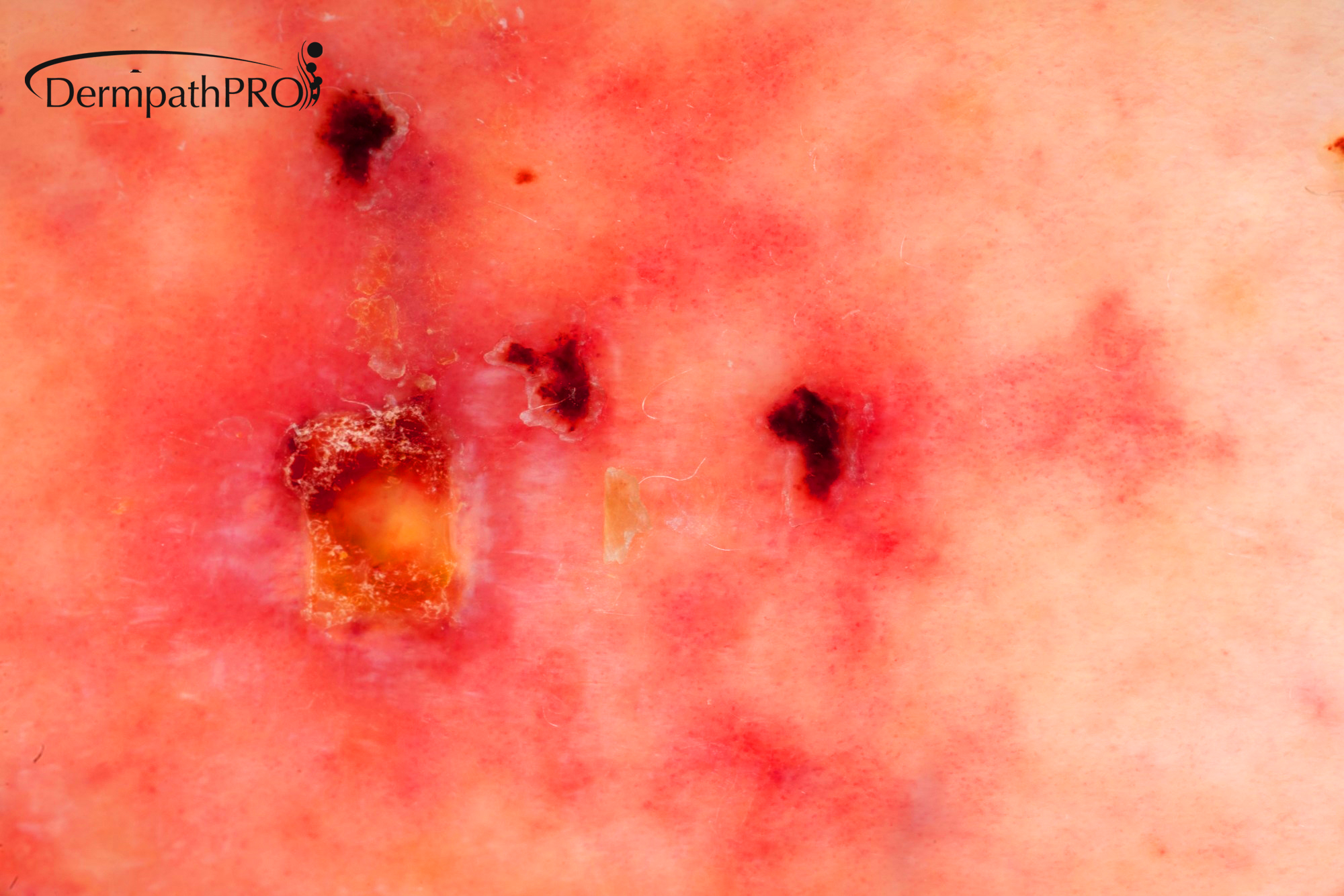
79M, erythematous changes and superficial ulceration right lower leg.


  • Like 2
  Report Record

User Feedback


Meenakshi Batrani

Posted

Seems like occlusive vasculopathy- livedoid vasculopathy. There is also significant involvement with lymphocytic infiltrate in arteriole-small artery sized vessels in deep dermal/subcutis- will also consider lymphocytic/thrombophilic macular arteritis (considered by some as an indolent variant of cutaneous PAN). 

Share this comment


Link to comment
share_externally

vincenzo

Posted

What about a Tromboangiitis Obliterans ( Buerger )?


Share this comment


Link to comment
share_externally

Krishnakumar subramanian

Posted

vascular damage with occlusion with lymphocytes and histiocytes with arrangement of fibrosis around vessels. the pathology is happening at deep dermis and SC level. there is no evidence of stasis

Lymphocytic thrombophilic arteritis-usually young women-and rare to ulcerate.


Share this comment


Link to comment
share_externally

Anil Patki

Posted

Is the patient hypertensive? If yes, one should consider Martorell hypertensive ischemic ulcers.

Share this comment


Link to comment
share_externally

Saleem Taibjee

Posted

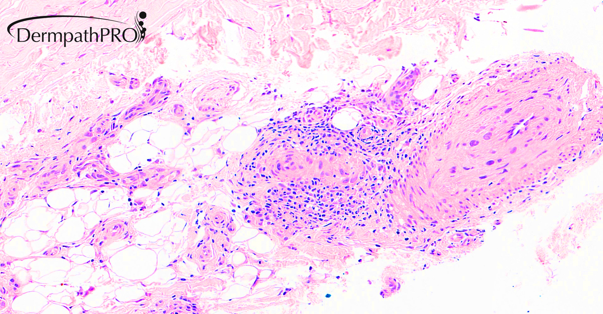
Thanks. this is a case of cutaneous polyarteritis nodosa. The livedoid lesions clinically would fit very well.

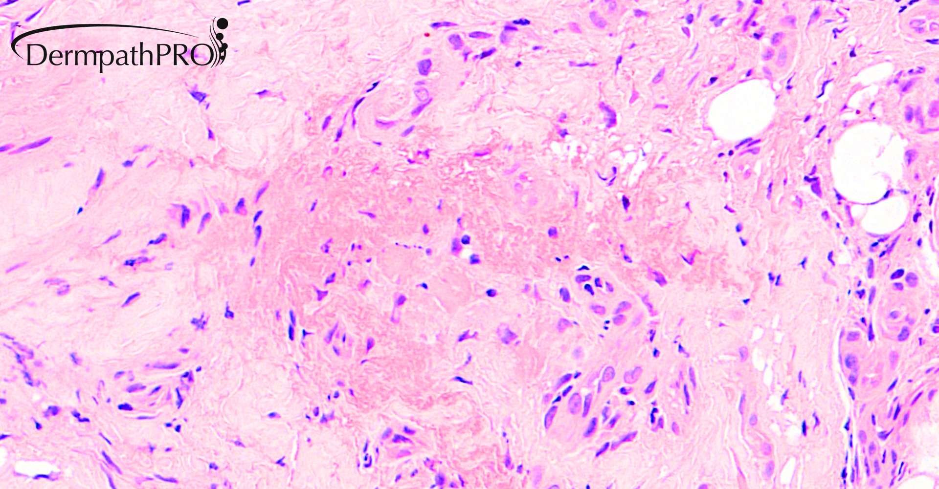
The case nicely highlights the pitfalls of histology in chronic cutaneous PAN, and reflects the ‘lives of lesions’. Here a relatively late stage was biopsied by the clinician first seeing the patient, and hence we see lymphocytic inflammation, lacking neutrophils, absence of fibrinoid necrotizing vasculitis. The images above also show the re-canalisation of previously involved vessels in this later stage. PAN may require examination of multiple levels to reveal the most florid pathology, and repeat biopsies. To reflect the comments from others above, the differential diagnosis would include cutaneous lymphocytic thrombophilic (macular) arteritis (probably on the same spectrum as cPAN), systemic polyarteritis nodosa, microscopic polyangiitis and thrombophlebitis.
By the time I met the patient, the lesions were more in the active phase, and repeat biopsy (see images below) showed more convincing vasculocentric inflammation, also including neutrophils, and even a focal hint of fibrinoid change.
I wish to acknowledge 2 people here:
Firstly, Dr Eduardo Calonje, who had suggested the diagnosis of cPAN based on the more subtle first biopsy above.
Secondly, Dr Craig Wakeham, the General Practitioner first seeing the patient. Dr Wakeham sadly died of COVID-19 recently. He was a wonderful doctor and person, and great loss to our community in Dorset, England.
09799_20.0xb.jpg
09799_20.0x.jpg

Share this comment


Link to comment
share_externally


Join the conversation

You can post now and register later. If you have an account, sign in now to post with your account.

Guest
Add a comment...

×   Pasted as rich text.   Paste as plain text instead

  Only 75 emoji are allowed.

×   Your link has been automatically embedded.   Display as a link instead

×   Your previous content has been restored.   Clear editor

×   You cannot paste images directly. Upload or insert images from URL.

Loading...

×
×
  • Create New...