Jump to content
In this section we have spot diagnoses posted on a daily basis since June 2010, now over 4000! You can review the archived cases and read the suggested diagnoses by users and the final comment by the contributors.
Case are uploaded each week day by 10 am UK time with the correct diagnosis will generally be posted at 8 pm UK time. Why not view the most recent spot diagnosis and proffer a diagnosis?

Case Number : Case 2694 - 03 November 2020 Posted By: Iskander H. Chaudhry

Please read the clinical history and view the images by clicking on them before you proffer your diagnosis.
Submitted Date :
   (0 reviews)

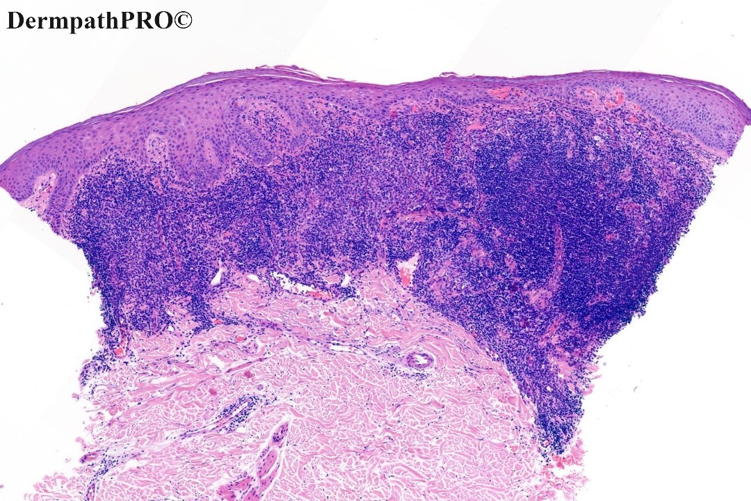
75M, Biopsy upper back - pink patch gradually increasing. ?Bowen's


  Report Record

User Feedback


Richard Logan

Posted

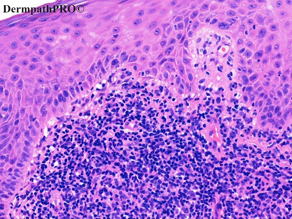
These sections show a dense, sheeted, predominantly lymphocytic infiltrate occupying the upper dermis.  The infiltrate also includes histiocytes, plasmacytoid cells and a few rather superficially located neutrophils .  There is only trivial lymphocytic epidermal exocytosis and lymphocyte cytology is not obviously atypical.  There is a minor degree of basal vacuolation.

Immunohistochemistry is essential to determine whether this is a lymphomatous, or pseudolymphomatous process.

Share this comment


Link to comment
share_externally

Admin_Dermpath

Posted

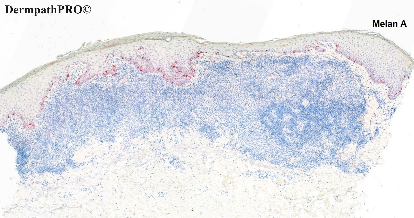
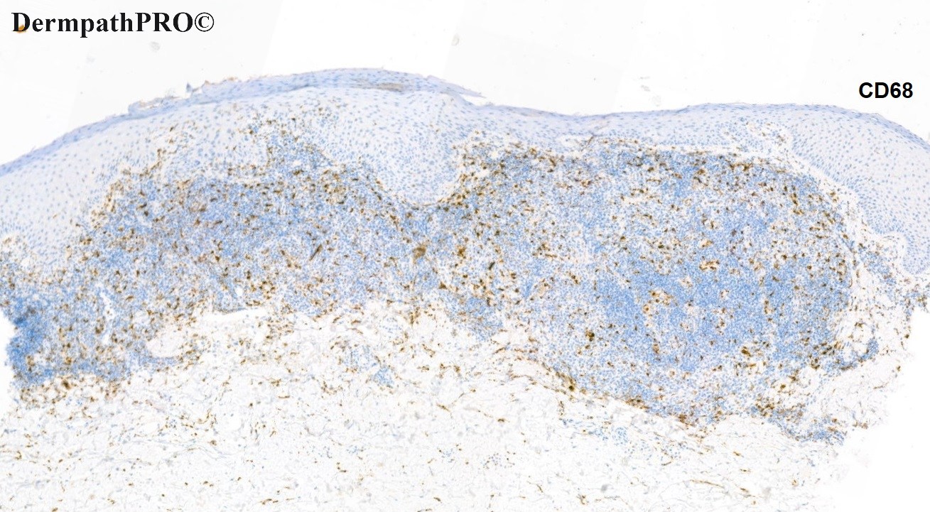
Case updated with Immunohistochemistry stains.

Share this comment


Link to comment
share_externally

My first thought was similar to Vincenzo's but I thought there was basal keratinocyte atypia so I thought it could be inflamed actinic keratosis. However I think keratinocyte atypia can also be seen in lupus. I don't know for sure. Mucin in the deep dermis? 

Share this comment


Link to comment
share_externally

Richard Logan

Posted

Iskander - may we have your thoughts on this one please?

Share this comment


Link to comment
share_externally

Iskander H. Chaudhry

Posted

Dear All. My comments below:

Skin with acute on chronically inflamed lesion with acanthosis and dense dermal inflammation. No
junctional nesting is seen. The lesion is negative for Melan A and scattered CD68 cells are seen in the dermis. The epidermis focally shows mild reactive atypia, however, the main feature is the intense dermal inflammation with possible lymphoid aggregates. Immunochemistry shows a mixed T and B cell infiltrate (lymphoid aggregate)  with surrounding T cell infiltrate (CD4>CD8). CD30 was negative. On clinical discussion the appearances were most in keeping with a pseudo-lymphoma. 

Share this comment


Link to comment
share_externally


Join the conversation

You can post now and register later. If you have an account, sign in now to post with your account.

Guest
Add a comment...

×   Pasted as rich text.   Paste as plain text instead

  Only 75 emoji are allowed.

×   Your link has been automatically embedded.   Display as a link instead

×   Your previous content has been restored.   Clear editor

×   You cannot paste images directly. Upload or insert images from URL.

Loading...

×
×
  • Create New...