Jump to content
In this section we have spot diagnoses posted on a daily basis since June 2010, now over 4000! You can review the archived cases and read the suggested diagnoses by users and the final comment by the contributors.
Case are uploaded each week day by 10 am UK time with the correct diagnosis will generally be posted at 8 pm UK time. Why not view the most recent spot diagnosis and proffer a diagnosis?

Case Number : Case 2696 - 05 November 2020 Posted By: Saleem Taibjee

Please read the clinical history and view the images by clicking on them before you proffer your diagnosis.
Submitted Date :
   (0 reviews)

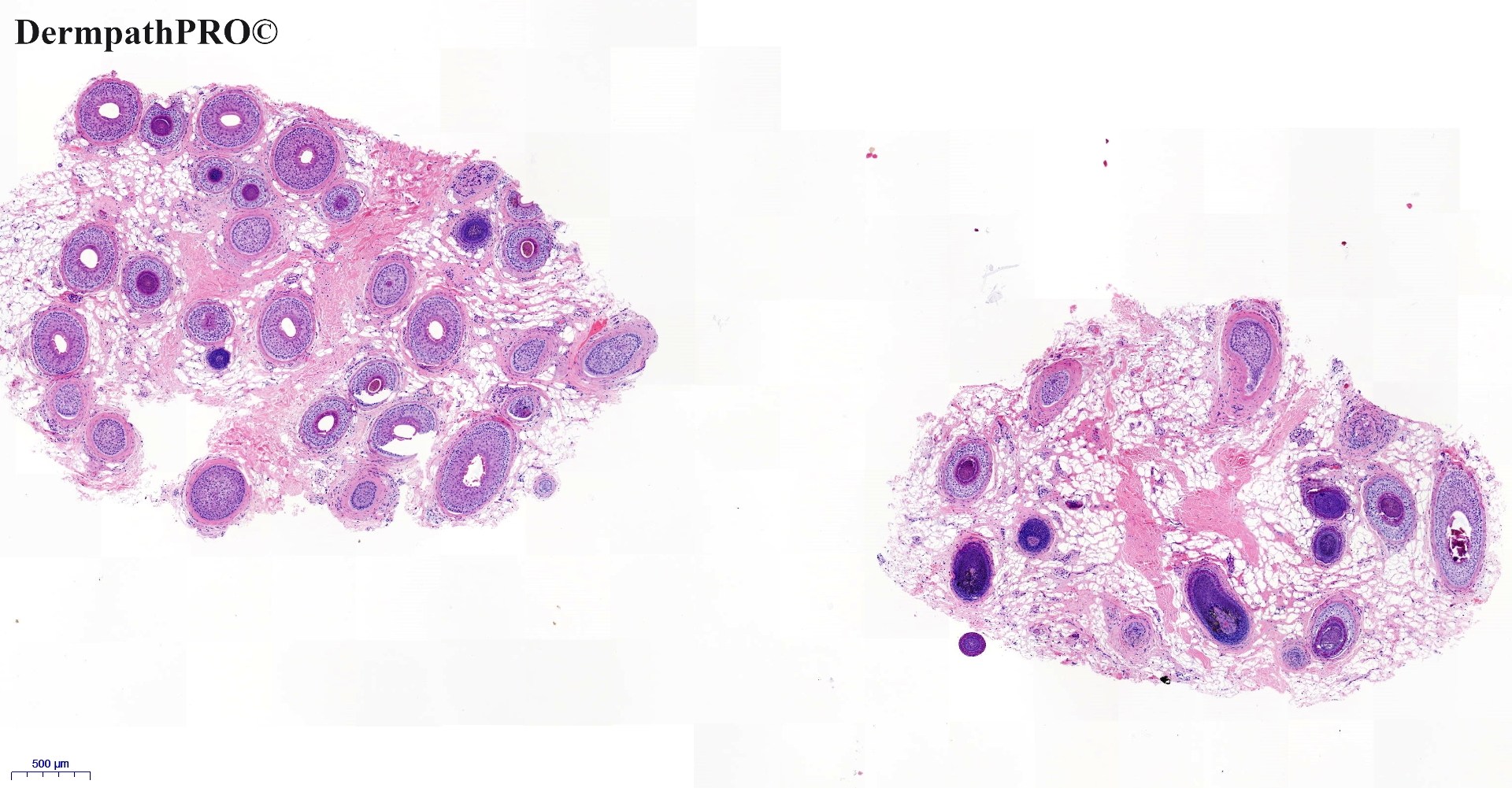
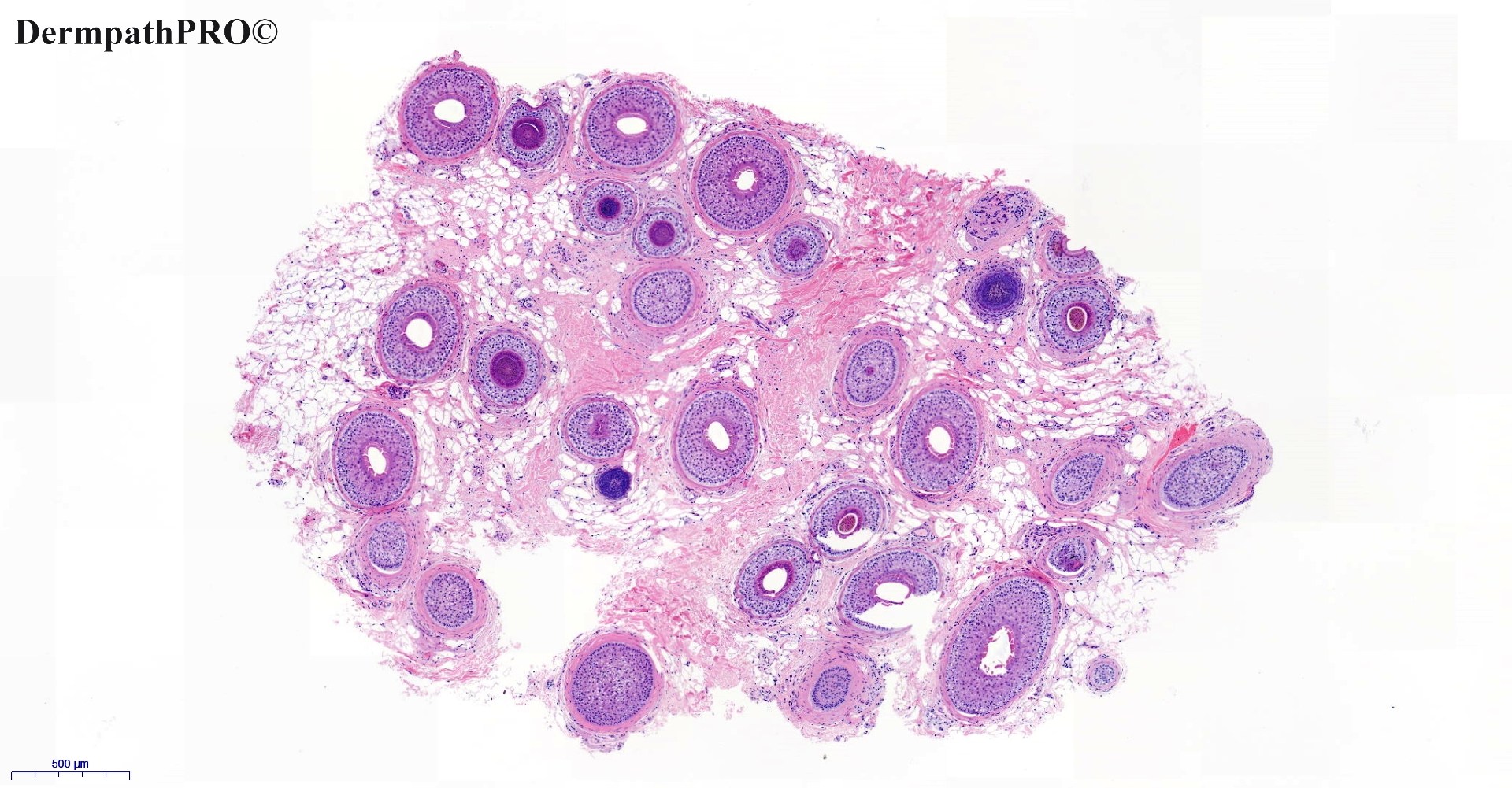
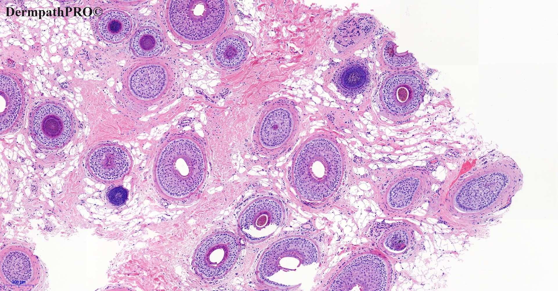
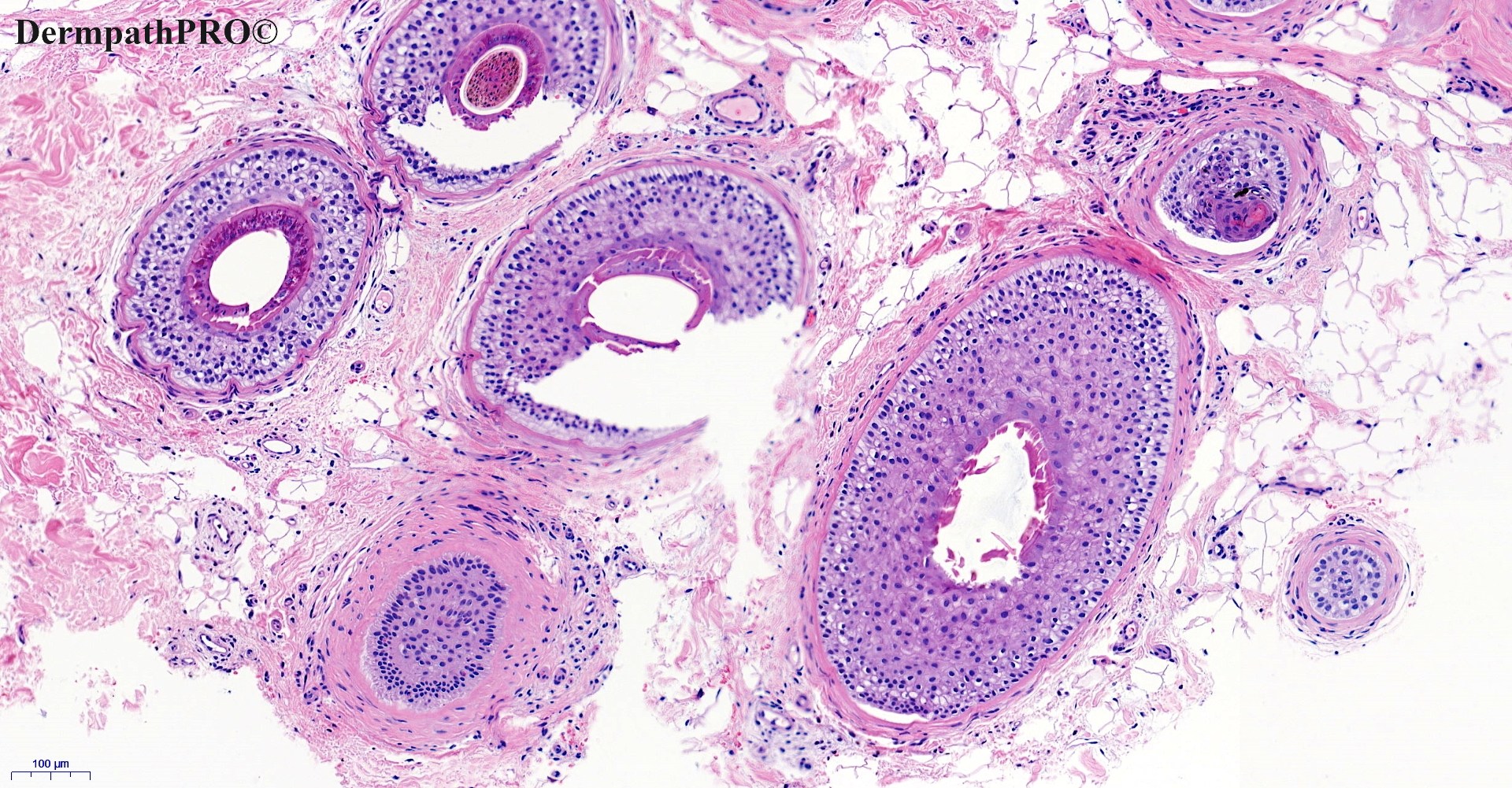
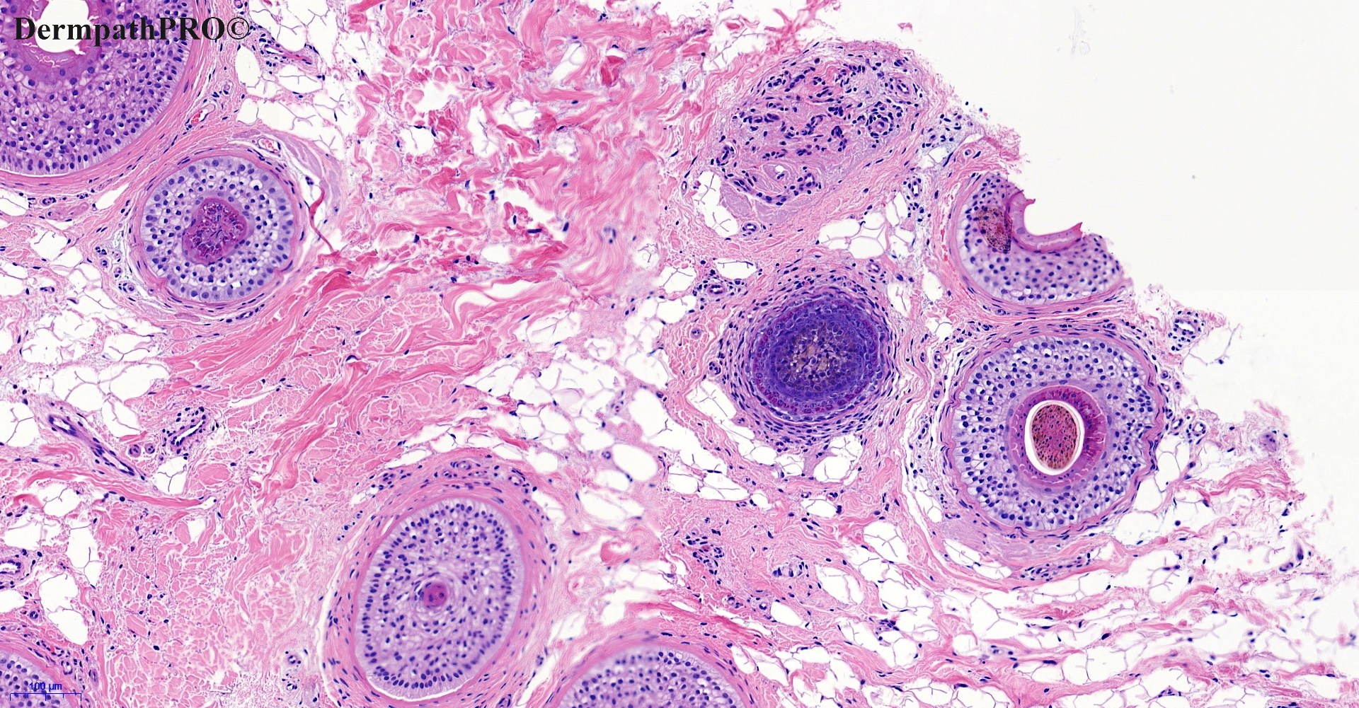
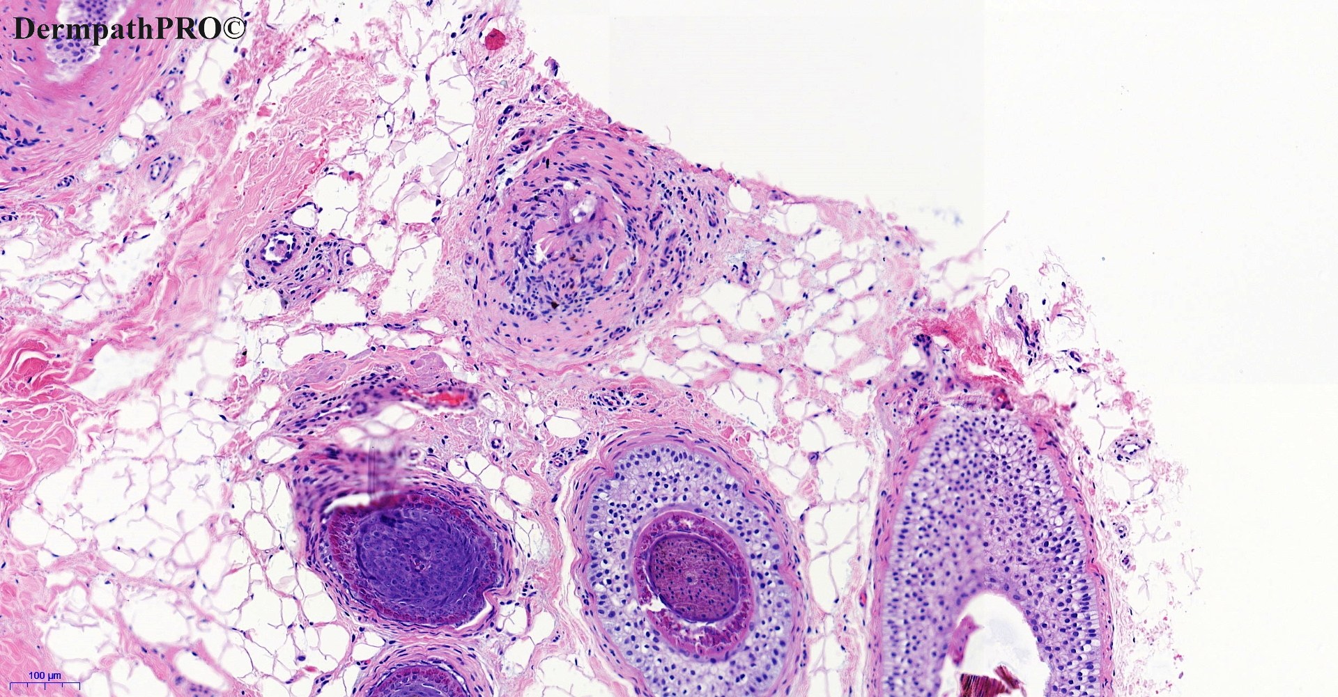
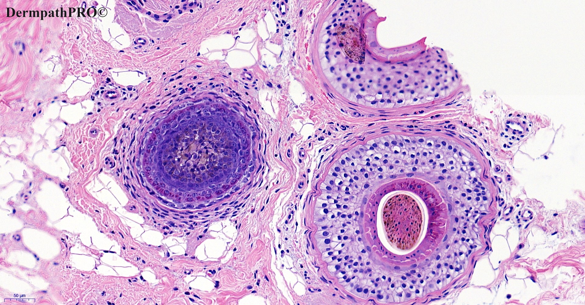
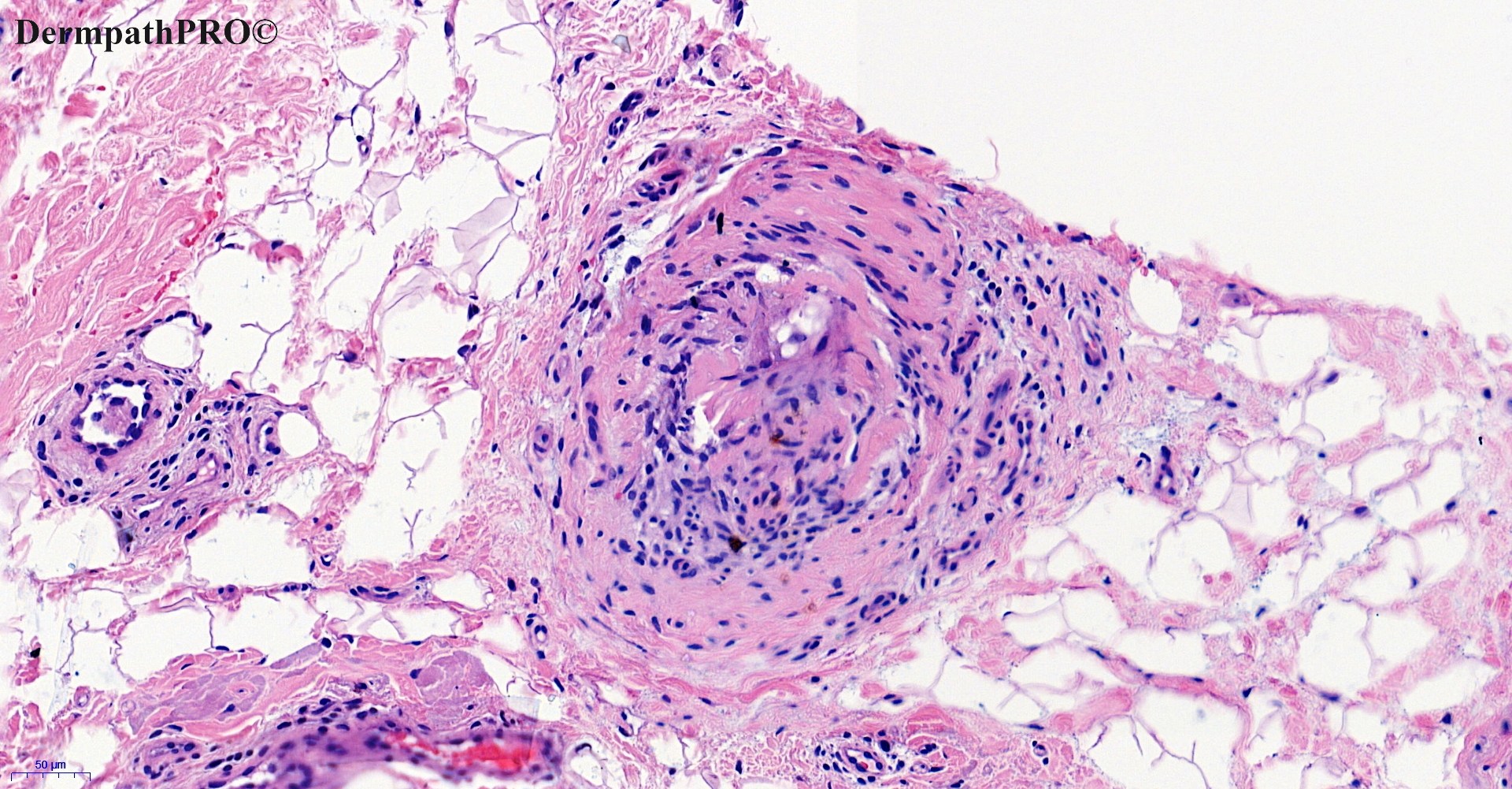
45M, Hair loss – patchy. Scalp feels hot. No pruritus or pain.


  • Thanks 1
  Report Record

User Feedback


Krishnakumar subramanian

Posted

hair follicles  show terminal hairs, hairs showing follicular miniaturization and some hair follicles in telogen/catagen phase. there is fibrosis around follicles with lymphocytes

Alopecia areata

Share this comment


Link to comment
share_externally

I wish I saw a swarm of bees, but: 1) I guess this isn’t the acute phase 2) I’m sure I’m not familiar with this pathological chapter. So I agree with Colleagues. 

Share this comment


Link to comment
share_externally

Saleem Taibjee

Posted

A few more images taken from additional levels, as well as CD3

29583_10.0x L4-6.jpg

29583_20.0x L7-9.jpg

29583_20.0x CD3 b labelled.jpg

29583_20.0x CD3 labelled.jpg

29583_10.0x L10-12b.jpg

29583_20.0x L10-12b.jpg

29583_10.0x L10-12c.jpg

29583_20.0x L10-12d.jpg


Share this comment


Link to comment
share_externally

Saleem Taibjee

Posted

Hi. Yes this is alopecia areata (and correlates with the clinical description of patchy hair loss). I really like this recent case because it is presumably a semi-acute stage, and combines so many of the textbook features of AA in one biopsy.

In the initial images at the level of subcutaneous fat/dermis interface, as others have pointed out, the low-power clue is the combination of miniaturisation with hair cycle shift (with increased number of catagen and telogen follicles). We also see a subtle peri-bulbar lymphocytic infiltrate, and this is then nicely highlighted by the subsequent CD3. There are not really any other good explanations for peri-bulbar inflammation.

The additional levels are at higher levels in the biopsy, and show other features such as pigment casts (not entirely specific for AA, but nonetheless a feature). There is also a combination of miniaturisation, as well as an odd cornification evident within some follicles (but failing to produce hair shafts). The papers suggest that this is likely to be as a result of rapid and abnormal hair cycling (anagen-catagen-telogen) in AA, and can be termed as ‘nanogen’ follicles. Such follicles correspond to empty infundibula or yellow dots on dermoscopic examination.

Of course, this is a good illustration of how horizontal sections can facilitate assessment of non-scarring alopecia.

Share this comment


Link to comment
share_externally


Join the conversation

You can post now and register later. If you have an account, sign in now to post with your account.

Guest
Add a comment...

×   Pasted as rich text.   Paste as plain text instead

  Only 75 emoji are allowed.

×   Your link has been automatically embedded.   Display as a link instead

×   Your previous content has been restored.   Clear editor

×   You cannot paste images directly. Upload or insert images from URL.

Loading...

×
×
  • Create New...