Jump to content
In this section we have spot diagnoses posted on a daily basis since June 2010, now over 4000! You can review the archived cases and read the suggested diagnoses by users and the final comment by the contributors.
Case are uploaded each week day by 10 am UK time with the correct diagnosis will generally be posted at 8 pm UK time. Why not view the most recent spot diagnosis and proffer a diagnosis?

Case Number : Case 2699 - 10 November 2020 Posted By: Uma Sundram

Please read the clinical history and view the images by clicking on them before you proffer your diagnosis.
Submitted Date :
   (0 reviews)

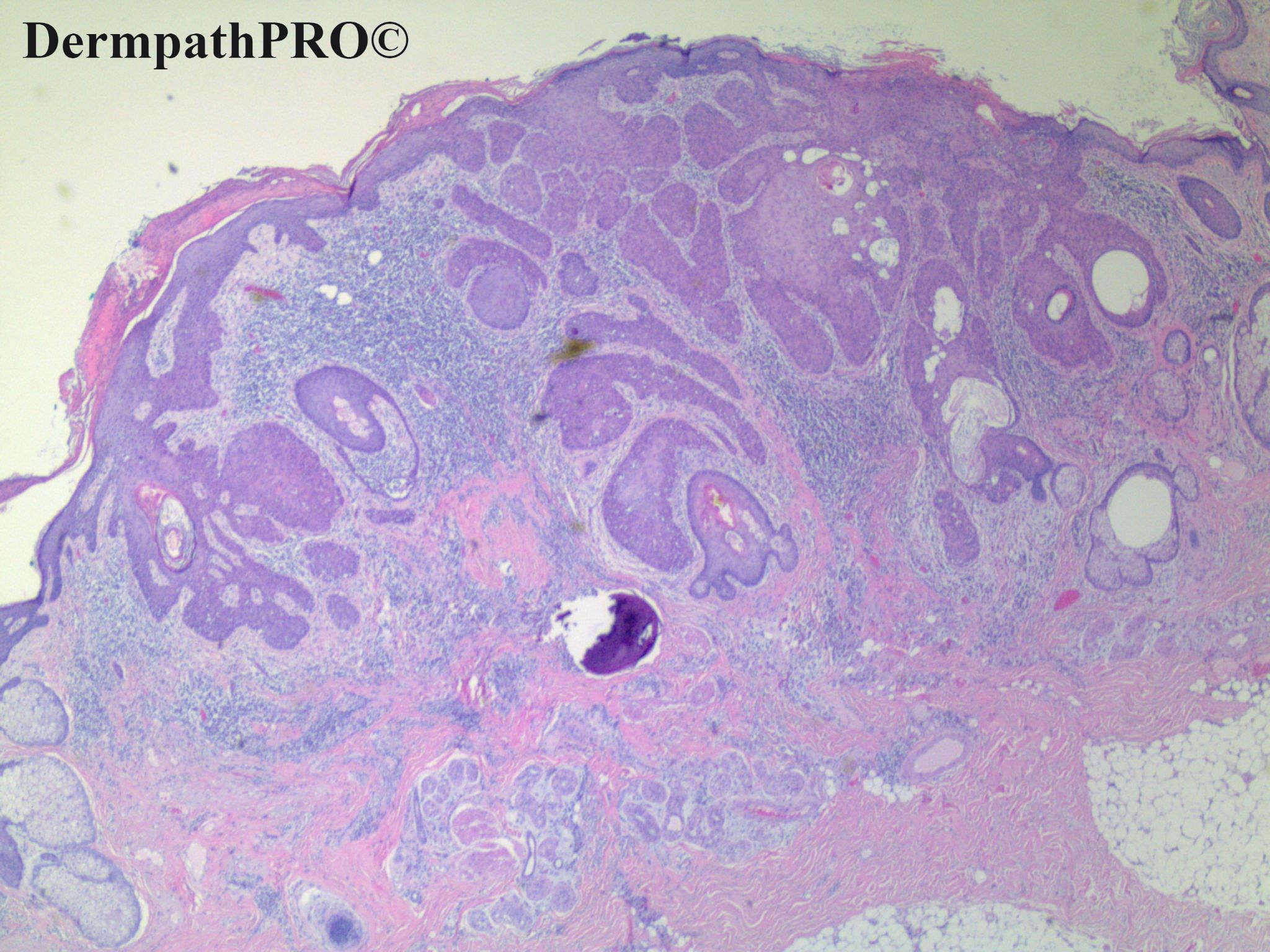
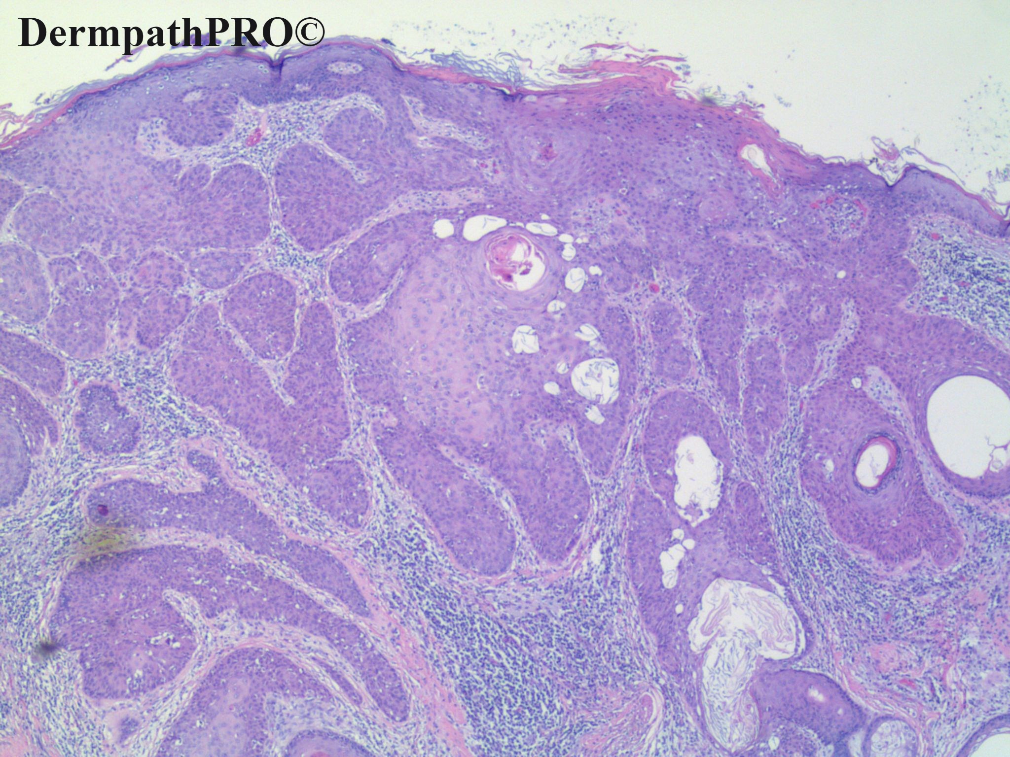
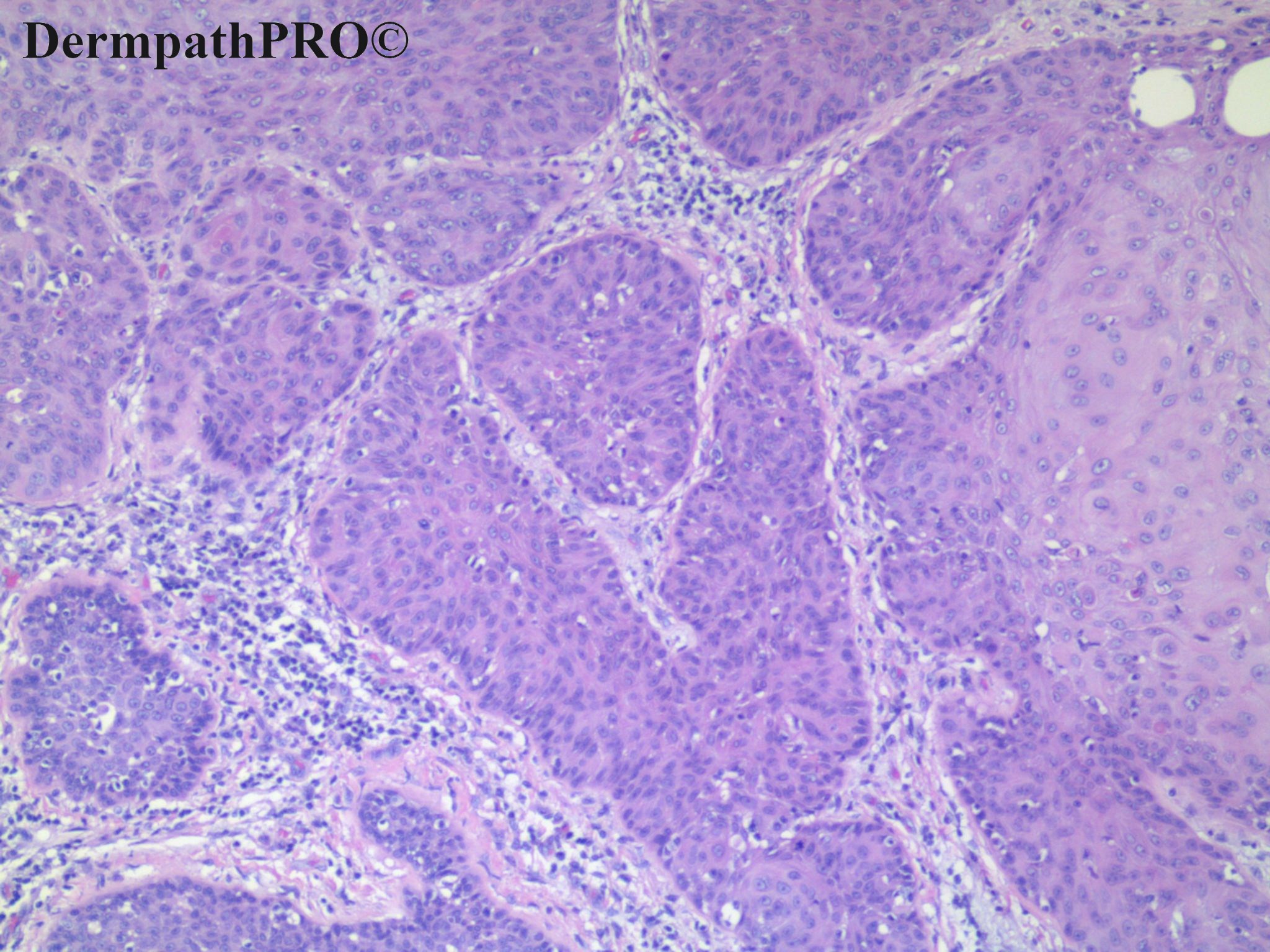
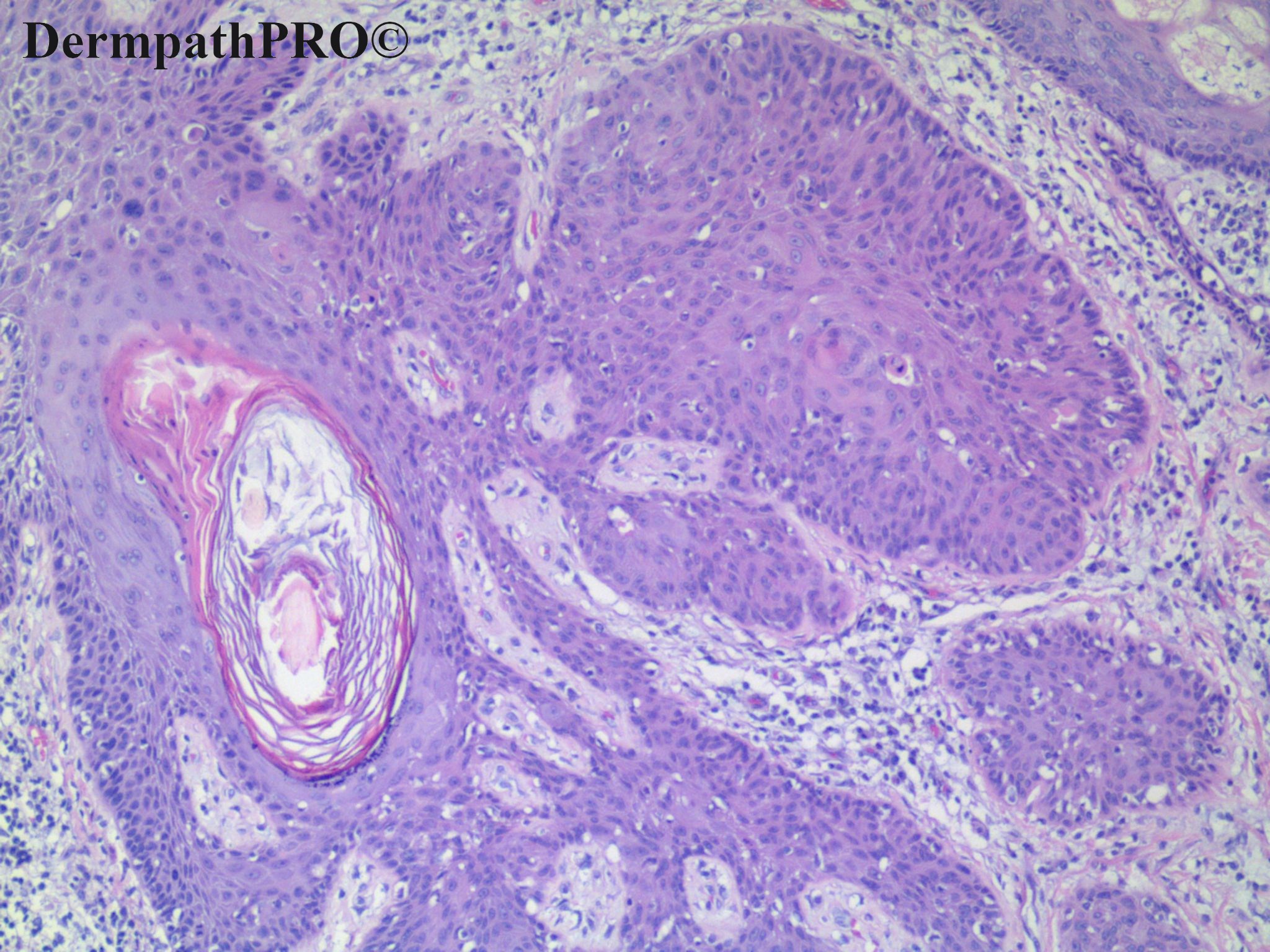
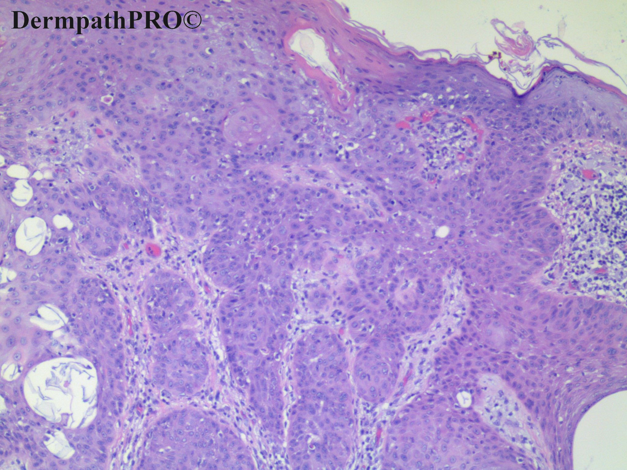
85 year old male with incidental lesion on left cheek and history of non melanoma skin cancer.


  Report Record

User Feedback


Follicle-based growth pattern, with some worrisome feature (  low grade atypia, pseudo-infiltration and trichilemmal differentiation with inverted follicular keratosis )...Difficult!

?Low grade trichilemmal carcinoma.  ?An odd desmoplastic trichilemmoma. ?Low grade fSCC ( as I learned thanks to Richard Carr )-


Share this comment


Link to comment
share_externally

Meenakshi Batrani

Posted

Agree with follicular differentiation. Not sure what to call it. The overall architectural features somewhat appear like a Pilar sheath acanthoma to me. But there is definitely atypia which doesn't go with it. 

Share this comment


Link to comment
share_externally

Uma Sundram

Posted

Hmm... Am surprised there aren't more comments. We too were somewhat stumped for an answer. We finally landed on follicular tumor with trichilemmal differentiation. We did not call it an outright malignancy. It was excised and we did not recommend further therapy.

Share this comment


Link to comment
share_externally

Krishnakumar subramanian

Posted

Thanks a lot Dr, just stumped, did not know how to label it

Share this comment


Link to comment
share_externally

Dr MONA MOHSEN

Posted

On 12/11/2020 at 17:09, Uma Sundram said:

Hmm... Am surprised there aren't more comments. We too were somewhat stumped for an answer. We finally landed on follicular tumor with trichilemmal differentiation. We did not call it an outright malignancy. It was excised and we did not recommend further therapy.

Cant this be BCC.. peripheral palisading is obvious in some nests.?? 

Share this comment


Link to comment
share_externally


Join the conversation

You can post now and register later. If you have an account, sign in now to post with your account.

Guest
Add a comment...

×   Pasted as rich text.   Paste as plain text instead

  Only 75 emoji are allowed.

×   Your link has been automatically embedded.   Display as a link instead

×   Your previous content has been restored.   Clear editor

×   You cannot paste images directly. Upload or insert images from URL.

Loading...

×
×
  • Create New...