Jump to content
In this section we have spot diagnoses posted on a daily basis since June 2010, now over 4000! You can review the archived cases and read the suggested diagnoses by users and the final comment by the contributors.
Case are uploaded each week day by 10 am UK time with the correct diagnosis will generally be posted at 8 pm UK time. Why not view the most recent spot diagnosis and proffer a diagnosis?

Case Number : Case 2701 - 12 November 2020 Posted By: Saleem Taibjee

Please read the clinical history and view the images by clicking on them before you proffer your diagnosis.
Submitted Date :
   (0 reviews)

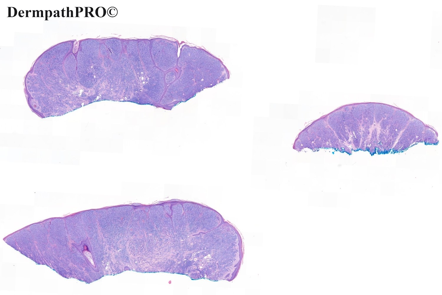
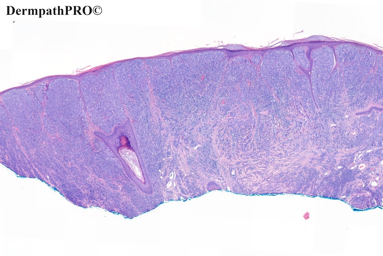
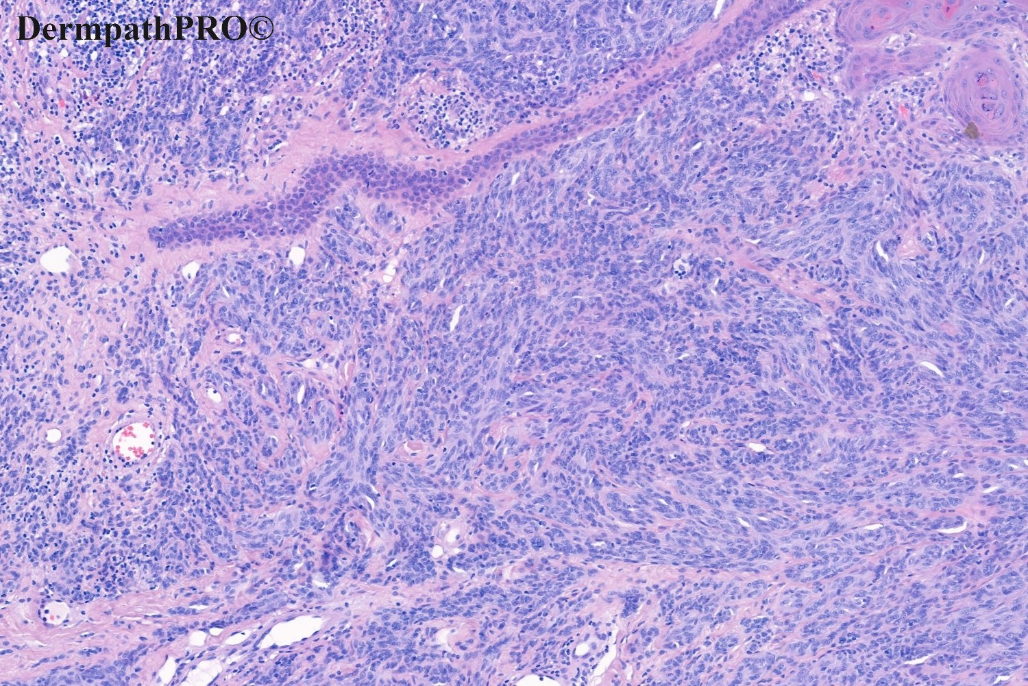
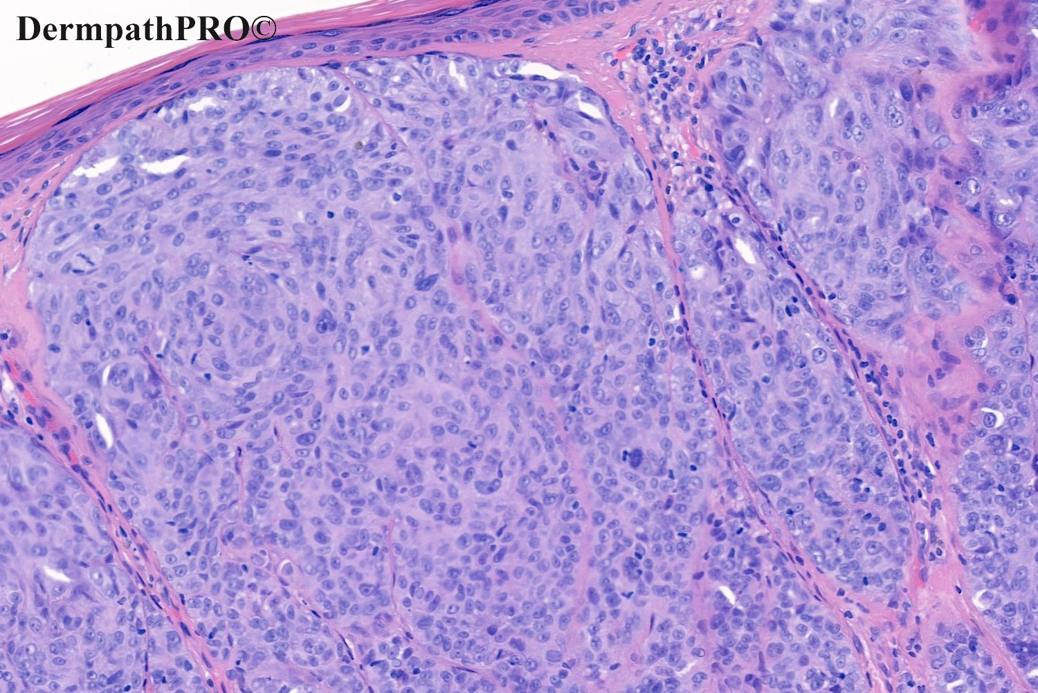
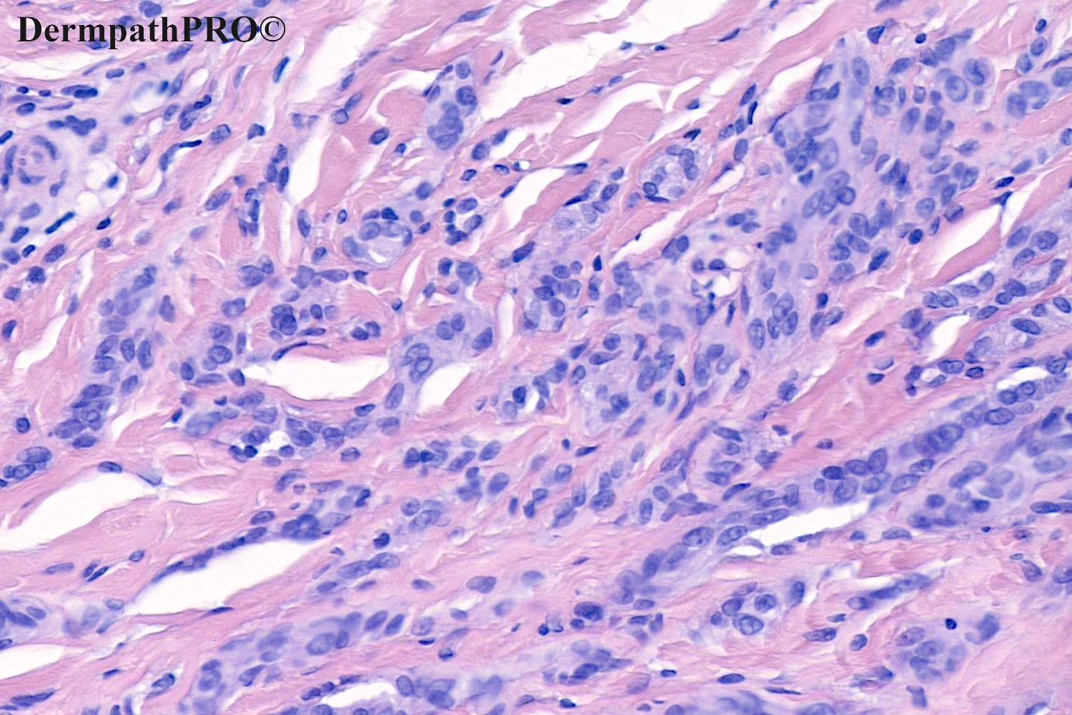
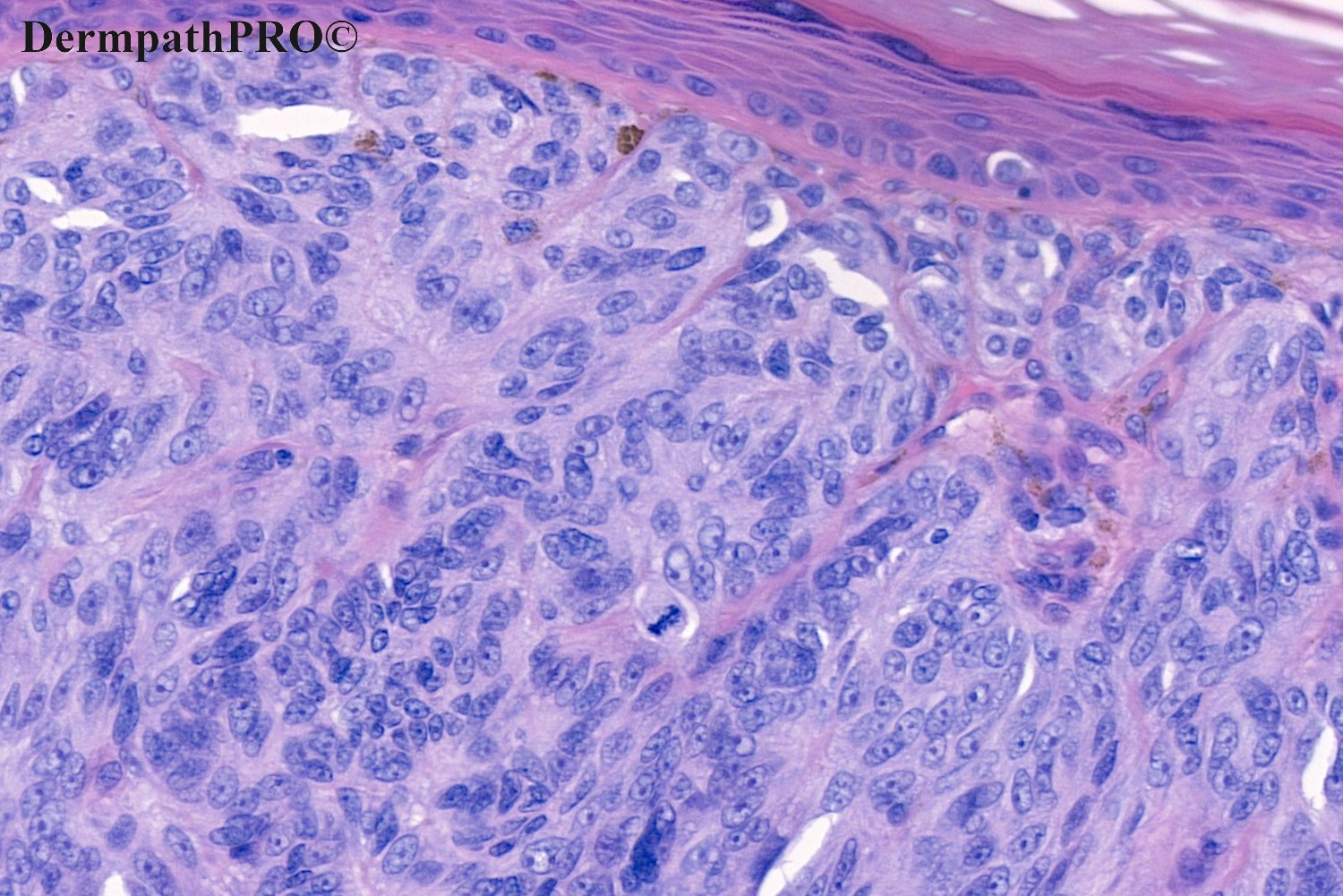
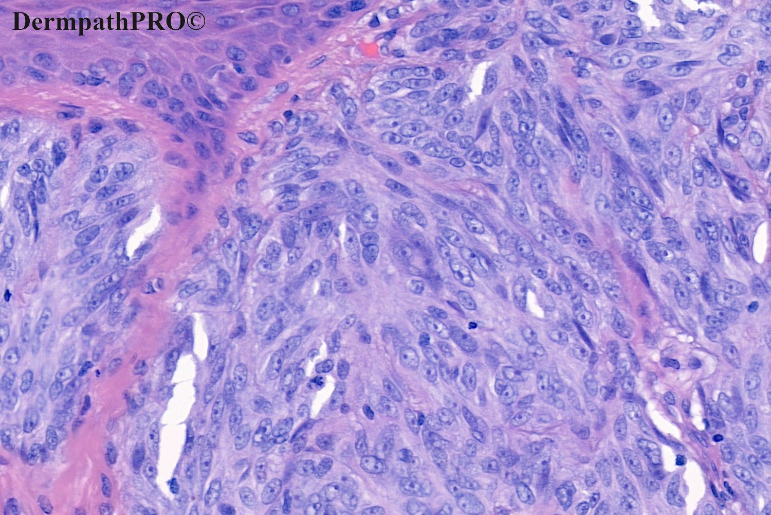
65M, Pedunculated red nodule vertex scalp ?Seb K ?Naevus.


  • Thanks 1
  Report Record

User Feedback


Richard Logan

Posted

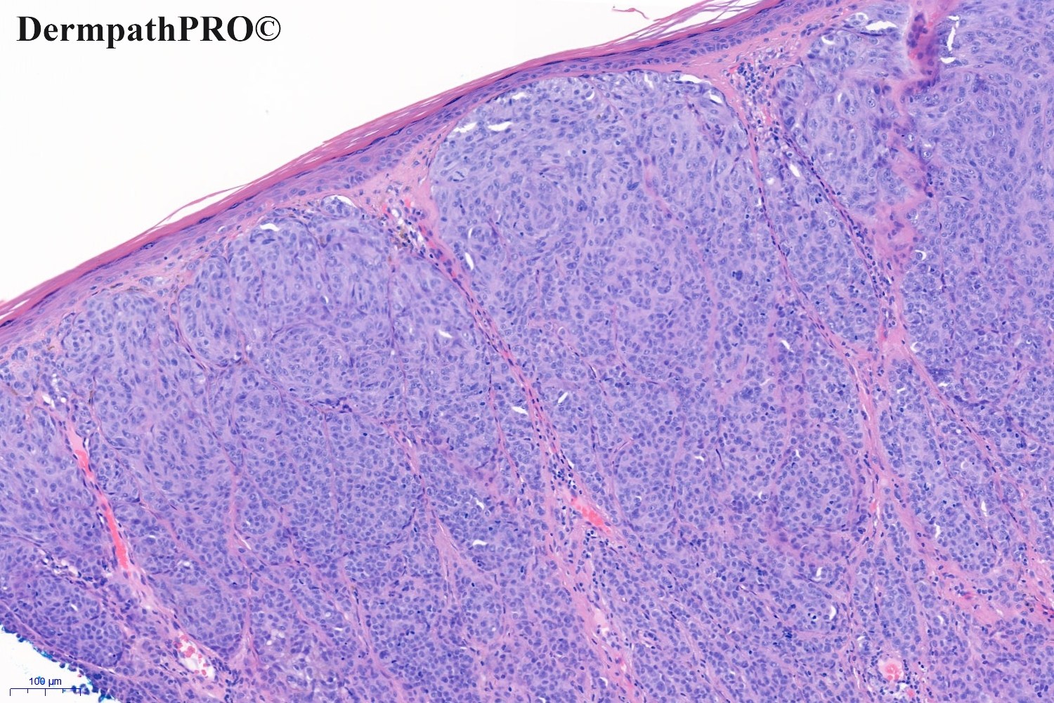
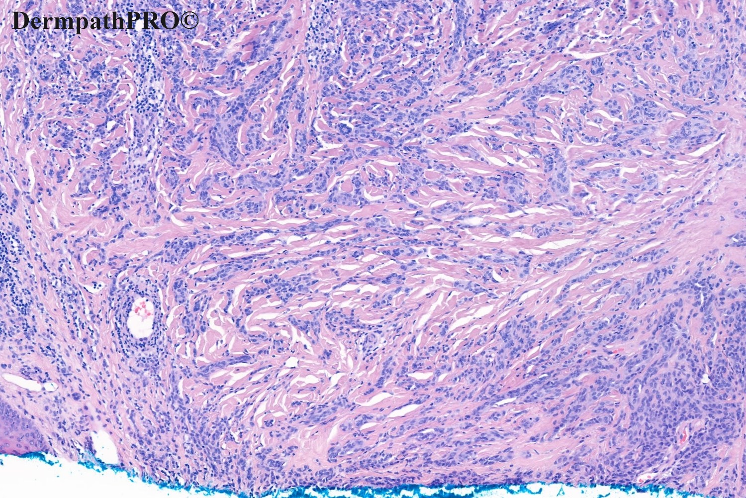
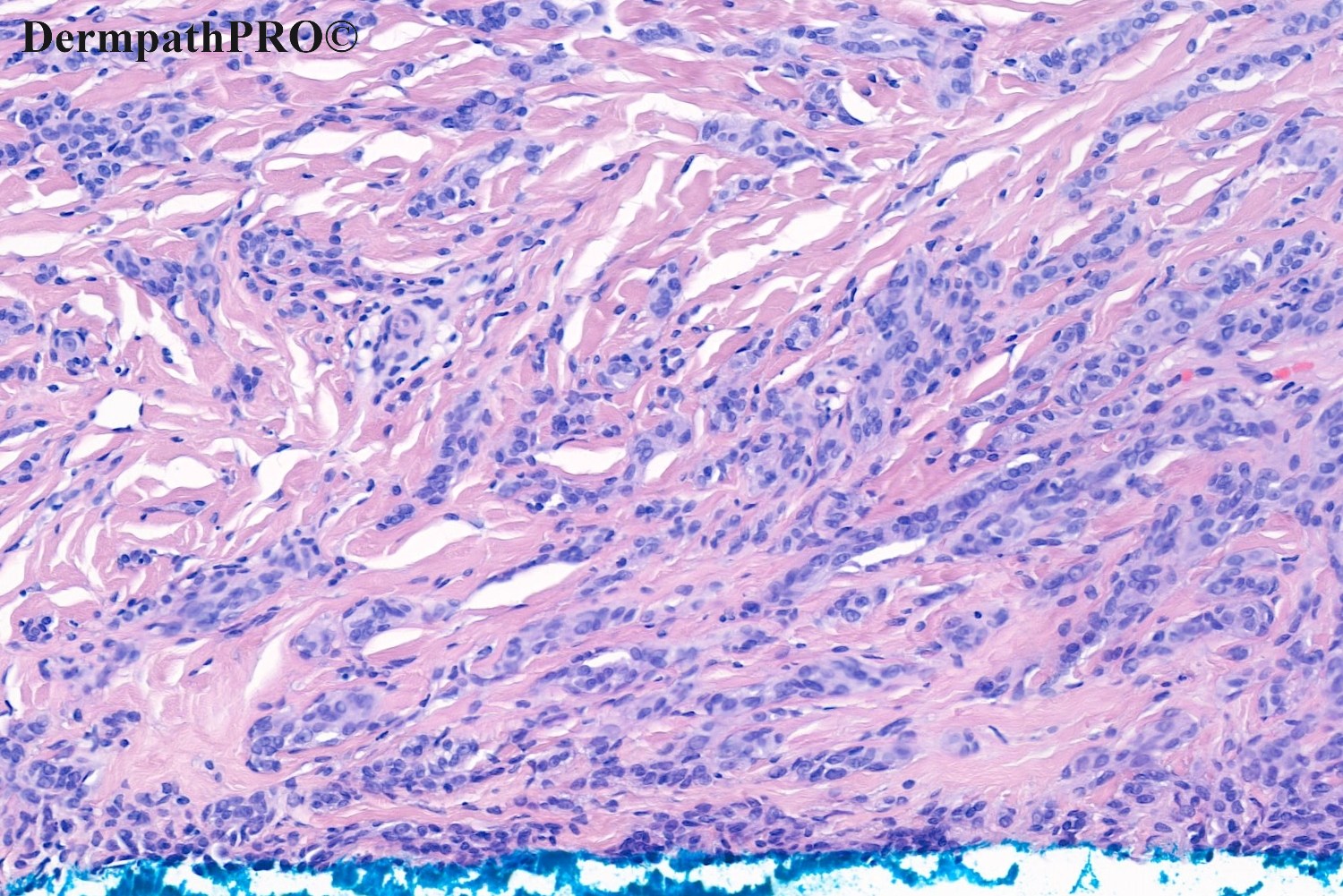
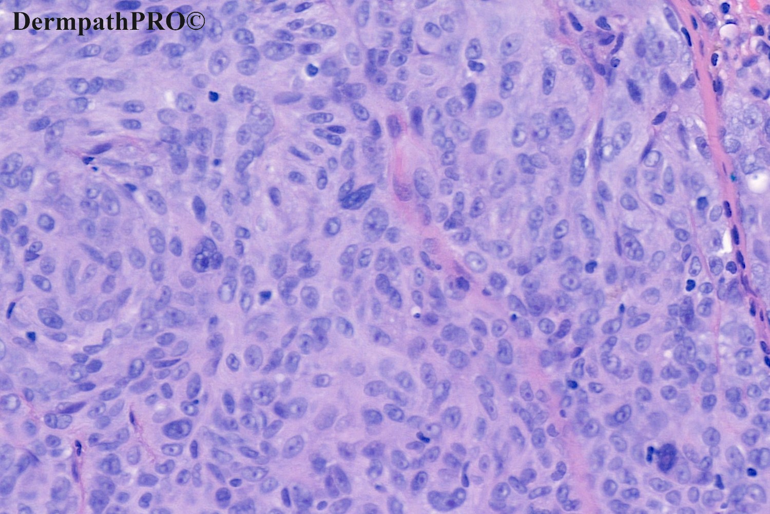
The pleomorphic nuclear cytology, mitotic activity and collagen dissection suggest this is a malignant process.  There is no apparent connection with the epidermis, so I would go for a metastasis, especially given the anatomical location.  Immunohistochemistry needed.

Share this comment


Link to comment
share_externally

A tricky maturation in this melanocytic tumor. The growth in papillary and mid dermis is expansive and stuffing. Favor melanoma. 

Share this comment


Link to comment
share_externally

Victor Delgado

Posted

Atypical Spitzoid melanoytic tumor, favor Melanoma. 

Share this comment


Link to comment
share_externally

Saleem Taibjee

Posted

Hi. There seems to be general consensus that this is a worrying melanocytic lesion. In fact my bottom line histology report was almost verbatim as for Victor i.e. “Spitzoid tumour of uncertain malignant potential, but probably best managed as for melanoma”. The nests superficially are rather expansile. There was mitotic activity. However, a very good impression of maturation with depth, and no deep dermal mitoses seen. Hence I did not feel I could make a clear-cut diagnosis of melanoma based on histology alone, although I would not be at all surprised if the lesion were to metastasize.

In terms of ancillary investigations, BRAFV600E was negative, as we might expect if a true Spitz lineage. I had the impression of fascicular areas on the histology, and interestingly there was quite striking expression of ALK in this lesion, especially within these areas. This could support the possibility of an ALK fusion as a driver genetic event in this lesion, and indirectly further support Spitz lineage. As we learn more about the genetics, we realise that this in itself does not allow us to predict prognosis, because there are lesions along the whole spectrum of Spitz (Spitz naevus, STUMP, Spitz melanoma) which may have underlying ALK fusions (by the way, note that the term Spitz melanoma is now preferred to Spitzoid melanoma in this context, the latter is a morphological term which also encompasses some examples of BRAF-mutated superficial spreading melanoma which may have Spitzoid-appearing cells). See https://pubmed.ncbi.nlm.nih.gov/32701692/

Here is a nice recent review article https://www.ncbi.nlm.nih.gov/pmc/articles/PMC6300473/

Of interest, PRAME was also positive in this lesion.

See images below.

BW

Saleem

07059_5.0x ALK labelled.jpg

07059_10.0x ALK labelled.jpg

29956_2.0x PRAME labelled.jpg

29956_10.0x PRAME labelled.jpg


Share this comment


Link to comment
share_externally


Join the conversation

You can post now and register later. If you have an account, sign in now to post with your account.

Guest
Add a comment...

×   Pasted as rich text.   Paste as plain text instead

  Only 75 emoji are allowed.

×   Your link has been automatically embedded.   Display as a link instead

×   Your previous content has been restored.   Clear editor

×   You cannot paste images directly. Upload or insert images from URL.

Loading...

×
×
  • Create New...