Jump to content
In this section we have spot diagnoses posted on a daily basis since June 2010, now over 4000! You can review the archived cases and read the suggested diagnoses by users and the final comment by the contributors.
Case are uploaded each week day by 10 am UK time with the correct diagnosis will generally be posted at 8 pm UK time. Why not view the most recent spot diagnosis and proffer a diagnosis?

Case Number : Case 2712 - 27 November 2020 Posted By: Dr. Richard Carr

Please read the clinical history and view the images by clicking on them before you proffer your diagnosis.
Submitted Date :
   (0 reviews)

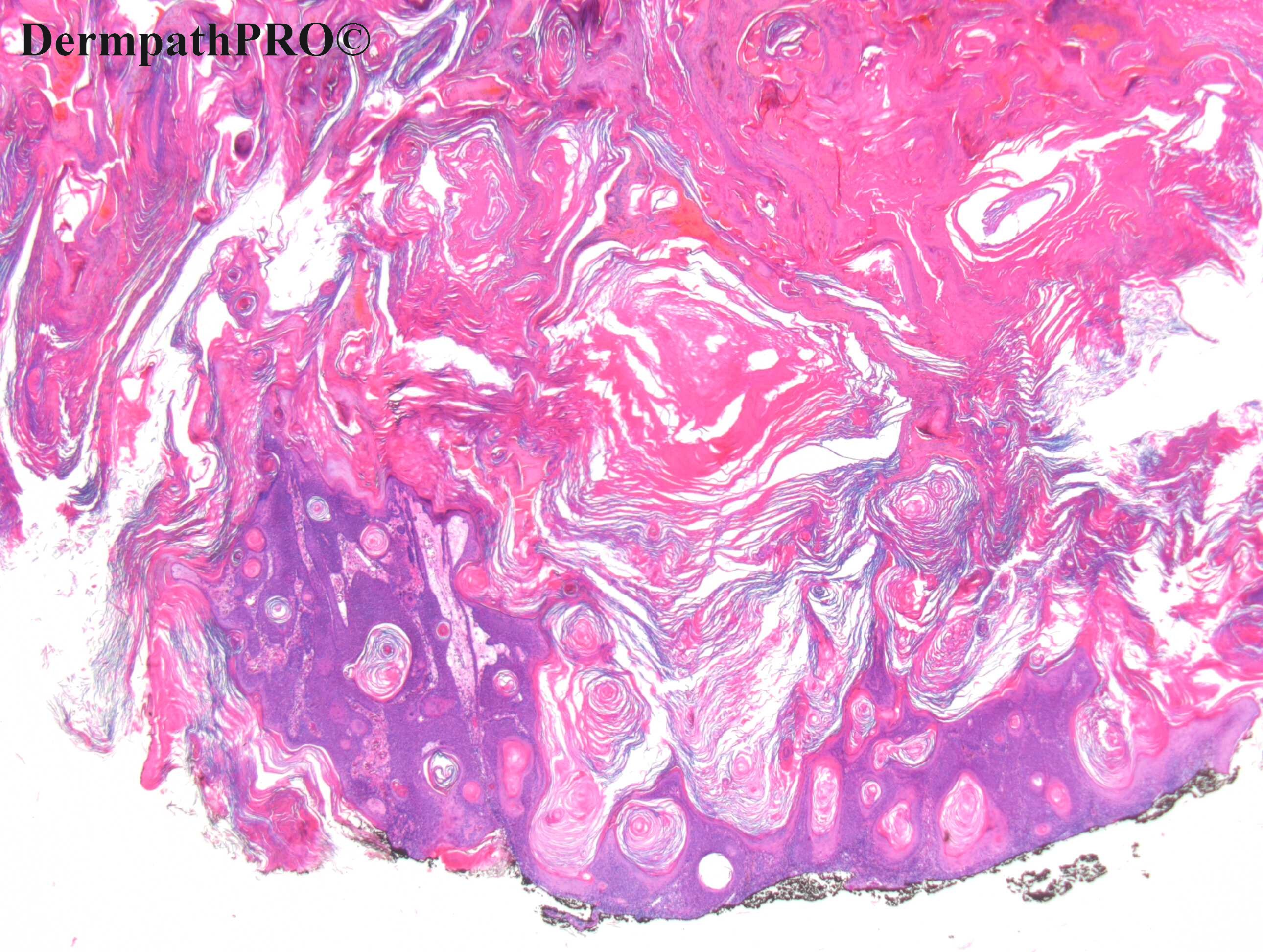
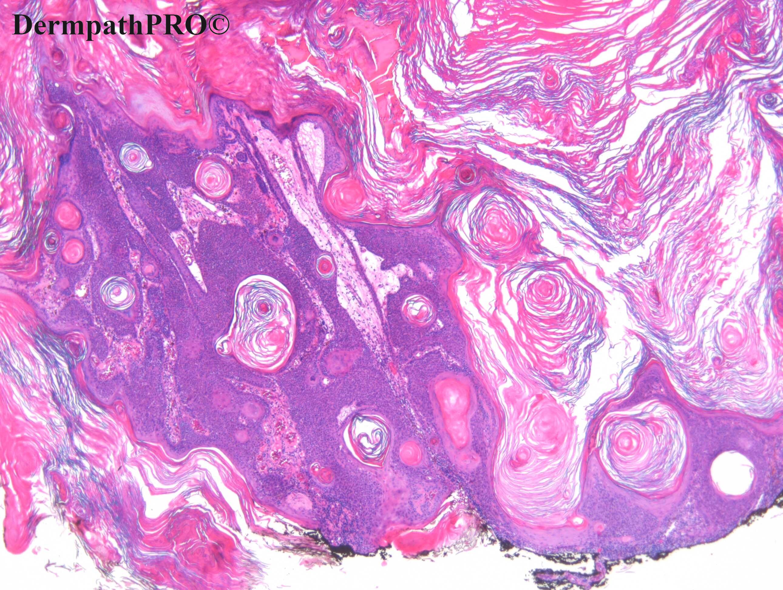
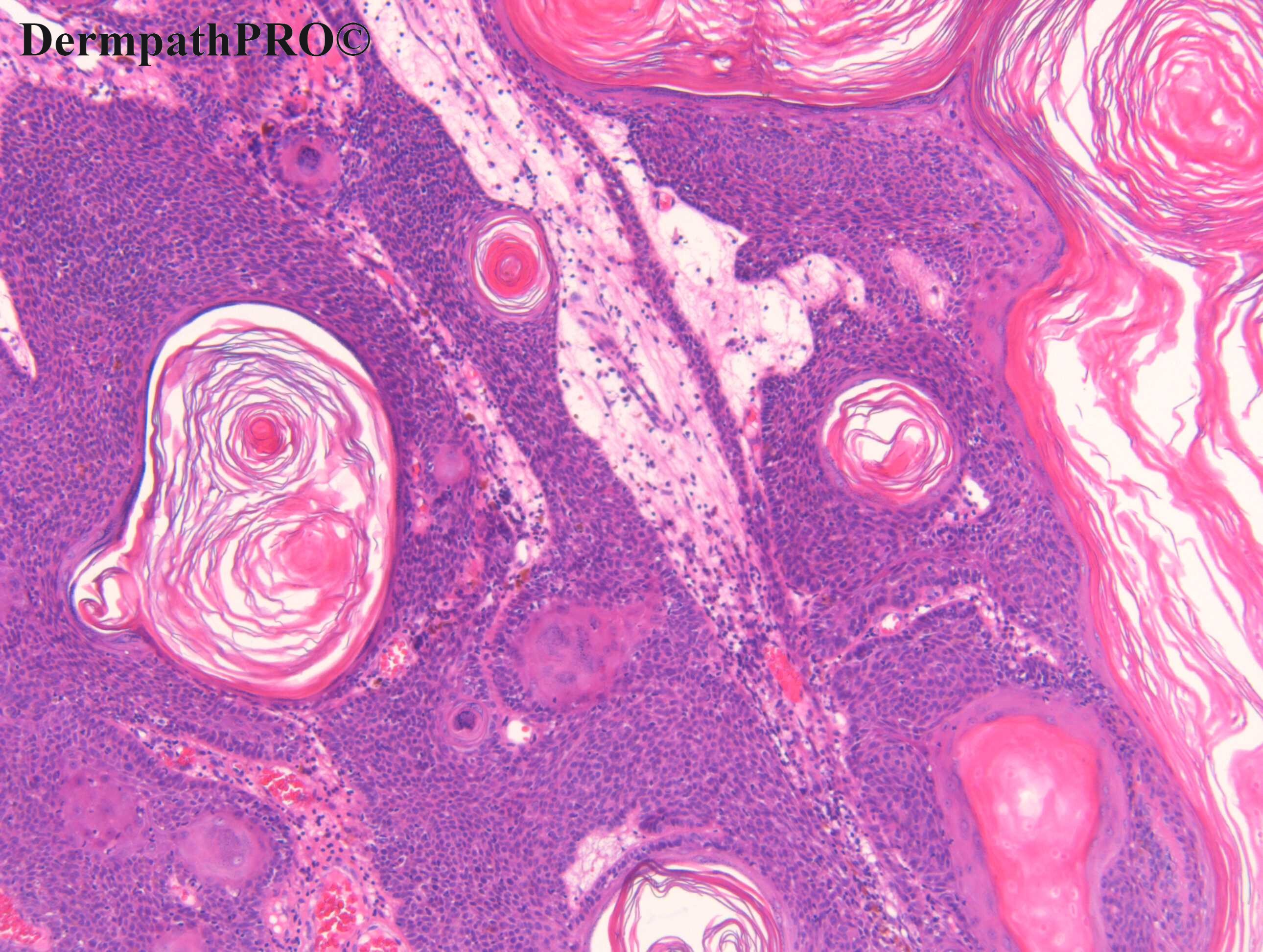
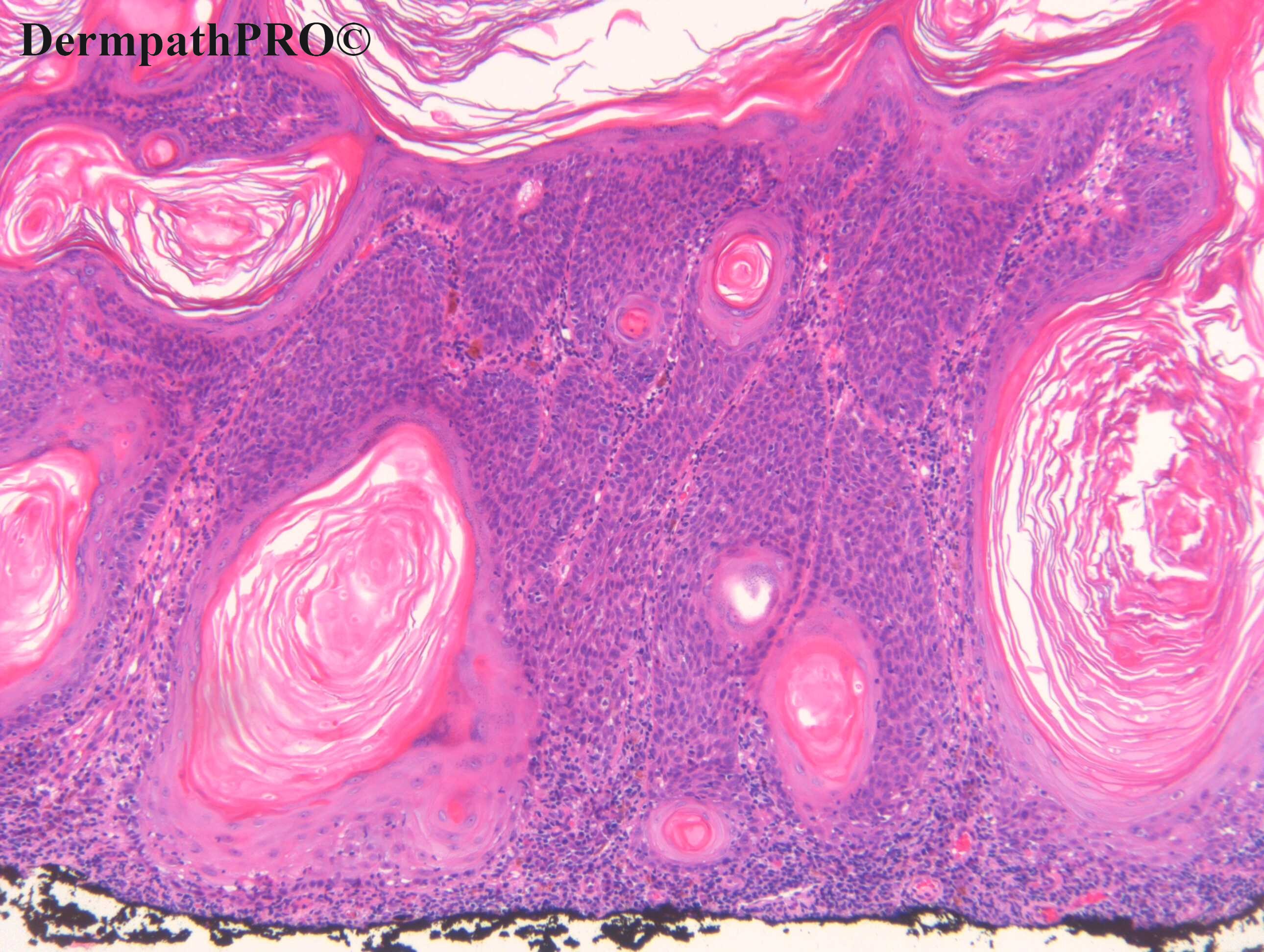
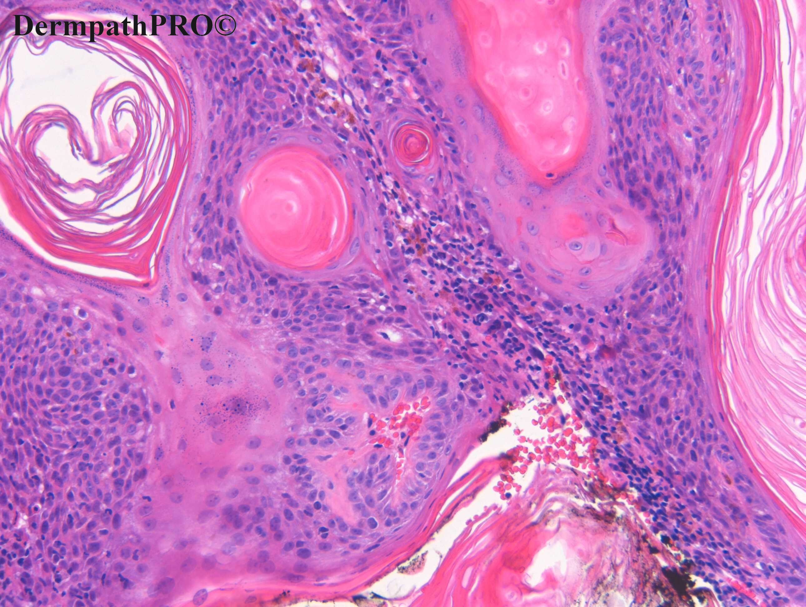
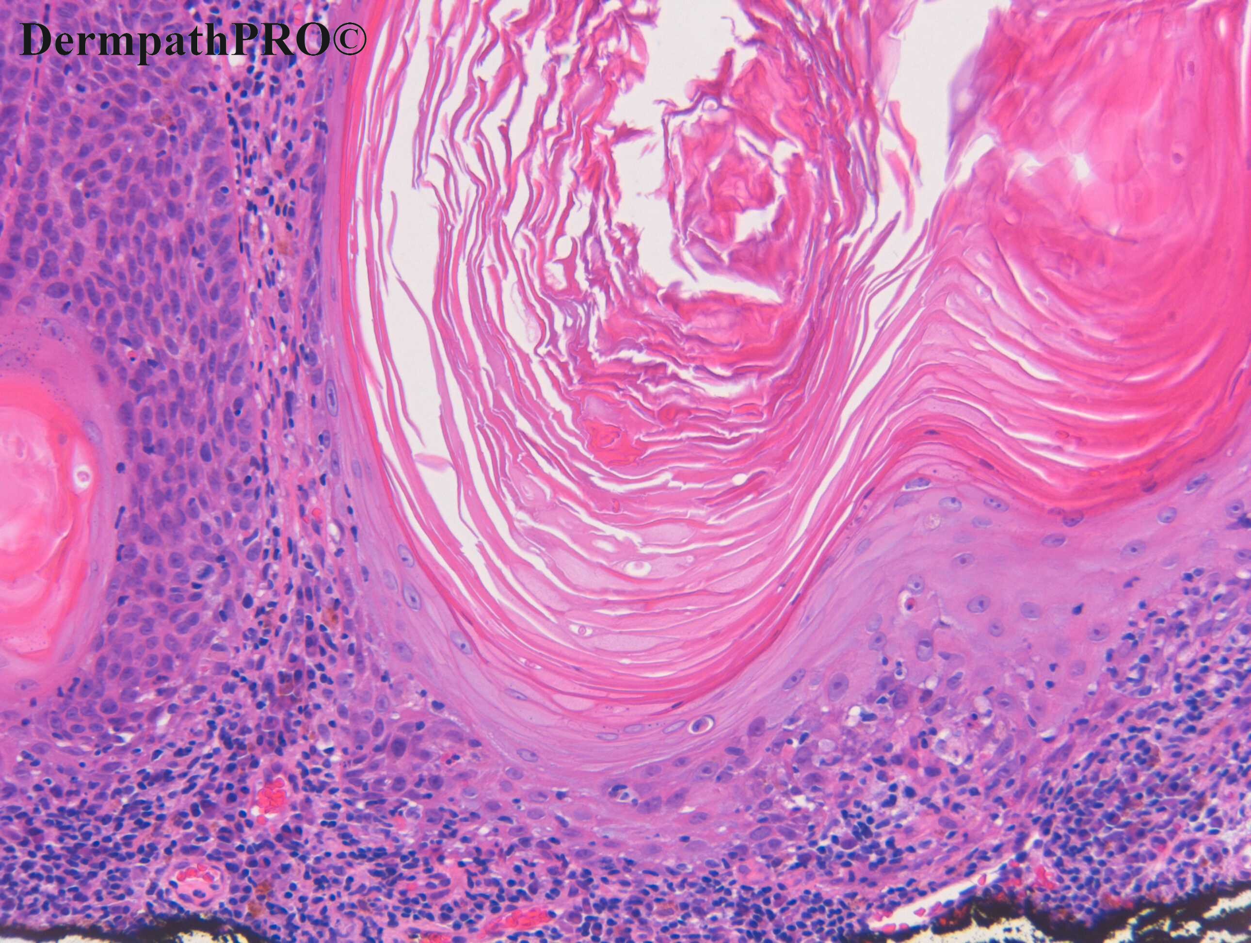
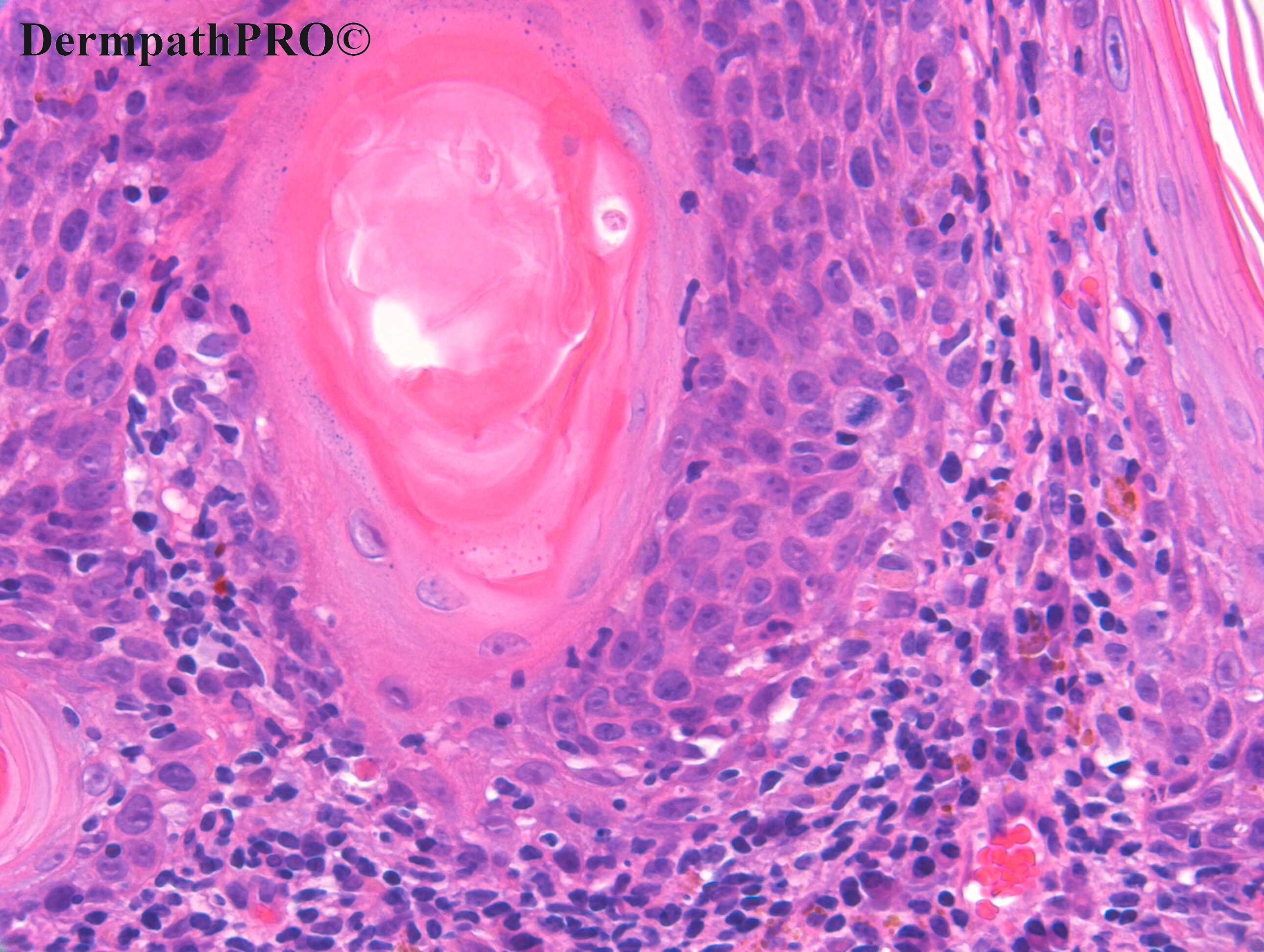
F90, Central chest. 3/12 hyperkeratotic nodule ?SEBK, ?BAK, ?resolving KA


  • Like 1
  Report Record

User Feedback


Krishnakumar subramanian

Posted

seb keratosis but anything from Dr Carr i am scared  whether i am missing SCC

Share this comment


Link to comment
share_externally

Meenakshi Batrani

Posted

Looks SK to me. Let's see what Dr. Carr tells us about this case. 

Share this comment


Link to comment
share_externally

Dr. Richard Carr

Posted

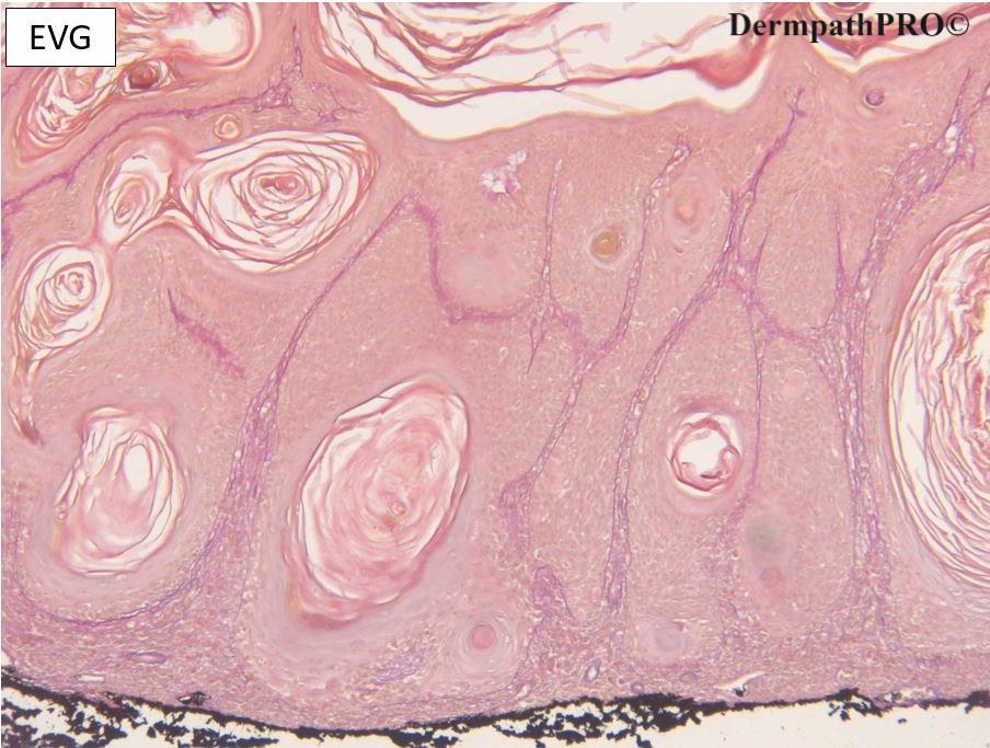
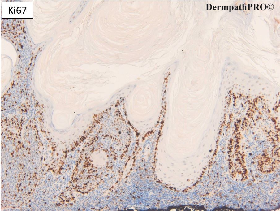
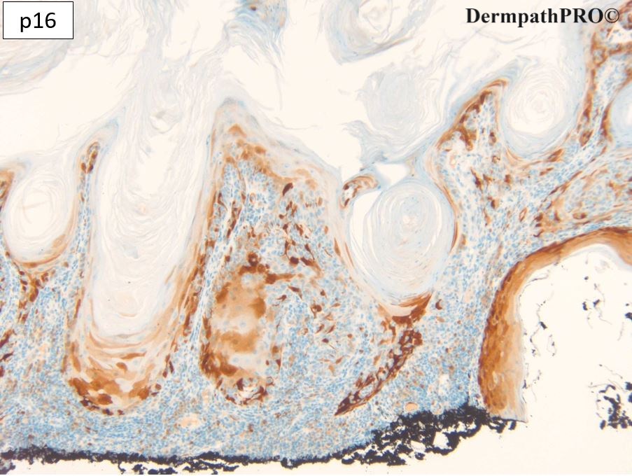
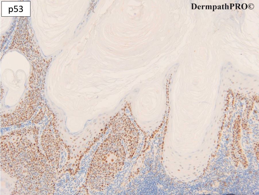
EVG - no entrapment is typical of in situ lesions and SEBK. p16 is mosaic - often a little patchy in SEBK. p53 is wild type. Note lack of moderate or strong staining in the mature squamous component. Ki67 can be quite high and appear thickened in SEBK (a basal cell proliferation) but completely spares the maturation in the squamous component.

I reported this as SEBK. The nuclear cytoplasmic ratio was a little high so just to be sure I did the IHC to r/o SEBK-like bowenoid lesion. The laminated orthokeratin is very typical and I suspect the lesion was previously traumatised (note the more compact horn superficially with blood extravasation.

Share this comment


Link to comment
share_externally

Krishnakumar subramanian

Posted

Dear sir, p53 wild type i understand, is p16 null type or reduced, can you please clarify

Share this comment


Link to comment
share_externally

Dr. Richard Carr

Posted

p16 is mosaic (wild). Can be a little patchy in SEBK and benign lesions.

Share this comment


Link to comment
share_externally


Join the conversation

You can post now and register later. If you have an account, sign in now to post with your account.

Guest
Add a comment...

×   Pasted as rich text.   Paste as plain text instead

  Only 75 emoji are allowed.

×   Your link has been automatically embedded.   Display as a link instead

×   Your previous content has been restored.   Clear editor

×   You cannot paste images directly. Upload or insert images from URL.

Loading...

×
×
  • Create New...