Jump to content
In this section we have spot diagnoses posted on a daily basis since June 2010, now over 4000! You can review the archived cases and read the suggested diagnoses by users and the final comment by the contributors.
Case are uploaded each week day by 10 am UK time with the correct diagnosis will generally be posted at 8 pm UK time. Why not view the most recent spot diagnosis and proffer a diagnosis?

Case Number : Case 2671 - 01 October 2020 Posted By: Saleem Taibjee

Please read the clinical history and view the images by clicking on them before you proffer your diagnosis.
Submitted Date :
   (0 reviews)

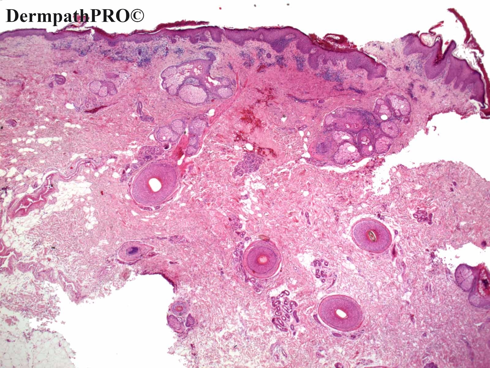
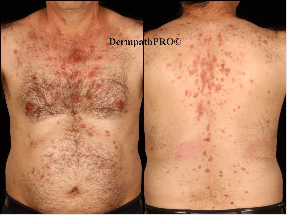
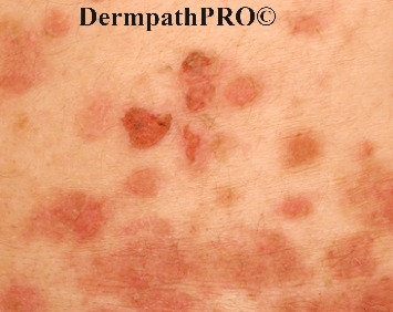
60M, eroded plaques scalp and trunk.

  Report Record

User Feedback


Richard Logan

Posted

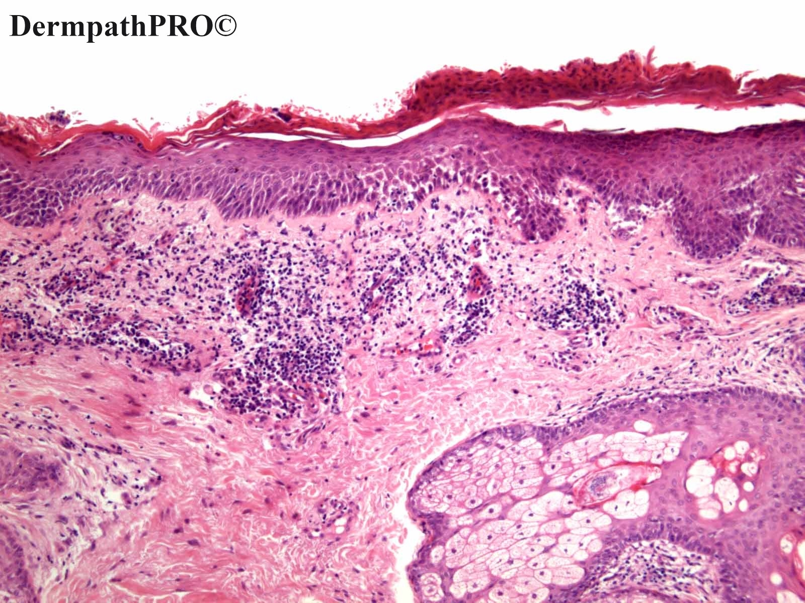
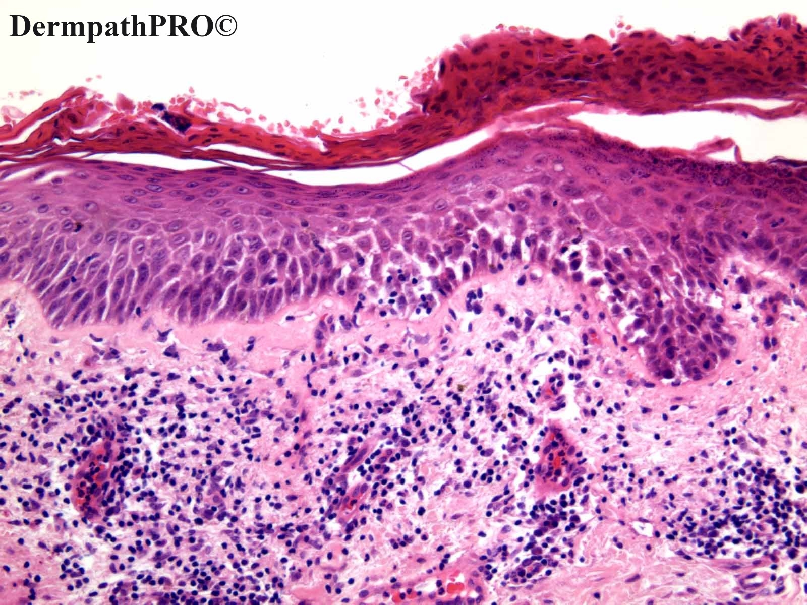
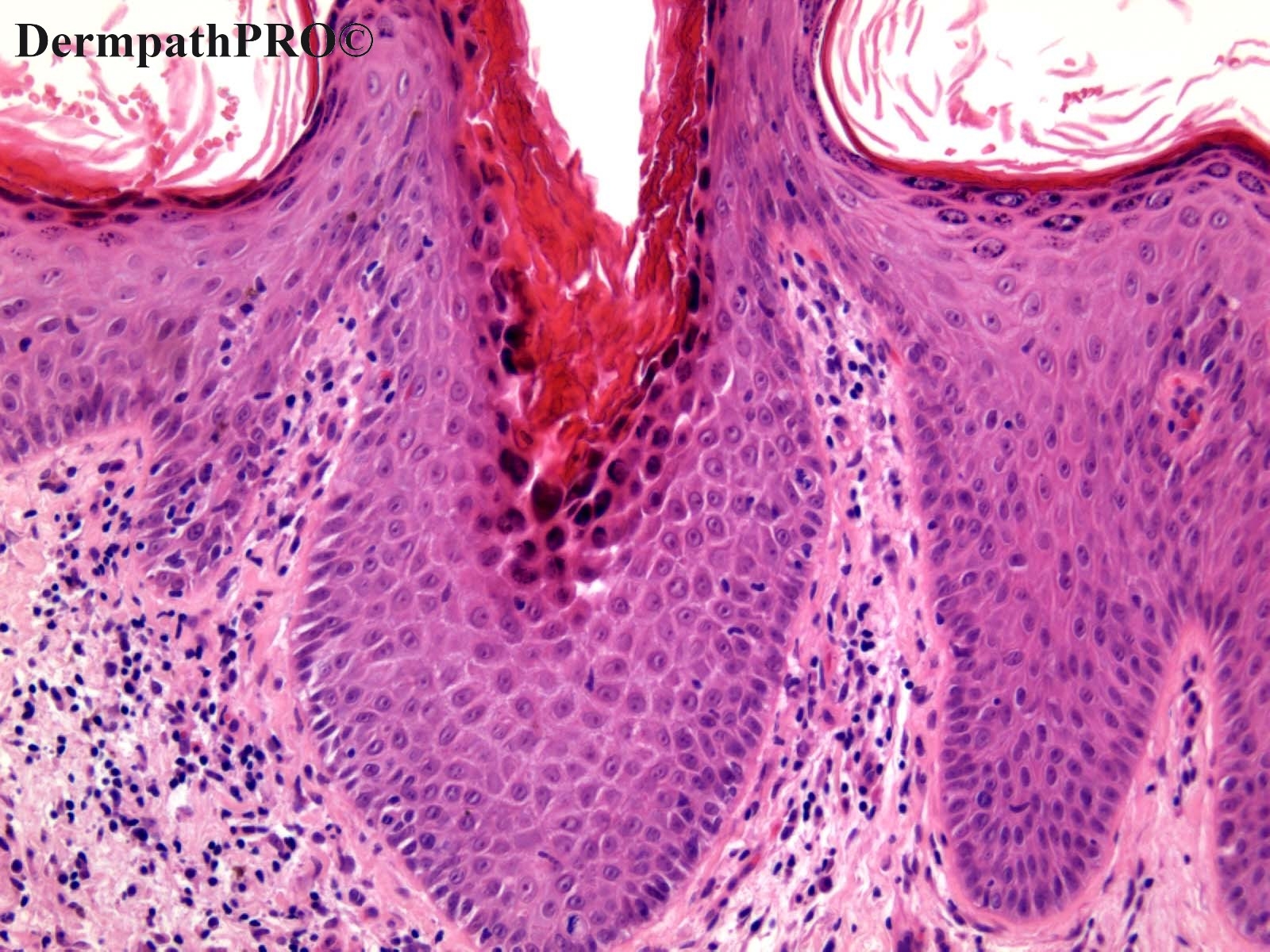
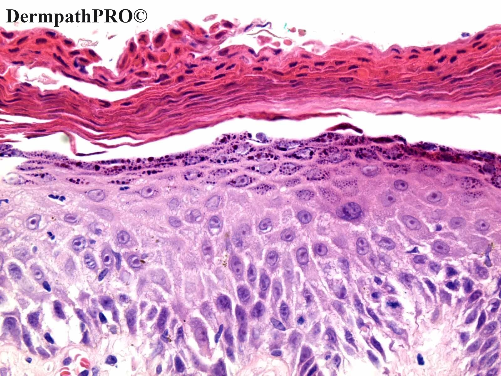
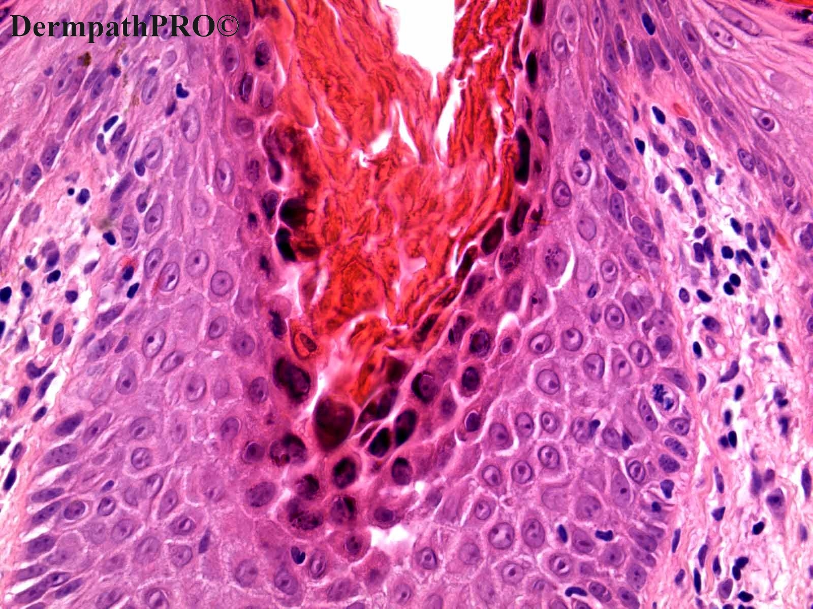
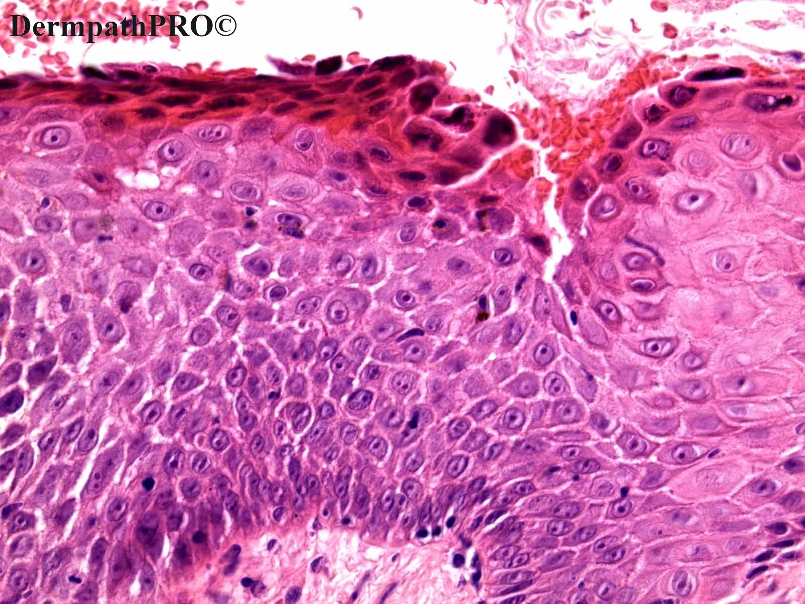
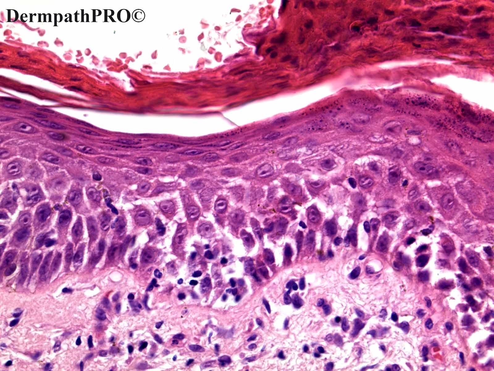
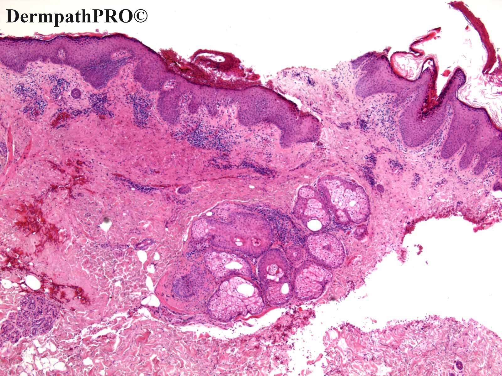
Pemphigus.  The clinical pictures suggest foliaceous, but the level of the split (at least in some of the photomicrographs) is low down in the epidermis, so I think it is Pemphigus vulgaris.

Share this comment


Link to comment
share_externally

Saman Fatah

Posted

Agree with Richard that the level of the split is variable depending on which photomicrographs you focus on, some looks like subcorneal/granular others are supra-basal. There is a lift and tilt at the edge of biopsy in image 1& 2, not sure if this is a true split or a processing issue.

Clinical photos shows relatively superficial erosions suggestive of PF unless it was taken whilst patient already on steroids and these are in the healing phase. Knowing mucosal involvement or not is useful.

DIF +/- ELISA for DSG1/DSG3 is necessary with correlation with clinical finding to establish if it is PF or PV. 

Share this comment


Link to comment
share_externally

Something pemphigus vulgaris-like, something Foliaceous-like, something lupus-like, in a patient with scalp involvement ( face? ) and clinical pictures suggestive of PF; there is some intraepidermal and dermal eosinophil...I don’t know what this is, but thought of Pemphigus erythematous. 

Share this comment


Link to comment
share_externally

Meenakshi Batrani

Posted

Pemphigus- Level of split is both superficial and suprabasal- tough to classify which type. Morphological as well as immunological overlap and shifts can occur between pemphigus vulgaris and pemphigus foliaceous.

Share this comment


Link to comment
share_externally

Krishnakumar subramanian

Posted

suprabasal cleft with villa seen-pemphigus vulgaris

Share this comment


Link to comment
share_externally

Saleem Taibjee

Posted

Thanks for the interesting comments.

Yes, this is a case of pemphigus. Overall I favoured foliaceous subtype given mostly superficial acantholysis (subtle), but in particular correlating with the clinical features of rather subtle erosions (in contrast to flaccid blisters in vulgaris) and absence of mucosal involvement. Furthermore, the clinical course was of improvement with relatively low doses of prednisolone (although things did grumble on several months as I recall). See table below which summarises the distinguishing features between PF and PV.

But there is no doubt that in areas the histology demonstrates clefting at lower levels of the epidermis. As Meenakshi states, there are some cases with overlap. It is quite an old case, the direct immunofluorescence showed intracellular IgG deposition. I don’t think ELISA for Dsg1 vs Dsg3 was ascertained, but I would not be surprised if both are involved here. It is also worth remembering that in cases with a mixed picture to consider paraneoplastic or drug aetiology, although neither was implicated in this particular case.

I also included the case to highlight an important low power clue to pemphigus foliaceous. As Saman points out, there are areas where the stratum corneum has separated from the epidermis which we can term ‘floating stratum corneum sign’. This is rather conspicuous and not just artefact, as confirmed by the presence of occasional acantholytic cells within both the roof and floor of the clefting within some foci.

foliaceous vs vulgaris.jpg

Share this comment


Link to comment
share_externally

Saman Fatah

Posted

Thanks Saleem for educating us about this useful observation and how to differentiate from an artefact.  

Share this comment


Link to comment
share_externally


Join the conversation

You can post now and register later. If you have an account, sign in now to post with your account.

Guest
Add a comment...

×   Pasted as rich text.   Paste as plain text instead

  Only 75 emoji are allowed.

×   Your link has been automatically embedded.   Display as a link instead

×   Your previous content has been restored.   Clear editor

×   You cannot paste images directly. Upload or insert images from URL.

Loading...

×
×
  • Create New...