Jump to content
In this section we have spot diagnoses posted on a daily basis since June 2010, now over 4000! You can review the archived cases and read the suggested diagnoses by users and the final comment by the contributors.
Case are uploaded each week day by 10 am UK time with the correct diagnosis will generally be posted at 8 pm UK time. Why not view the most recent spot diagnosis and proffer a diagnosis?

Case Number : Case 2682 - 16 October 2020 Posted By: Dr. Richard Carr

Please read the clinical history and view the images by clicking on them before you proffer your diagnosis.
Submitted Date :
   (0 reviews)

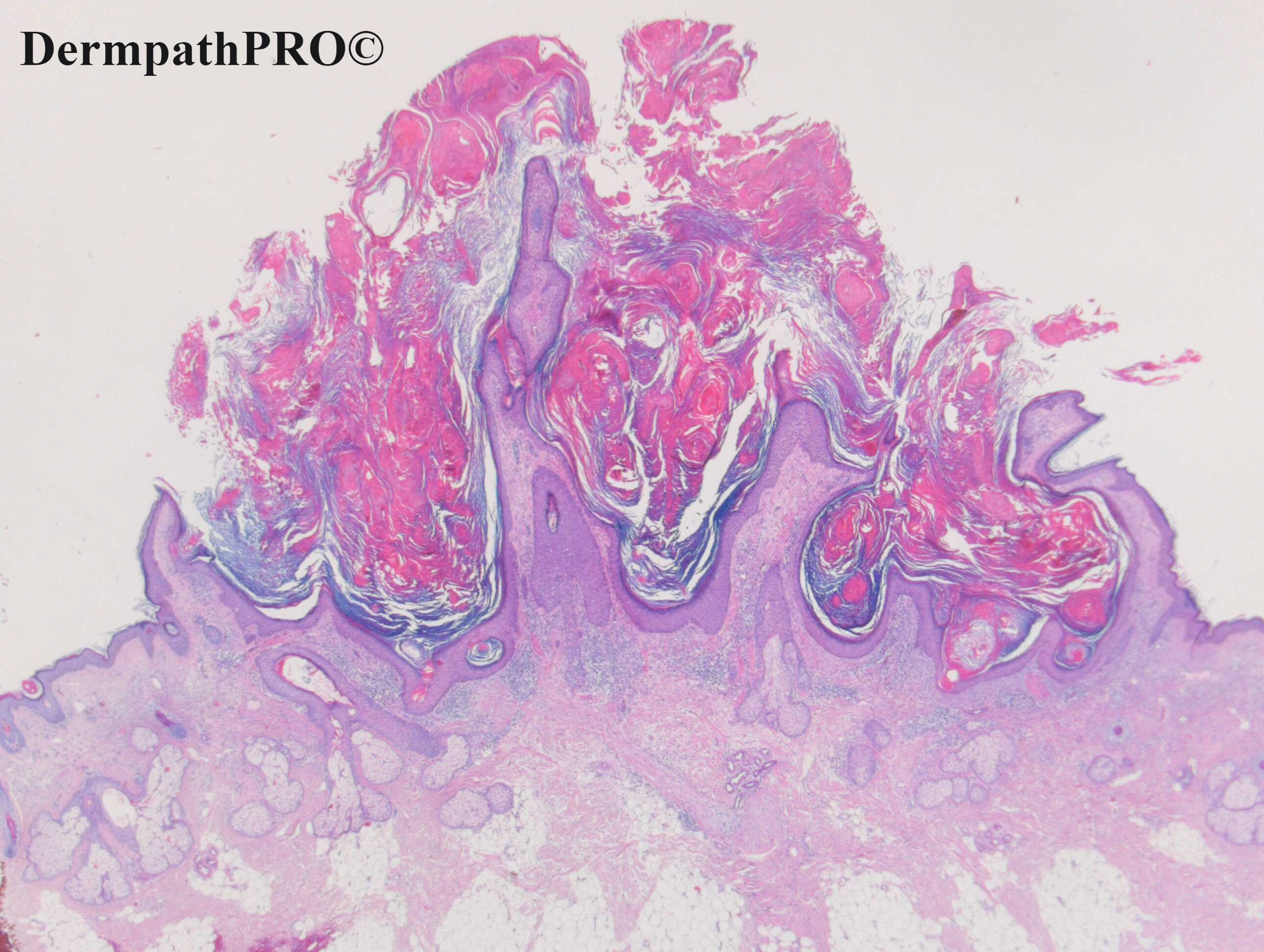
F45. Cheek. ? SCC.


  Report Record

User Feedback


Richard Logan

Posted

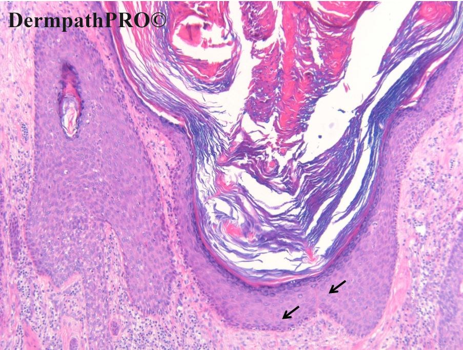
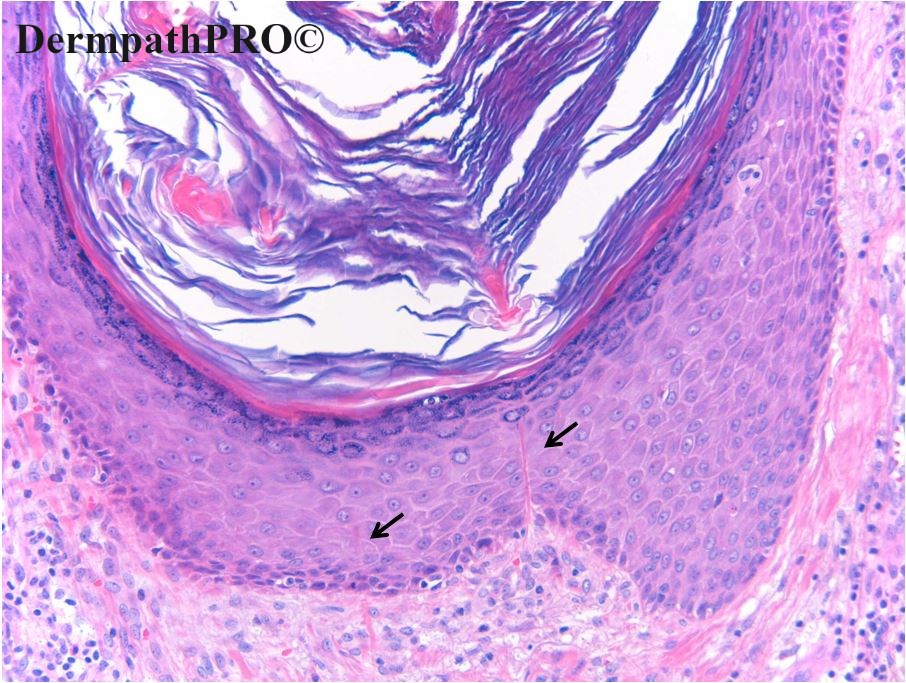
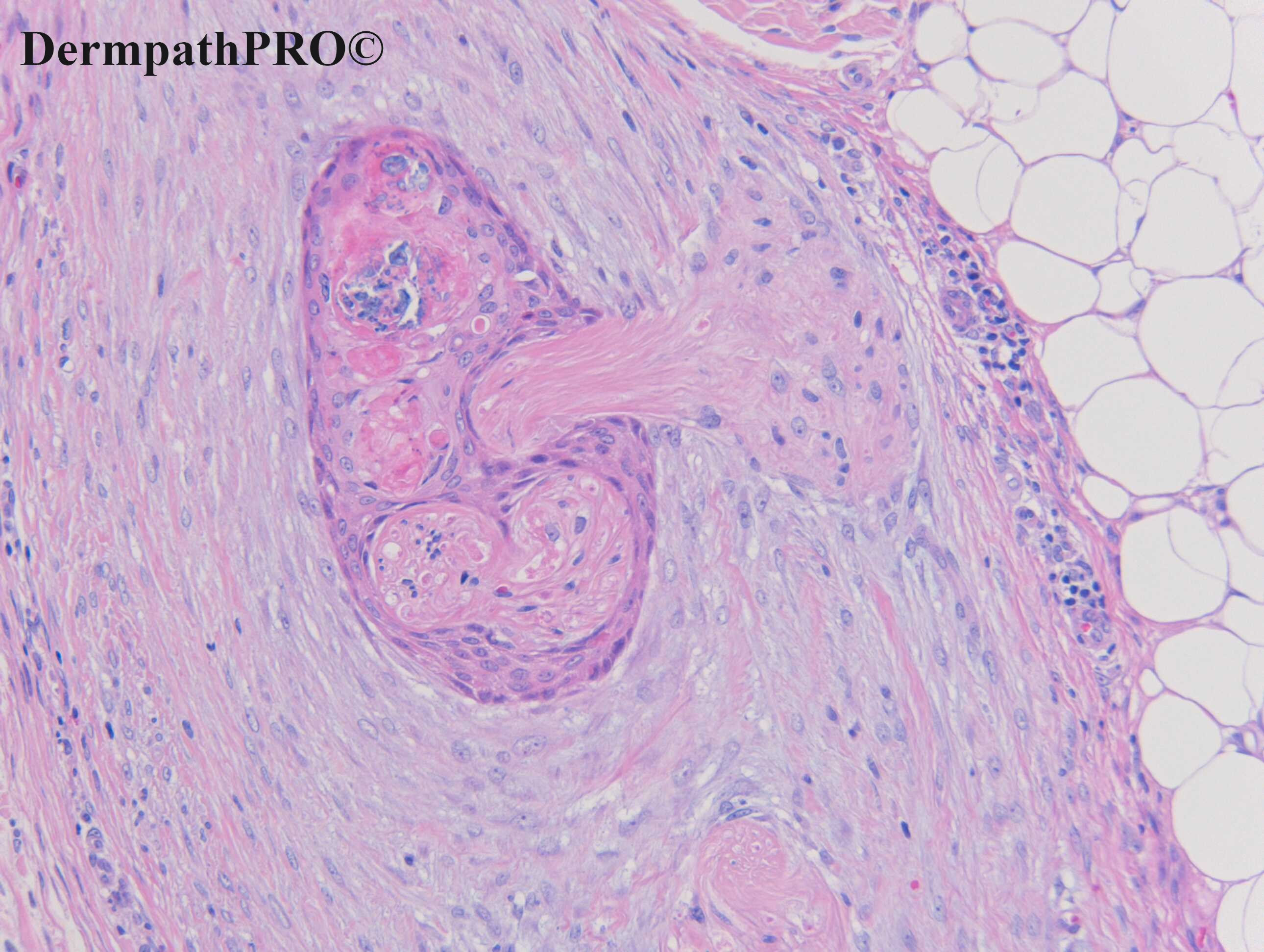
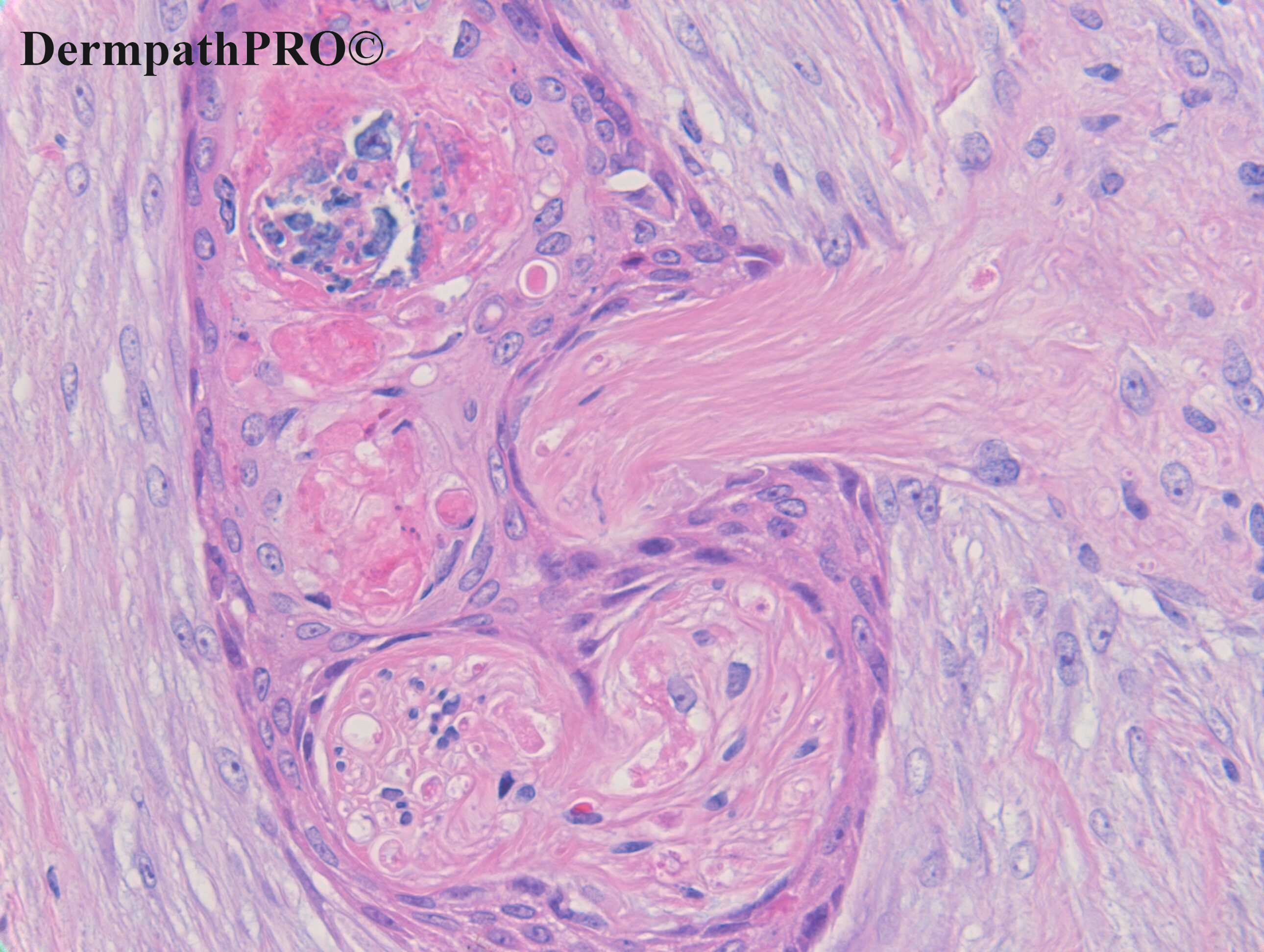
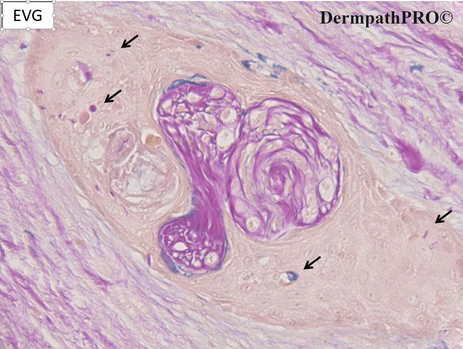
A benign-looking, lichenoid  squamous papilloma.  There are two small foci of perforating collagenosis which would have been easy to overlook were it not for the helpful black arrows!  I doubt whether these represent the main pathology as there isn't the usual overlying neutrophilic parakeratosis so perhaps this is an epiphenomenon.  There is a strange appearance in the hair follicle which I am struggling to interpret, with possible neutrophilic debris, dyskeratosis and what might be a form of extrusion from the follicular epithelium.  Perhaps a sign of viral or fungal infection?  I don't really know.

Share this comment


Link to comment
share_externally

?Late stage of Keratoacanthoma  
Collagen entrapment in mature squamous epithelium, desmo plastic  stroma around the deep squamoid nest, with peri neural growth could be clues to this diagnosis. 
However agree with difficulty of this case: it looks like a pathological tangle.  
I’m also thinking of follicular SCC. 

Share this comment


Link to comment
share_externally

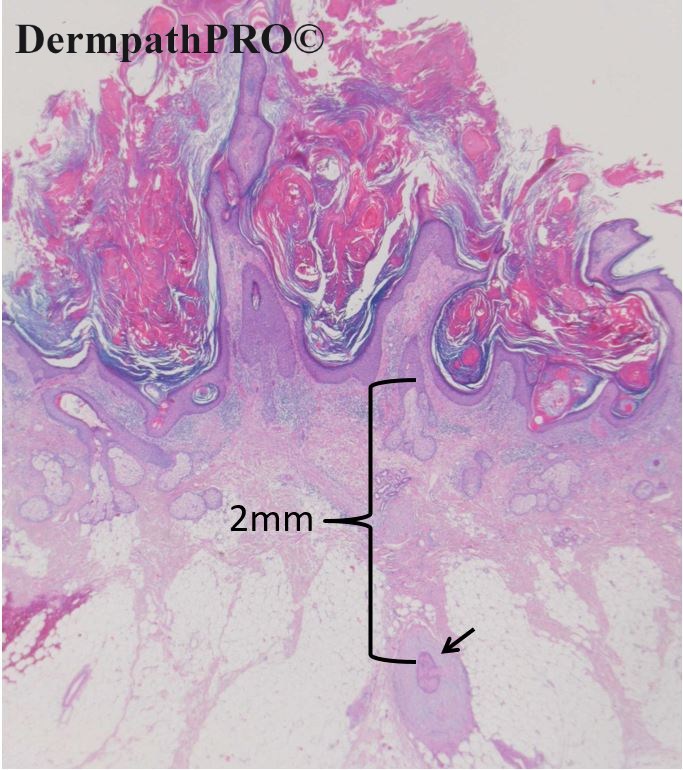
Interesting case. I agree that the lesion on top looks like a regressing keratoanthoma or a well differentiated SCC. The perineural invasion of the squamous nest in the subcutaneous tissue is an enigma,  particularly in the absence of any reactive changes in the 2mm intervening dermal and subcutaneous tissue. If one assumes that the perineural invasion is from the SCC, I would expect a connection between a nerve in the more superficial tissue above the focus of perineural invasion (deeper levels?). If one postulates that this is a keratoacanthoma  that was deep invading to the subcutaneous tissue associated with  perineural invasion and has regressed, I would expect fibrosis or some kind of reactive changes in the intervening tissue between the superficial KA and the focus of perineural invasion. I think this case is free for interpretation.  Can't wait for Dr. Carrie version.

Share this comment


Link to comment
share_externally

Favor regressing keratoacanthoma over SCC because of the lack of keratinocyte atypia. Anh summarizes really well. My understanding is that perineural invasion does not have prognostic implications in KA. Waiting to hear from Dr. Carr...

Share this comment


Link to comment
share_externally

Meenakshi Batrani

Posted

Favoring late KA. Dr. Karr has been enlightening us on these lesions previously as well, waiting for his insightful comments on this case. 

Share this comment


Link to comment
share_externally

Krishnakumar subramanian

Posted

Keratoacanthoma because of superficial location

Perineural invasion is rare in KA, but has been reported

Keratoacanthoma with perineural invasion: a report of 40 cases A M Godbolt 1, J J Sullivan, D Weedon anyway shall wait for Dr Carr's comments

Share this comment


Link to comment
share_externally

Dr. Richard Carr

Posted

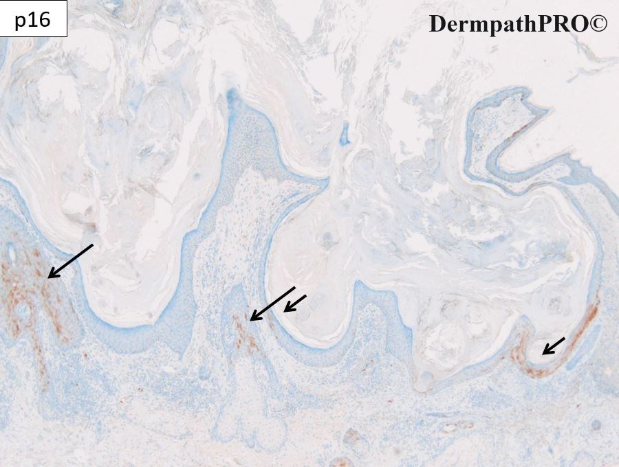
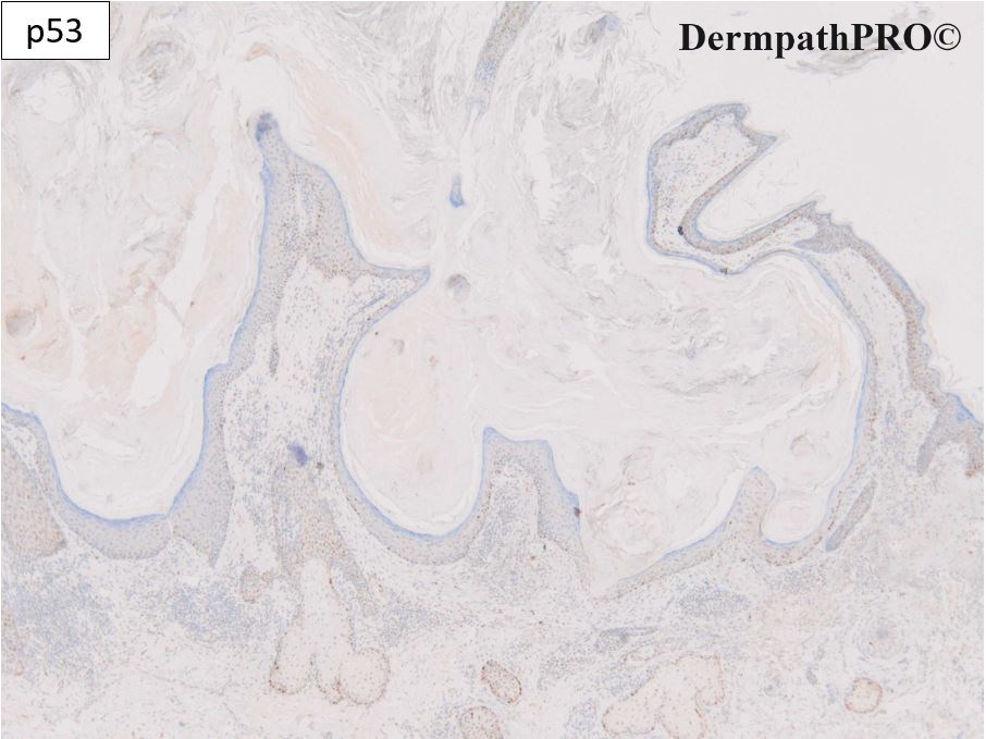
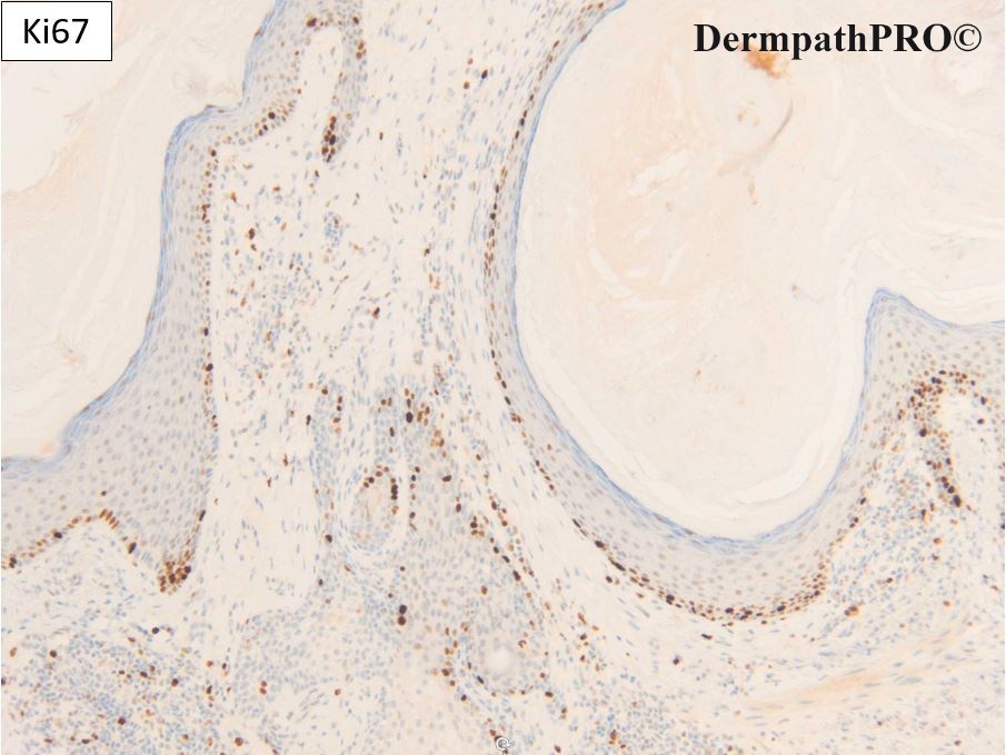
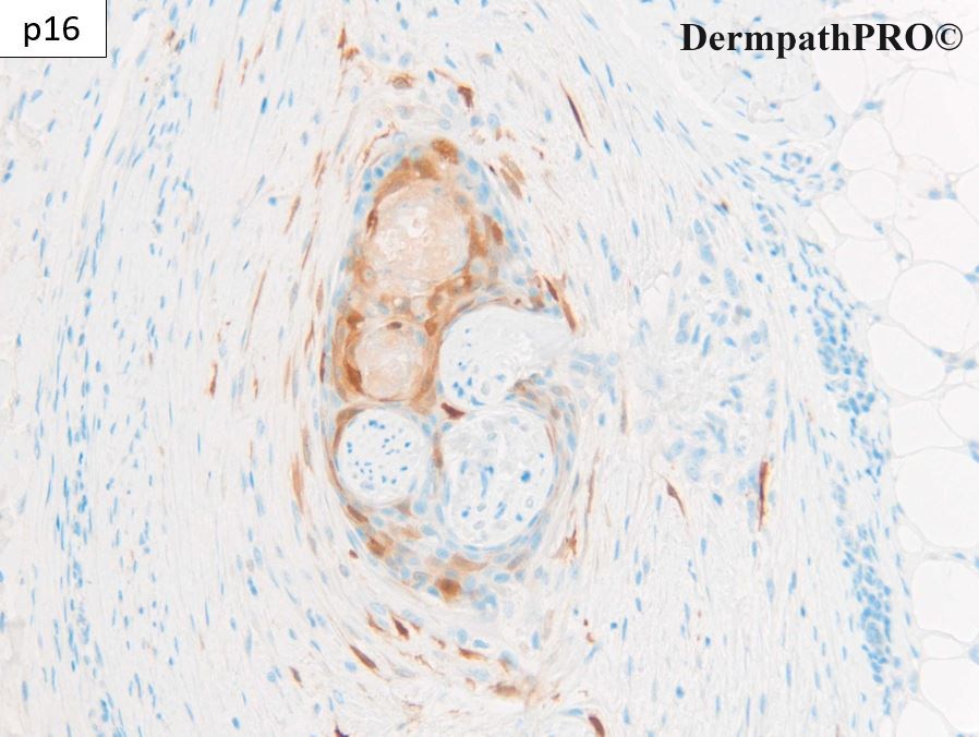
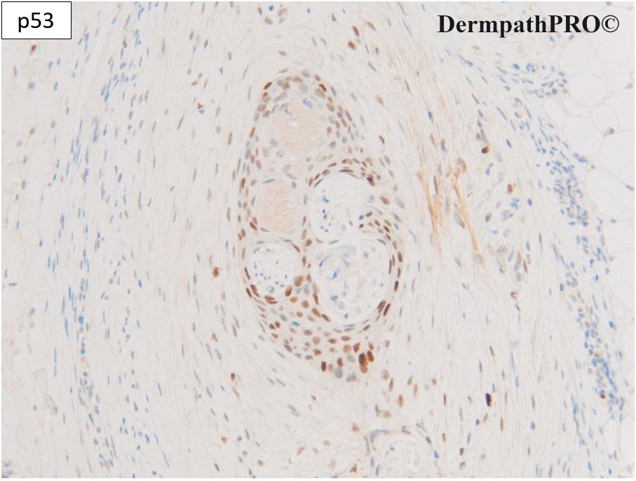
I reported this as in keeping with a regressed KA and residual banal squamous epithelium around the nerve presumably from when the KA was more active. The p16 is mosaic and p53 wild type which fits with KA or a benign / reactive proliferation. The EVG in the perineural focus shows evidence of the prior more infiltrative prolieration (foci of trapped collagen & elastic). I recommended watchful waiting.

KA with perineural invasion are particularly common in the central face and peri-oral location and they may also show venous invasion at this location too.

Weedon stated he'd seen around 129 cases of otherwise typical KA with perineural invasion and around 30 or 40 with venous invasion and not patient he was aware of came to harm. I've personally seen around 20 with perineural and a handful with venous invasion and so far no patient came to harm. I do wonder in the above case if the patient might develop a cyst around the nerve or if the immune-macrophage system might mop up the last of the lesion.

There are many pitfalls in the diagnosis of typical KA and KA-like SCC's. I hope our work will help you all in the future with these challenges but I have to give you a warning this diagnosis is fraught with difficulty based on clinical & H&E examination alone.

Share this comment


Link to comment
share_externally

Dr. Richard Carr

Posted

I'm currently doing an inter-observer study of 15 digitised cases. Please e-mail me at richard.carr@swft.nhs.uk if you'd like to participate. All the cases will have powerpoiint presentations with resutls of the EVG & IHC.


Share this comment


Link to comment
share_externally

Dr. Richard Carr

Posted

Forgot to mention at this young age you should enquire any possibility of Muir Torre syndrome. 

Share this comment


Link to comment
share_externally


Join the conversation

You can post now and register later. If you have an account, sign in now to post with your account.

Guest
Add a comment...

×   Pasted as rich text.   Paste as plain text instead

  Only 75 emoji are allowed.

×   Your link has been automatically embedded.   Display as a link instead

×   Your previous content has been restored.   Clear editor

×   You cannot paste images directly. Upload or insert images from URL.

Loading...

×
×
  • Create New...