Jump to content
In this section we have spot diagnoses posted on a daily basis since June 2010, now over 4000! You can review the archived cases and read the suggested diagnoses by users and the final comment by the contributors.
Case are uploaded each week day by 10 am UK time with the correct diagnosis will generally be posted at 8 pm UK time. Why not view the most recent spot diagnosis and proffer a diagnosis?

Case Number : Case 2691 - 29 October 2020 Posted By: Saleem Taibjee

Please read the clinical history and view the images by clicking on them before you proffer your diagnosis.
Submitted Date :
   (0 reviews)

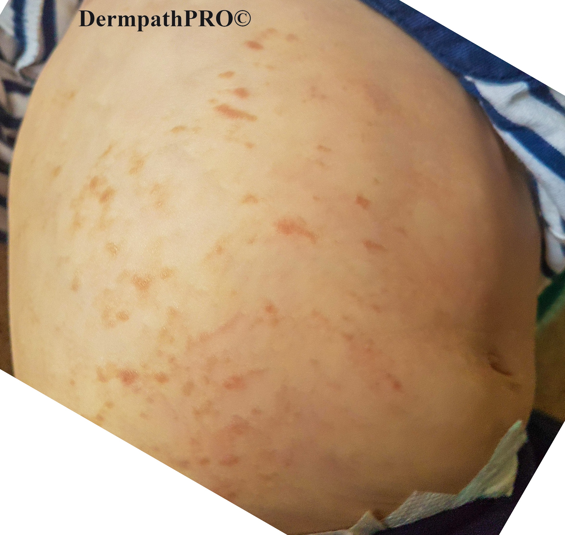
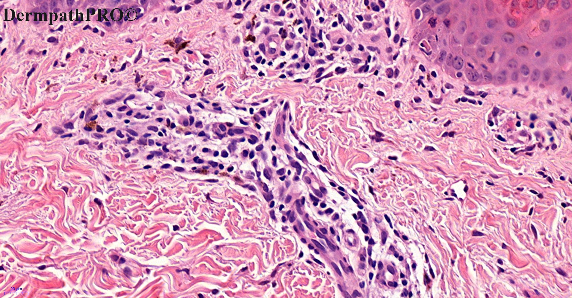
6-month-old baby girl, pigmented rash right side of abdomen.


  • Thanks 1
  Report Record

User Feedback


Mmh Terrible. I hope I’m wrong. Incontinentia pigmenti?


Share this comment


Link to comment
share_externally

Richard Logan

Posted

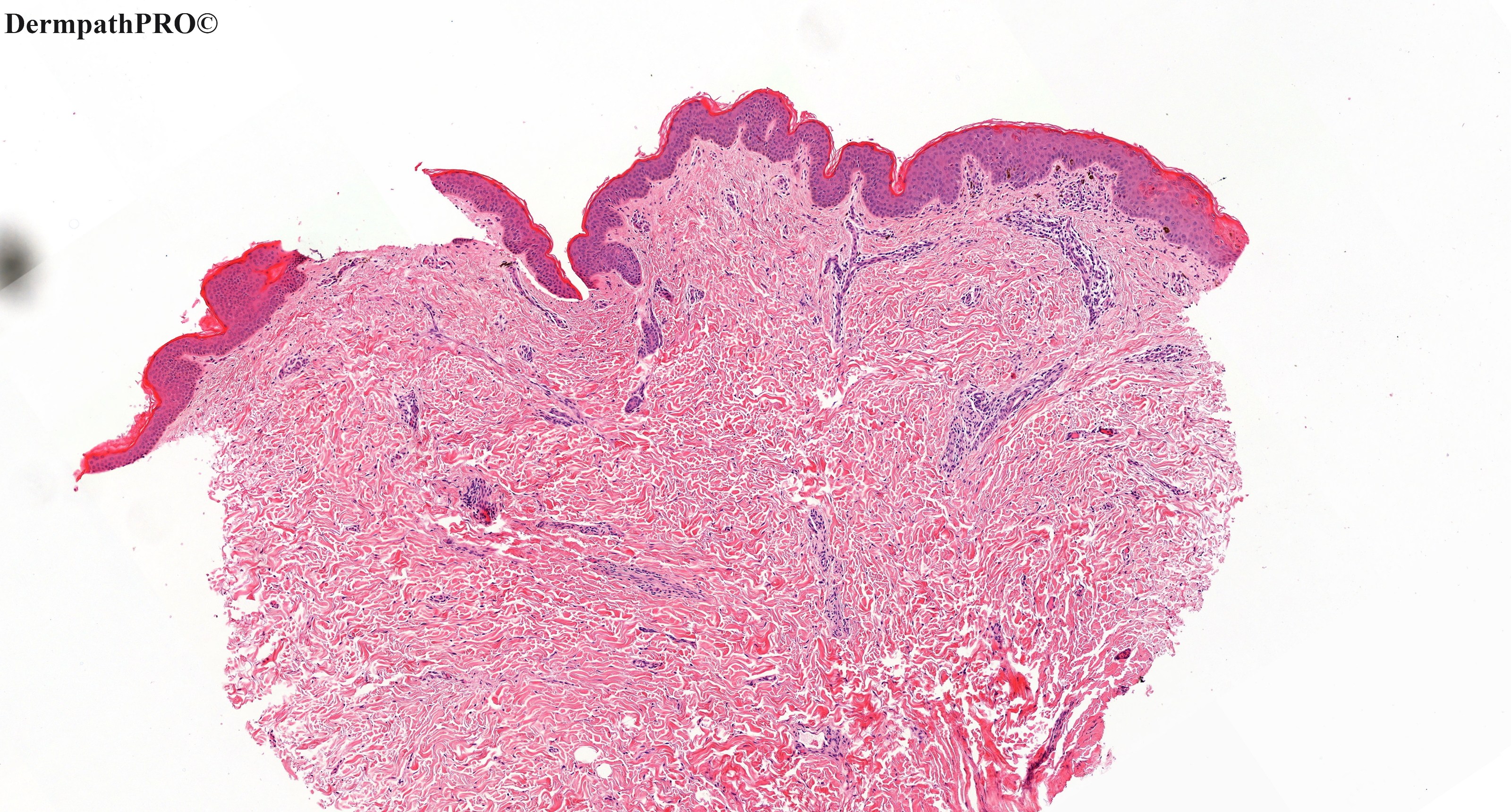
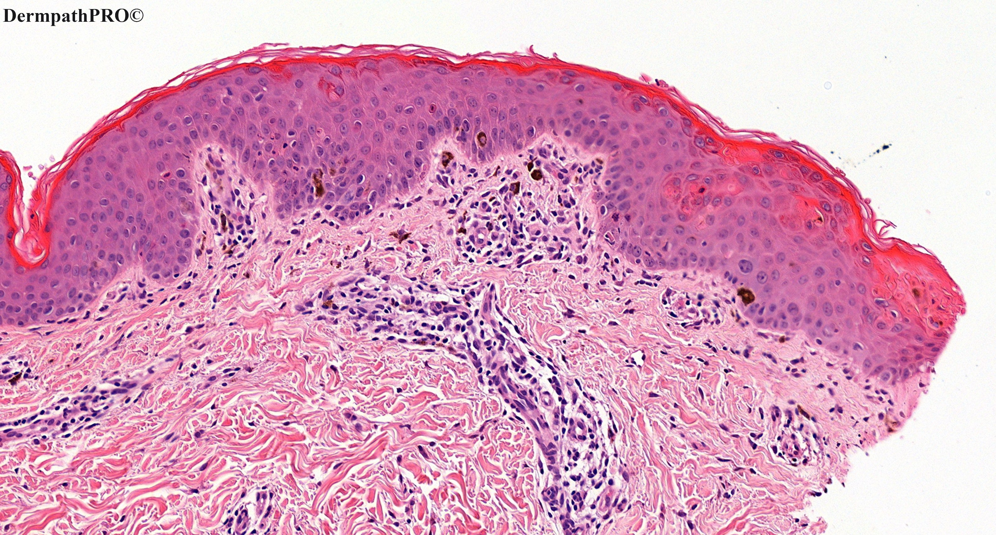
I agree that the histology would be compatible with a diagnosis of incontinentia pigmenti.  The unilateral nature of the rash is unusual I think, but perhaps IP can show mosaicism in this way - Saleem will enlighten us I'm sure.

Another possibility would be phytophotdermatitis, but this would be an odd distribution for that phenomenon as of course it usually occurs on exposed sites.  History crucial here.

Share this comment


Link to comment
share_externally

Krishnakumar subramanian

Posted

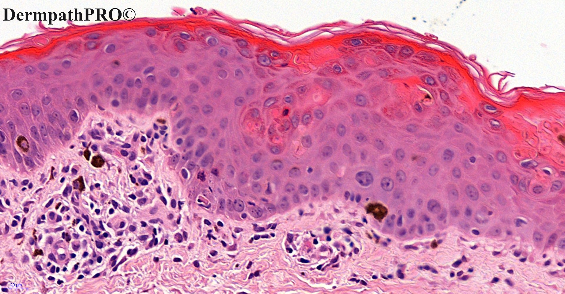
necrotic keratinocytes and pigmented incontinence

? drug rash

? photodermatitis

Share this comment


Link to comment
share_externally

Saman Fatah

Posted

The appearance of the eruption, age and histology suggests IP ? 3rd stage.

Share this comment


Link to comment
share_externally

Victor Delgado

Posted

Great case, agree with @Saman Fatah, may be 3rd  - end stage,  a little epidermal atrophy, whorled necrotic keratinocytes and drop of melanic pigment with a lot of melanophages.

Share this comment


Link to comment
share_externally

I think that staining for mast cells should be done to rule out telangiectasia macularis eruptiva perstans. Unilateral cases of this condition have been reported and histological changes can be very subtle.

Share this comment


Link to comment
share_externally

Saleem Taibjee

Posted

Some further information about this case:

The girl presented to the paediatricians with a blistering rash in the first week of life, most marked on the scalp and limbs (see below). 

However, the rash had completely settled by the time I saw her in the dermatology clinic the following week.

At that time I arranged genetic testing of the NEMO gene. The test was negative.

Ophthalmology screening at our local hospital was also negative.

20150921_105429-2.1.jpg

20150921_105509-2.1.jpg

20150921_105540-2.1.jpg


Share this comment


Link to comment
share_externally

Krishnakumar subramanian

Posted

Dear Dr  what is the final diagnosis, very interesting 


Share this comment


Link to comment
share_externally

Saleem Taibjee

Posted

Yes, this is incontinentia pigmenti.

This is a memorable case for me for several reasons.

As I mentioned, the original rash had completely settled by the time I saw her in the dermatology clinic in the second week of life. Even so, I was struck by the suggestion that the blisters were in a whorled pattern on the scalp, and linear on the limbs, and embarked on a difficult conversation with the parents that I thought this might be IP. I was then surprised when the genetic testing of the NEMO gene was negative, and began to doubt my diagnosis.

But when the child developed the new pigmented skin lesions (as shown) at 6 months of age localised to the right side of the abdomen, I was pretty sure of IP. I contacted the genetics lab. I was informed that the initial NEMO testing assessed for a more common deletion between exons 4 to 10 of the NEMO gene (Xq28) which is found in  65-70% of patients with IP. At my request, subsequent full sequencing showed a heterozygous pathogenic variant c184C>T p(Arg62Ter) confirming IP. This is an important learning point about genetic testing, to be aware of the testing which is undertaken, and sensitivity, and to discuss the testing with the genetics laboratory in such cases of an unexpected negative result.

The case is also interesting because, as Saman points out, the lesions at 6 months showed a pigmented morphology which we might classify as stage 3, but yet there were no obvious preceding inflammatory lesions at this location. This seems to be an interesting phenomenon in IP which I have come across previously. Of course, the histology of IP will parallel the clinical stage. Earlier stage blistering lesions may show eosinophilic spongiosis. Necrotic/apoptotic keratinocytes, as in this case, are a particular clue to IP.

Richard has asked about the patterns of cutaneous mosaicism pertinent to this case. There are, of course, various different cutaneous patterns of mosaicism depending on the tissue type/cell line affected, and the embryonic stage at which mosaicism arises. But in my limited experience of IP, this usually always manifests as cutaneous lesions arranged along Blaschko’s lines, first described by Alfred Blaschko in 1901 (see below). Blaschko’s lines arise due to the lateral migration of mosaic neuro-ectodermal cells from the neural crest in concert with the overall growth of the foetus. In this instance, the mosaic state is an expression of Lyonisation (skewed inactivation of the normal X chromosome in affected skin) in an X-linked dominant condition. The localisation on the right side of the abdomen is still consistent with this, because the lesions themselves are streaky.

Another important learning point is to ensure adequate and early ophthalmology assessment if IP is suspected. Initially I referred the patient to our local ophthalmology dept, and eye screening with standard fundoscopy was apparently normal. But fortunately around that time I had come across a presentation by Great Ormond Street Hospital emphasising the necessity for regular (3-monthly for 5 years, thereafter annual up to age 16 years) eye examination including fluorescent angiography, and after insisting on referral to the tertiary paediatric ophthalmology team, the child was subsequently confirmed to show retinal vascular changes of IP requiring laser therapy. She also now shows typical dental abnormalities.

Blaschko's lines.jpg

image.png


Share this comment


Link to comment
share_externally

Richard Logan

Posted

What a Tour de force".  Thank you Saleem.

Share this comment


Link to comment
share_externally


Join the conversation

You can post now and register later. If you have an account, sign in now to post with your account.

Guest
Add a comment...

×   Pasted as rich text.   Paste as plain text instead

  Only 75 emoji are allowed.

×   Your link has been automatically embedded.   Display as a link instead

×   Your previous content has been restored.   Clear editor

×   You cannot paste images directly. Upload or insert images from URL.

Loading...

×
×
  • Create New...