Jump to content
In this section we have spot diagnoses posted on a daily basis since June 2010, now over 4000! You can review the archived cases and read the suggested diagnoses by users and the final comment by the contributors.
Case are uploaded each week day by 10 am UK time with the correct diagnosis will generally be posted at 8 pm UK time. Why not view the most recent spot diagnosis and proffer a diagnosis?

Case Number : Case 2649 - 01 September 2020 Posted By: Uma Sundram

Please read the clinical history and view the images by clicking on them before you proffer your diagnosis.
Submitted Date :
   (0 reviews)

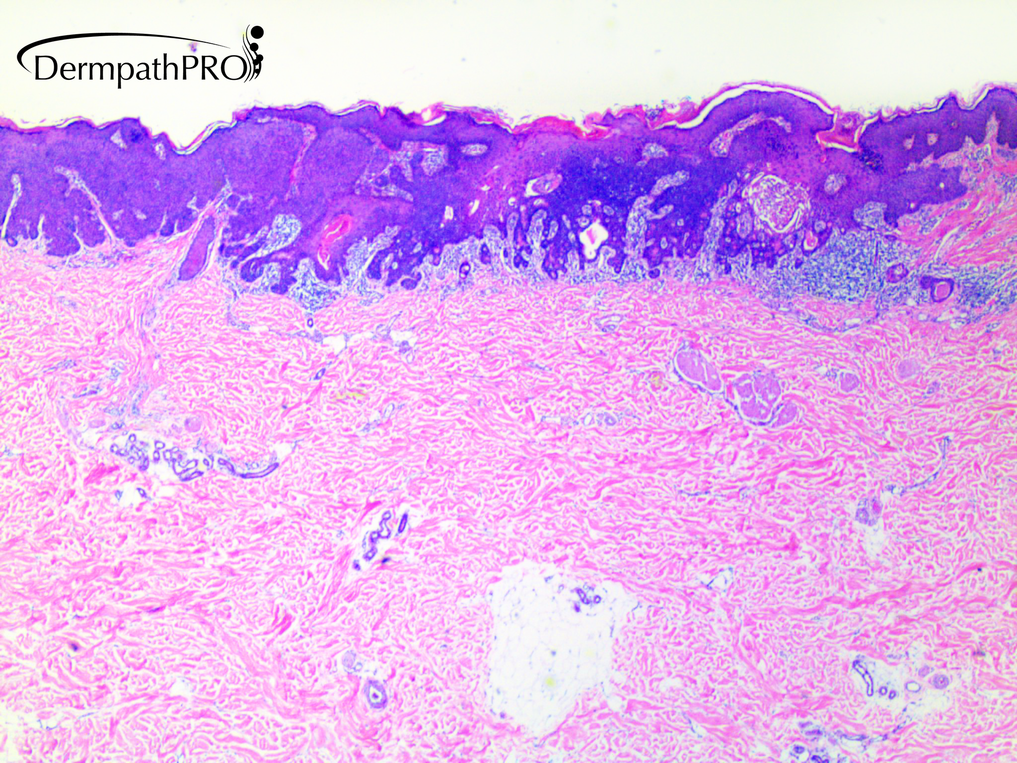
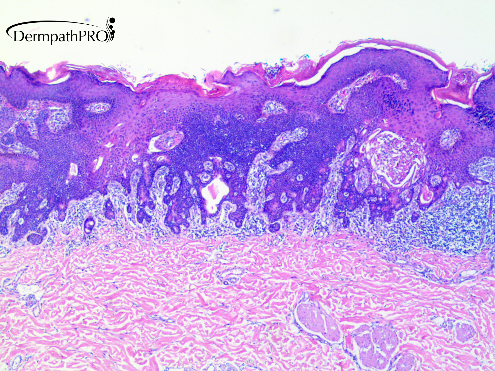
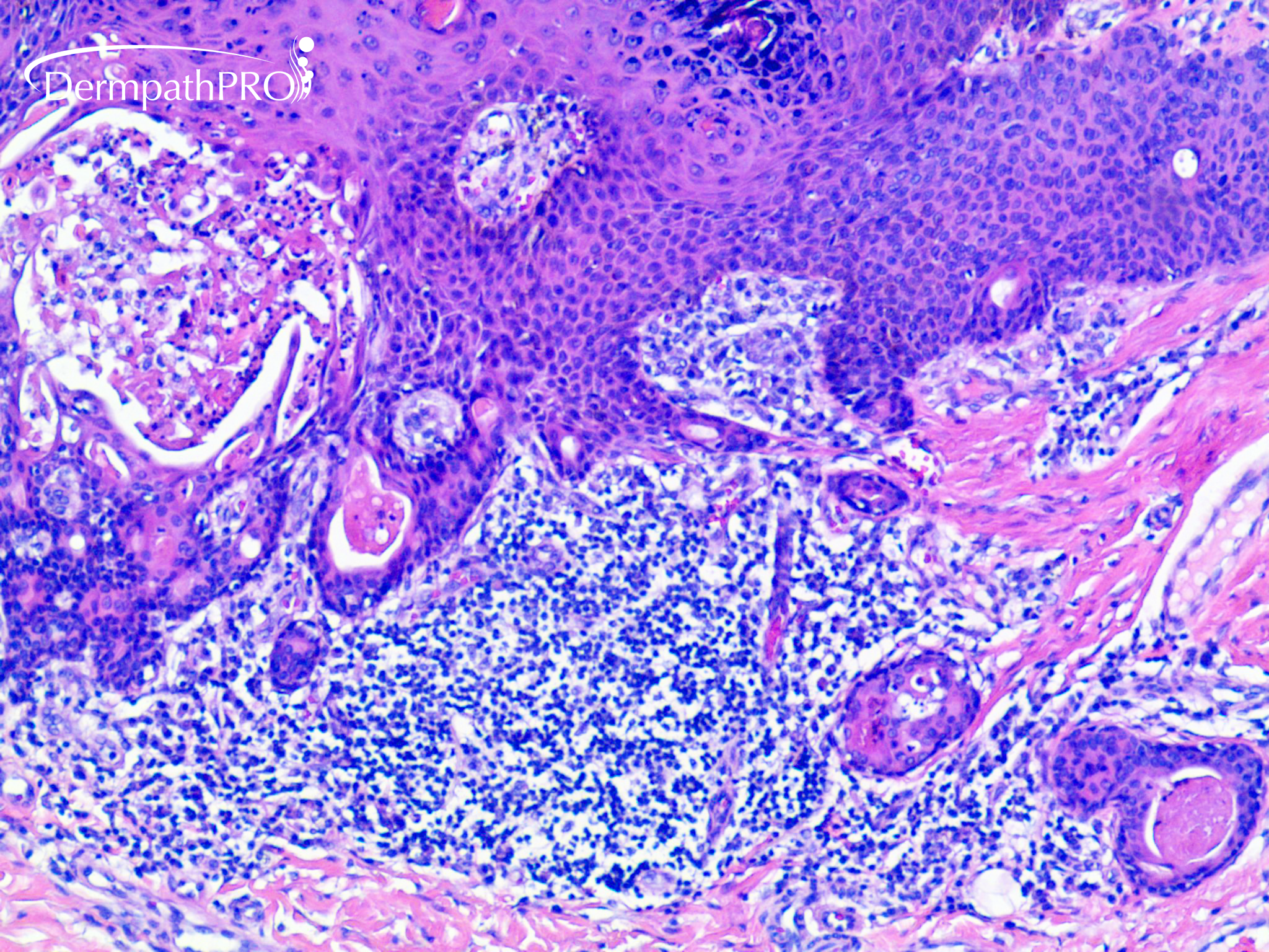
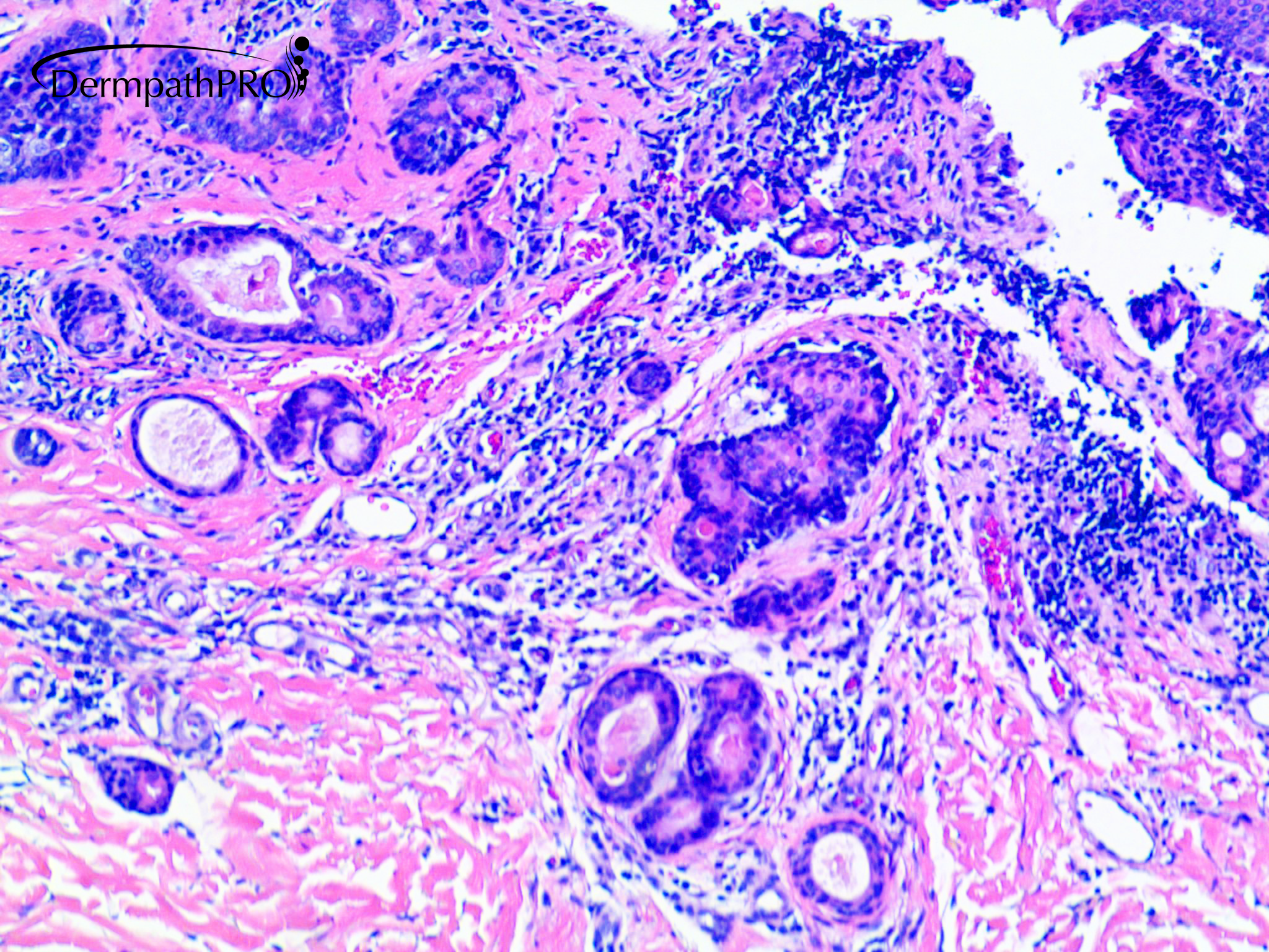
76 year old male with right upper arm lesion.


  • Like 1
  Report Record

User Feedback


Richard Logan

Posted

Looks like I'll have to open the batting, but, as happened frequently in my actual cricketting career, I risk getting out for a duck!

I see multiple anastomosing  bands of epidermally-derived cells, forming ductal structures and associated with an inflammatory dermal response.  There is evidence of necrosis in places.  The cytology of the tumour cells is difficult to see on these views.  However, the overall pattern suggests a diagnosis of low-grade eccrine porocarcinoma.

Share this comment


Link to comment
share_externally

7 hours ago, Richard Logan said:

Looks like I'll have to open the batting, but, as happened frequently in my actual cricketting career, I risk getting out for a duck!

I see multiple anastomosing  bands of epidermally-derived cells, forming ductal structures and associated with an inflammatory dermal response.  There is evidence of necrosis in places.  The cytology of the tumour cells is difficult to see on these views.  However, the overall pattern suggests a diagnosis of low-grade eccrine porocarcinoma.

Agree! A minimally invasive porocarcinoma. 

Share this comment


Link to comment
share_externally

Meenakshi Batrani

Posted

Agree with minimall inavsive porocarcinoma

Share this comment


Link to comment
share_externally

When I look at the "infiltr ative glands" in the superficial dermis, I think I still see 2 layers: the inner secretory columnar cells and the outer flat myoepithelial / basal cells which make me wonder whether the glands are benign.  So I would offer another possibility in the differential diagnosis: eccrine adenomatous metaplasia. I probably would do some stains to see whether the basal / myoepithelial layer is still present in those glands.


Share this comment


Link to comment
share_externally

Alex-Ventura-Leon

Posted

It looks poroid and invasive (minimal?) so agree with Porocarcinoma

Share this comment


Link to comment
share_externally

Uma Sundram

Posted

Yes, this was a difficult one. We thought we could see two layers in the dermis as well, so we went with poroma, rather than porocarcinoma. However, wide margins were taken of this lesion anyway, so both approaches are covered. 

Share this comment


Link to comment
share_externally

Richard Logan

Posted

Dear Dr. Sundram,

Have you seen a benign poroma excite such an inflammatory response before?

Share this comment


Link to comment
share_externally

Dr. Richard Carr

Posted

I think this is a case where the lesion failed to read the textbook. Looks benign to me though. "Benign acanthoma with tubular eccrine ductal differentiation"?

Share this comment


Link to comment
share_externally


Join the conversation

You can post now and register later. If you have an account, sign in now to post with your account.

Guest
Add a comment...

×   Pasted as rich text.   Paste as plain text instead

  Only 75 emoji are allowed.

×   Your link has been automatically embedded.   Display as a link instead

×   Your previous content has been restored.   Clear editor

×   You cannot paste images directly. Upload or insert images from URL.

Loading...

×
×
  • Create New...