Jump to content
In this section we have spot diagnoses posted on a daily basis since June 2010, now over 4000! You can review the archived cases and read the suggested diagnoses by users and the final comment by the contributors.
Case are uploaded each week day by 10 am UK time with the correct diagnosis will generally be posted at 8 pm UK time. Why not view the most recent spot diagnosis and proffer a diagnosis?

Case Number : Case 2652 - 04 September 2020 Posted By: Dr. Richard Carr

Please read the clinical history and view the images by clicking on them before you proffer your diagnosis.
Submitted Date :
   (0 reviews)

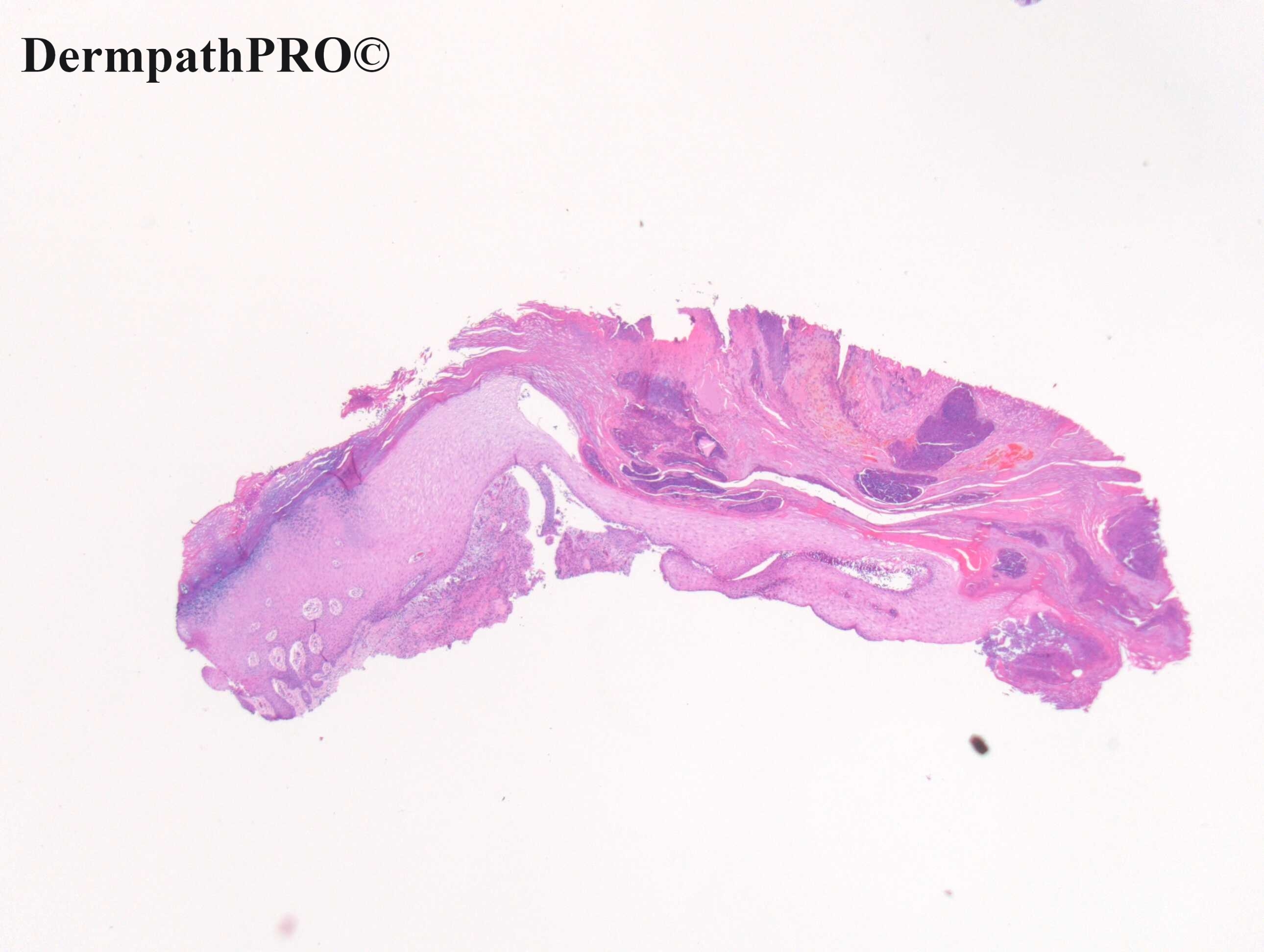
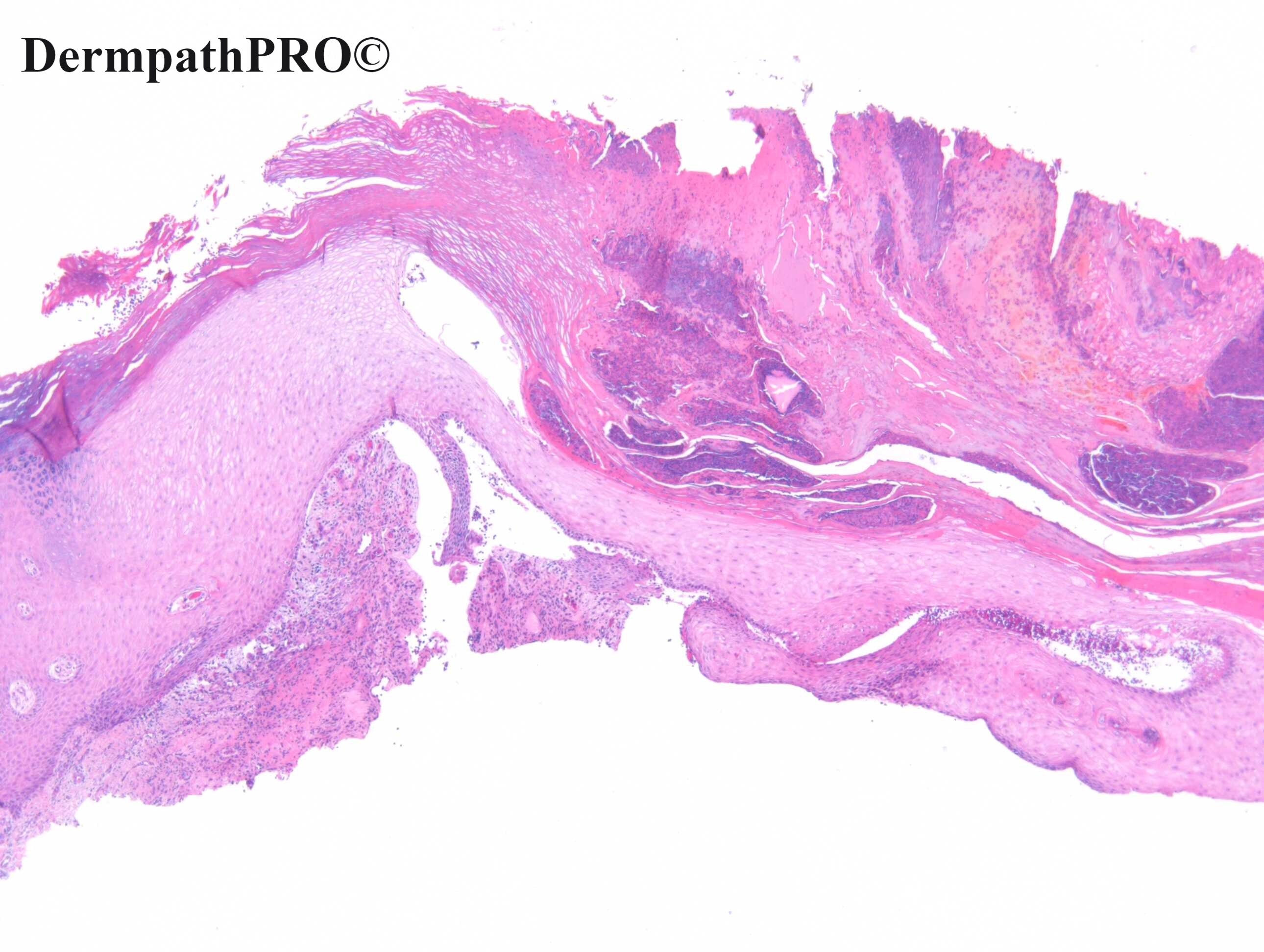
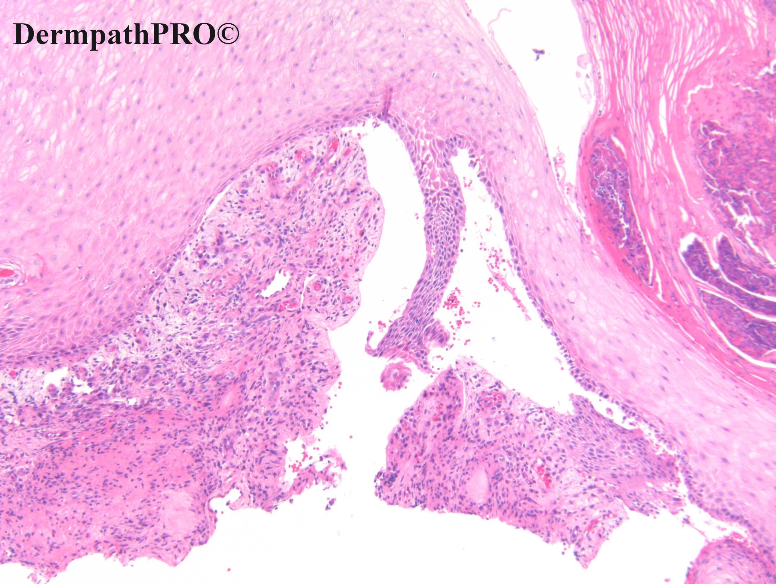
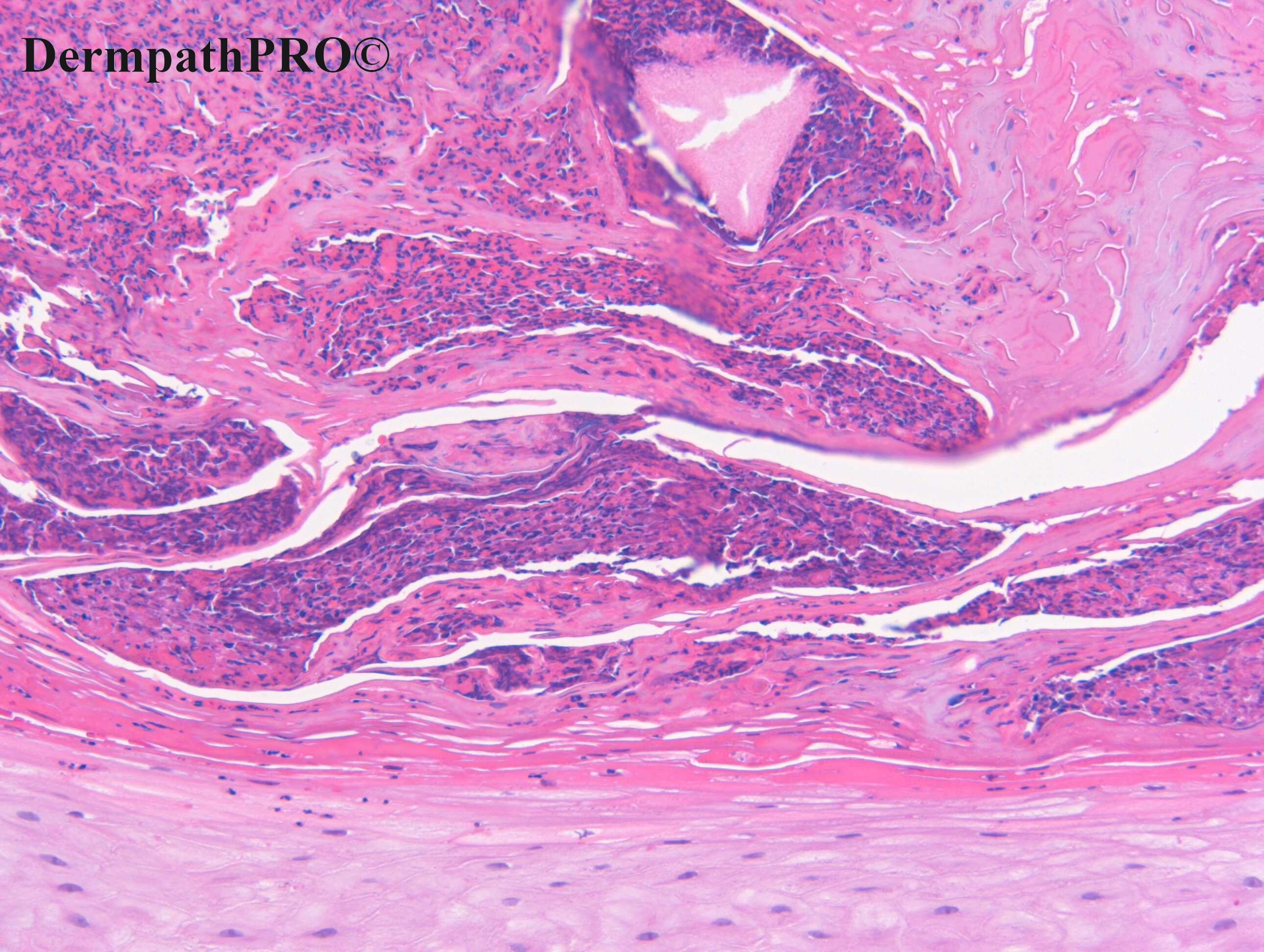
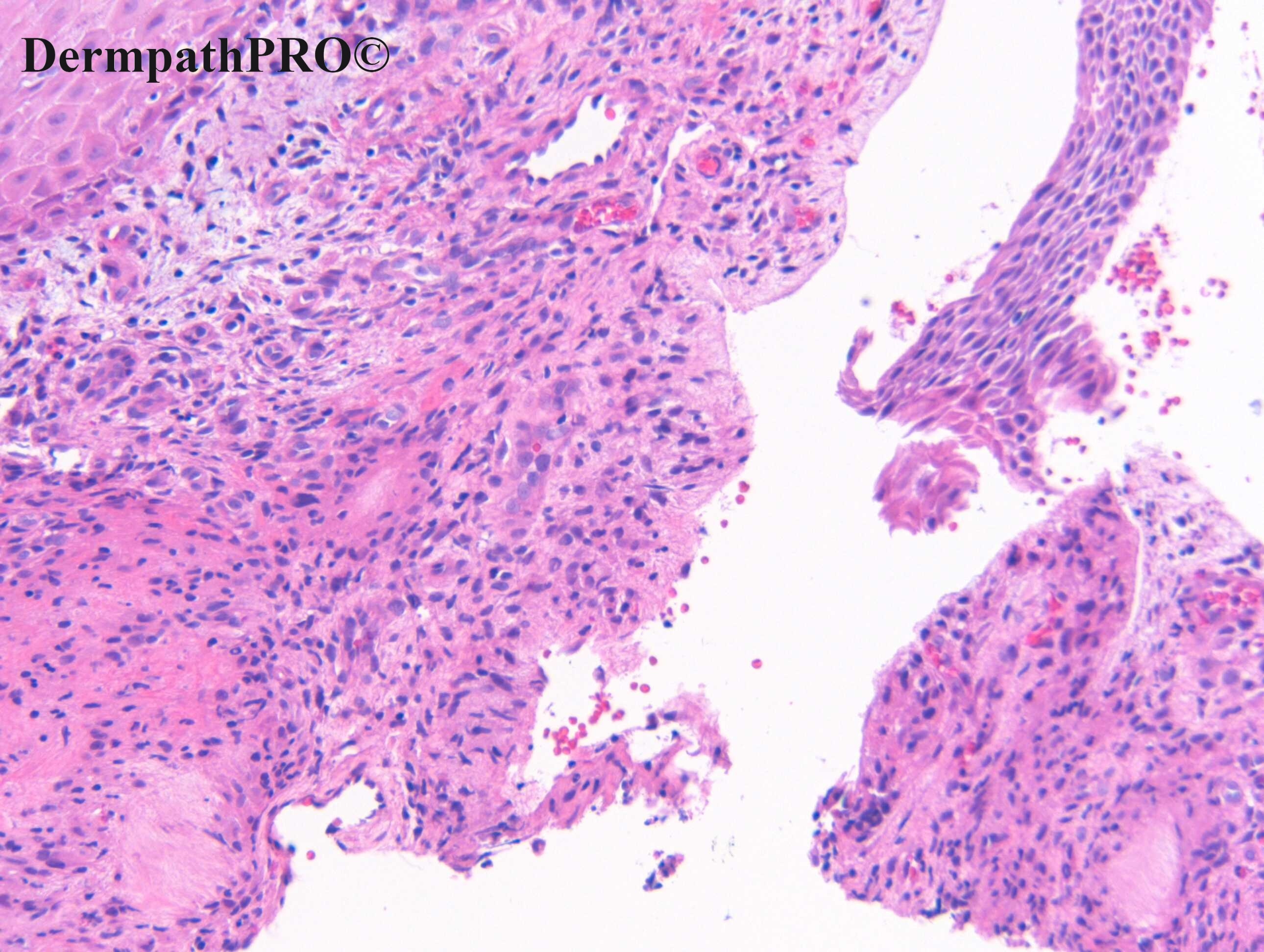
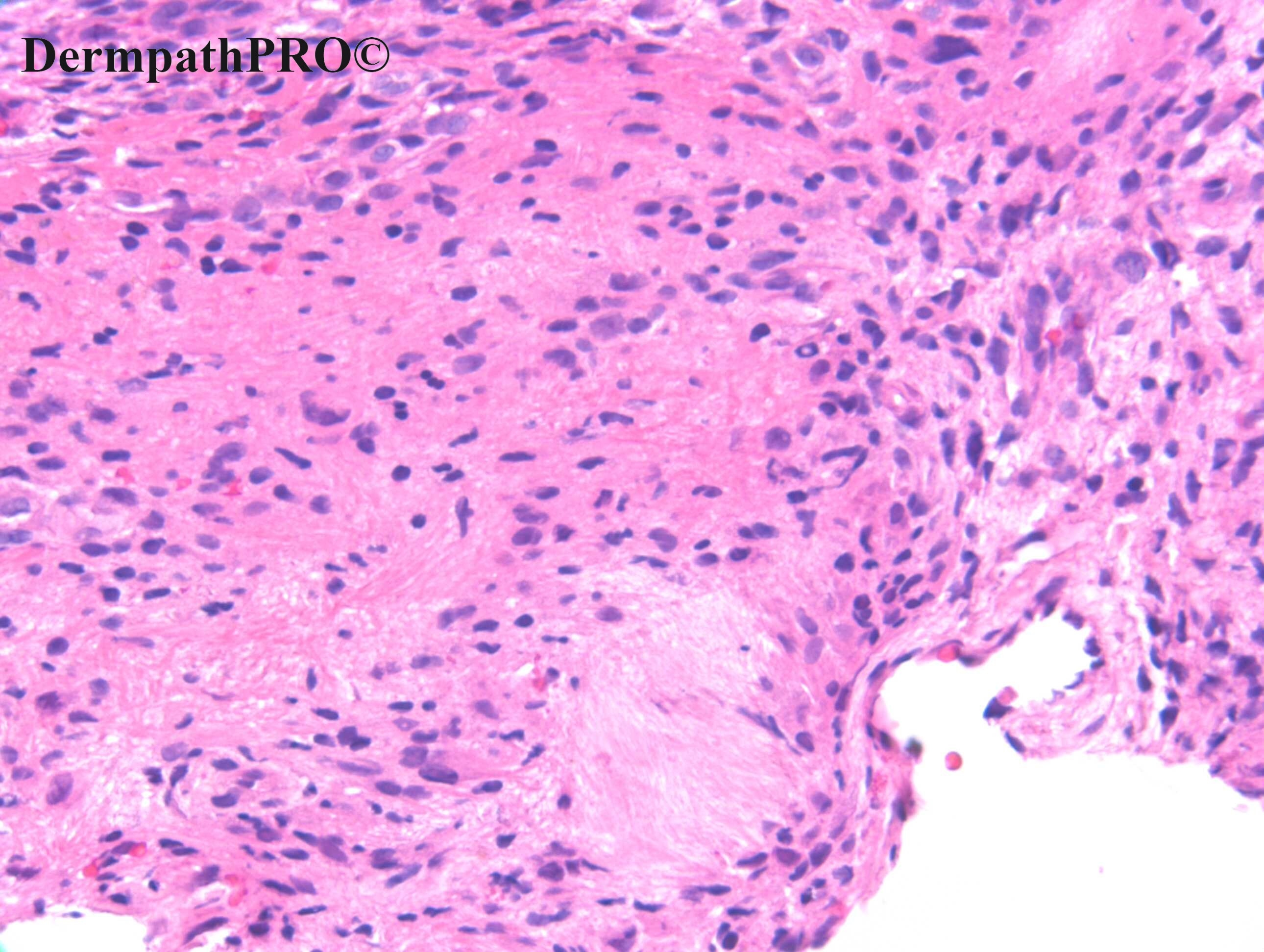
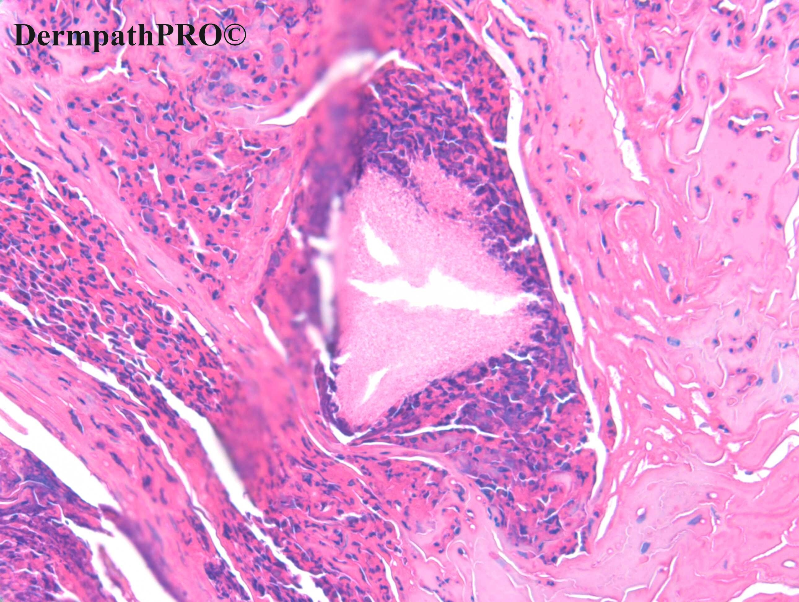
M85. Little finger. Ulcerated nodule, shave biopsy.


  • Thanks 1
  Report Record

User Feedback


Richard Logan

Posted

There is obviously a perforating disorder going on here, with quite a lot of neutrophils in the stratum corneum.  The small amount of dermis visible has a granulomatous look to it, perhaps with a hint of necrobiosis.  Given the site I would hazard a guess at perforating GA.  Palisaded neutrophilic granulomatous dermatitis or fish-tank granuloma would also have to be considered.

Share this comment


Link to comment
share_externally

Richard Logan

Posted

One more thought - the small "necrobiotic" dermal foci have a somewhat feathery look to them.  Gout comes into the equation.

Share this comment


Link to comment
share_externally

1 hour ago, Richard Logan said:

One more thought - the small "necrobiotic" dermal foci have a somewhat feathery look to them.  Gout comes into the equation.

Yes. Gout Tophus. 

Share this comment


Link to comment
share_externally

Alex-Ventura-Leon

Posted (edited)

Agree. Gout Tophus with transepidermal elimination

Edited by Alex-Ventura-Leon
Typo

Share this comment


Link to comment
share_externally

Meenakshi Batrani

Posted

Yes. Gout with transepidermal elimination

Share this comment


Link to comment
share_externally

Eman El-Nabarawy

Posted

Gout Tophus with transepidermal elimination is my thought.

Share this comment


Link to comment
share_externally

Dr. Richard Carr

Posted

Well done Richard. You rescued that one. If I'm right you mentioned you are a head & neck / oral pathologist so would not see gout often although I've seen a few cases on the pinna of the ear. I think this is a nice example of a pathology being quite distinctive even though the focus is tiny one can make a confident spot diagnosis. 

Share this comment


Link to comment
share_externally

Richard Logan

Posted

Actually I'm a retired Dermatologist, no special pathological training, as you can tell!

Share this comment


Link to comment
share_externally

16 hours ago, Richard Logan said:

Actually I'm a retired Dermatologist, no special pathological training, as you can tell!

Hello Richard Logan. I’m a retired ( general pathologist ) as well. 

Share this comment


Link to comment
share_externally

Dr. Richard Carr

Posted

Huge respect for getting this subtle pathology!

Share this comment


Link to comment
share_externally


Join the conversation

You can post now and register later. If you have an account, sign in now to post with your account.

Guest
Add a comment...

×   Pasted as rich text.   Paste as plain text instead

  Only 75 emoji are allowed.

×   Your link has been automatically embedded.   Display as a link instead

×   Your previous content has been restored.   Clear editor

×   You cannot paste images directly. Upload or insert images from URL.

Loading...

×
×
  • Create New...