Jump to content
In this section we have spot diagnoses posted on a daily basis since June 2010, now over 4000! You can review the archived cases and read the suggested diagnoses by users and the final comment by the contributors.
Case are uploaded each week day by 10 am UK time with the correct diagnosis will generally be posted at 8 pm UK time. Why not view the most recent spot diagnosis and proffer a diagnosis?

Case Number : Case 2655 - 09 September 2020 Posted By: Dr. Hafeez Diwan

Please read the clinical history and view the images by clicking on them before you proffer your diagnosis.
Submitted Date :
   (0 reviews)

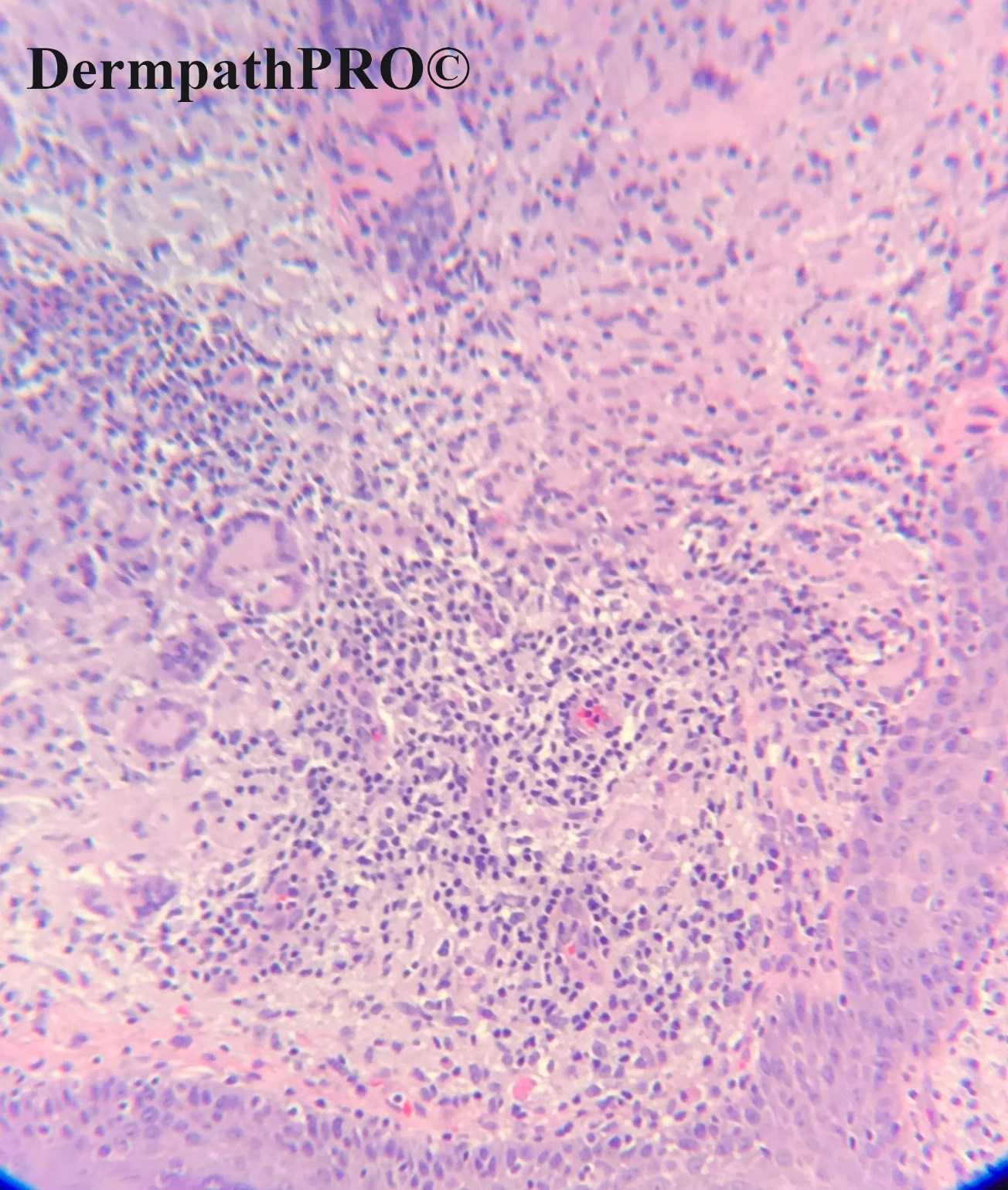
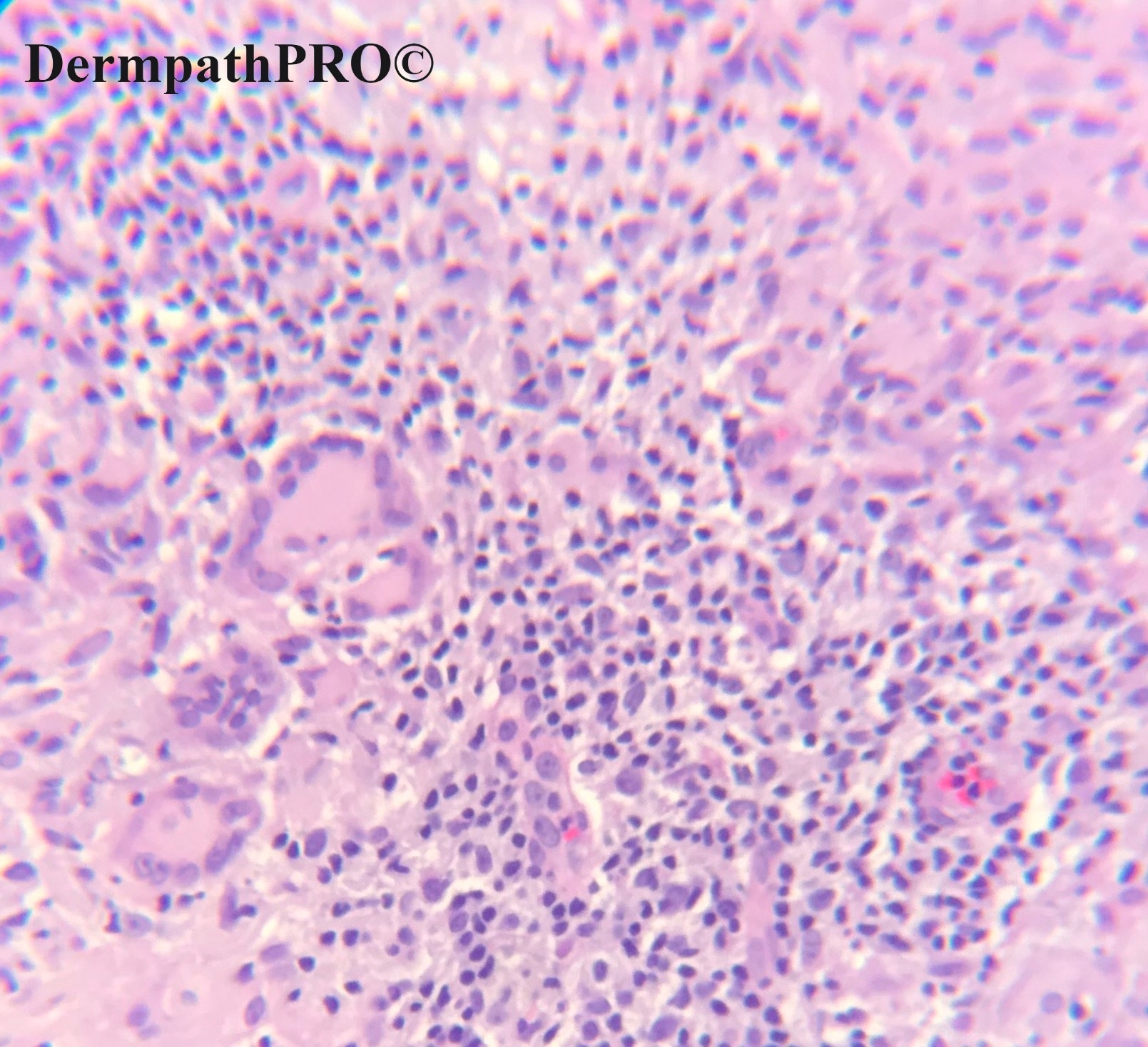
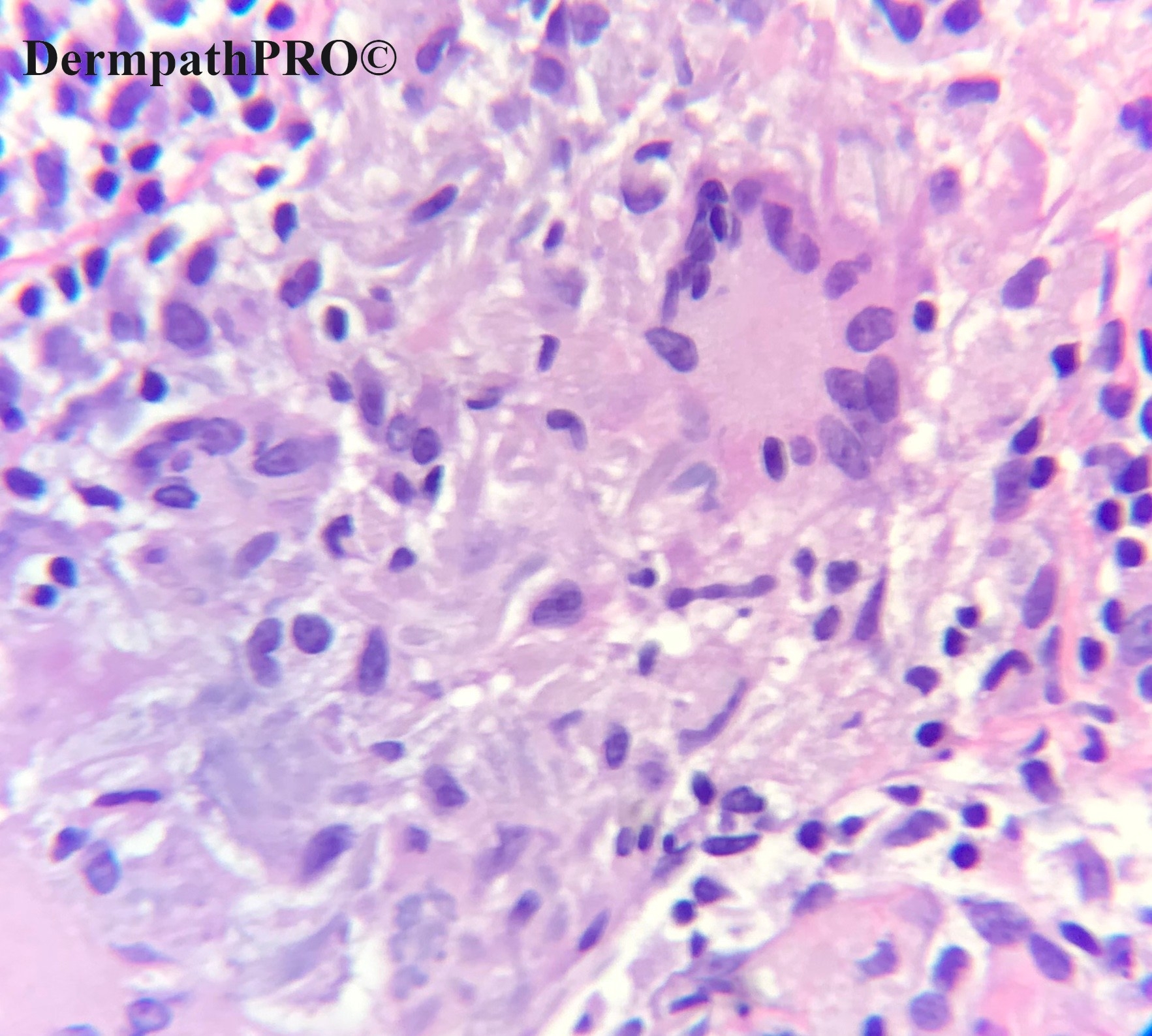
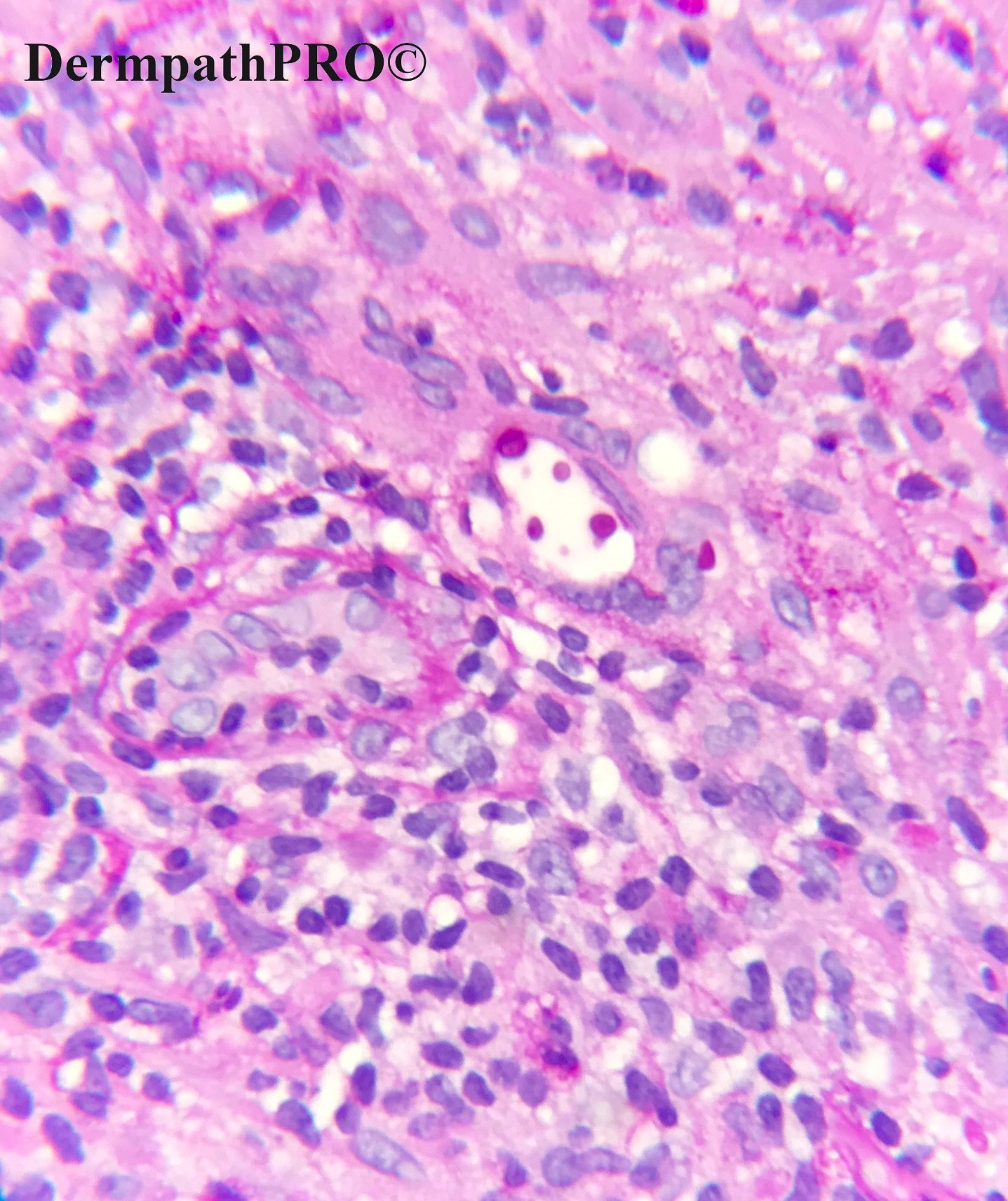
66 year-old female with right cheek lesion. Clinical impression: Rule out basal cell carcinoma.


  • Like 1
  Report Record

User Feedback


Richard Logan

Posted

This has a granulomatous look to it.  The admixture of inflammatory cells and vascular dilation with endothelial swelling suggest granuloma faciale.  Against this is the paucity, in fact absence of eosinophils, a very dubious Grenz zone and also giant cells, which I don't think are a feature of granuloma faciale.  If the last view is PAS then I'm not sure what it is highlighting.

The apposition of giant cells and solar elastosis would also make me consider actinic granuloma.

Share this comment


Link to comment
share_externally

Richard Logan

Posted

If I might be allowed to dig myself out of a hole, the giant cells are Langhans' type, and the stain possibly ZN or Wade-Fite, in which case lupus vulgaris and leprosy are better candidates.

Share this comment


Link to comment
share_externally

Alex-Ventura-Leon

Posted

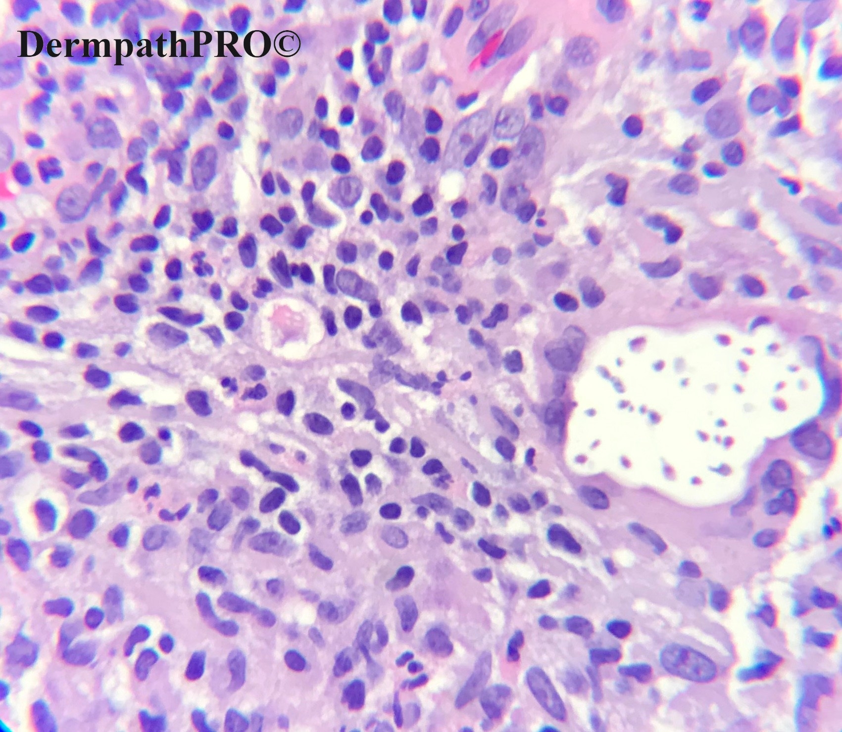
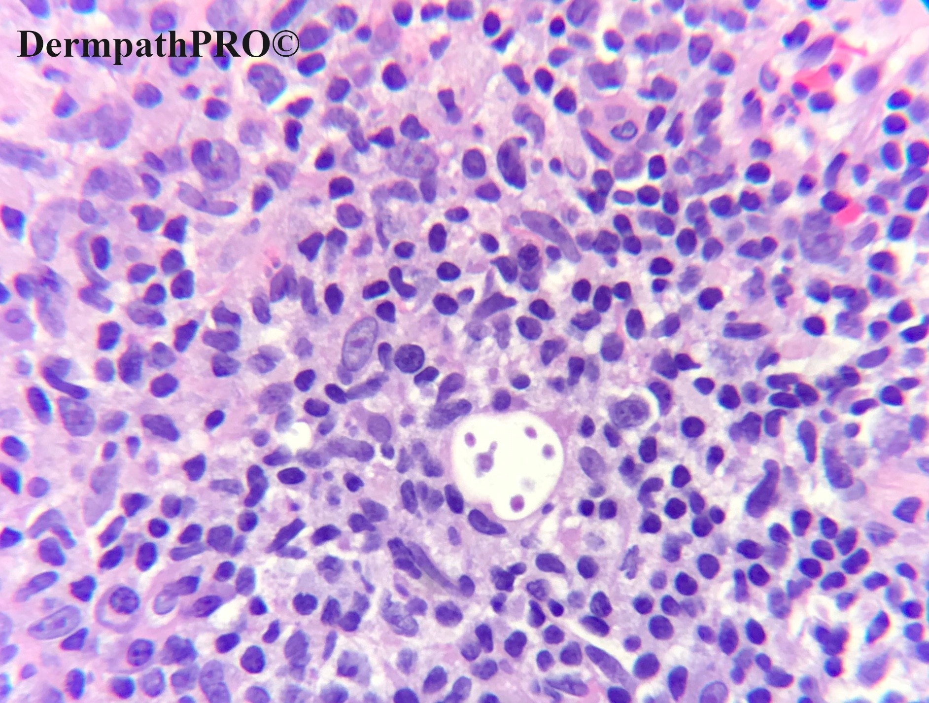
Granulomatous inflamation with fungal estructures inside of a multinucleated giant cell.

I´m thinking in Criptoccus but i´m not sure

Share this comment


Link to comment
share_externally

Never seen a similar microorganism in a cutaneous site. This reminds me of Giardiasis...but this isn’t pas-positive, as far as I know ( it should be Giemsa or toluidine blu-positive ). I don’t know, but the this “animal” reminds me of something...

Share this comment


Link to comment
share_externally

Krishnakumar subramanian

Posted

agree granulomatous inflammation with yeast like structures

? cryptococossis

GMS stain can help

Share this comment


Link to comment
share_externally

Meenakshi Batrani

Posted

I would keep differential of Histoplasma and Leishmaniasis. Tough to tell under H&E figure 3 and 4, it seems to have sheath and kinetoplast like structure. But if figure 4 is Giemsa/Toulidine blue and Figure 6 is PAS then it is PAS positive and does not takes up Giemsa, then it is Histoplasma. 

Share this comment


Link to comment
share_externally

Thought of histoplasmosis too, but don’t see any vacuole. 

Share this comment


Link to comment
share_externally

Maybe they are not microorganisms. Hamazaki Wesenberg bodies?

Share this comment


Link to comment
share_externally

Richard Logan

Posted

That's a tough one.  Were there any inflamed/occluded  follicles seen on other views?  Pityrosporum folliculitis of course is normally seen on the upper trunk in younger adults with numerous small follicular lesions.  It is not a diagnosis that one would normally think of for a solitary lesion on the face at this age.  Does that bring granuomatous rosacea into the equation?.

Share this comment


Link to comment
share_externally


Join the conversation

You can post now and register later. If you have an account, sign in now to post with your account.

Guest
Add a comment...

×   Pasted as rich text.   Paste as plain text instead

  Only 75 emoji are allowed.

×   Your link has been automatically embedded.   Display as a link instead

×   Your previous content has been restored.   Clear editor

×   You cannot paste images directly. Upload or insert images from URL.

Loading...

×
×
  • Create New...