Jump to content
In this section we have spot diagnoses posted on a daily basis since June 2010, now over 4000! You can review the archived cases and read the suggested diagnoses by users and the final comment by the contributors.
Case are uploaded each week day by 10 am UK time with the correct diagnosis will generally be posted at 8 pm UK time. Why not view the most recent spot diagnosis and proffer a diagnosis?

Case Number : Case 2656 - 10 September 2020 Posted By: Saleem Taibjee

Please read the clinical history and view the images by clicking on them before you proffer your diagnosis.
Submitted Date :
   (0 reviews)

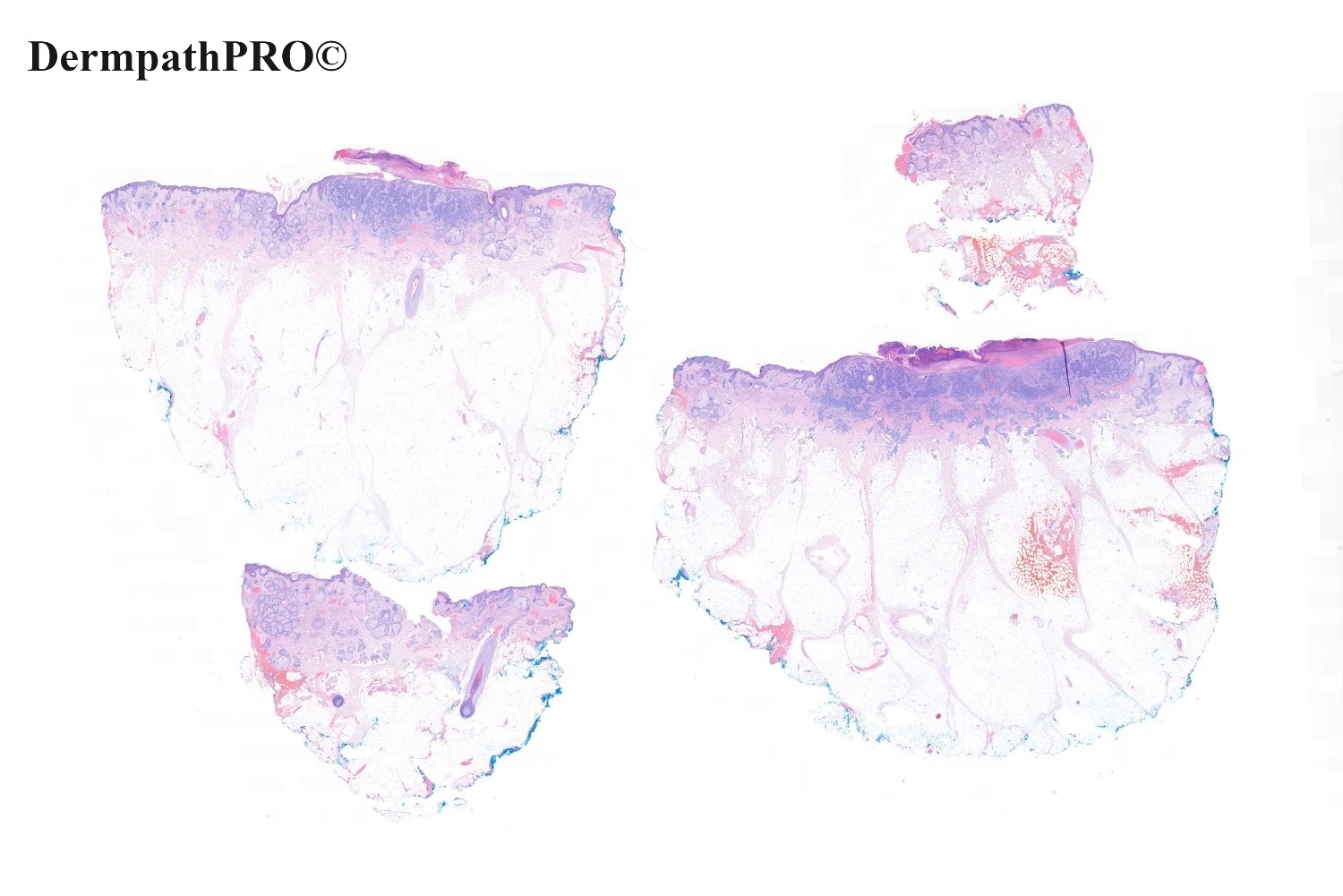
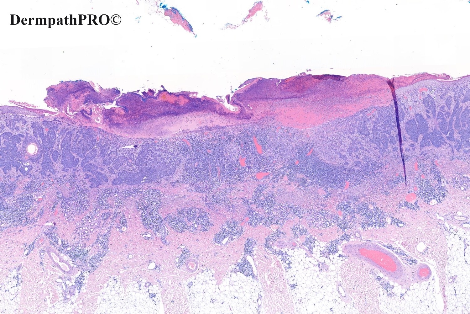
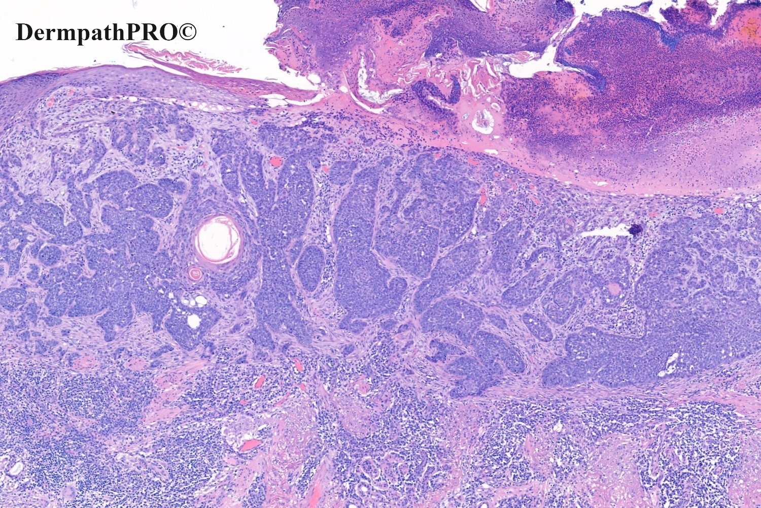
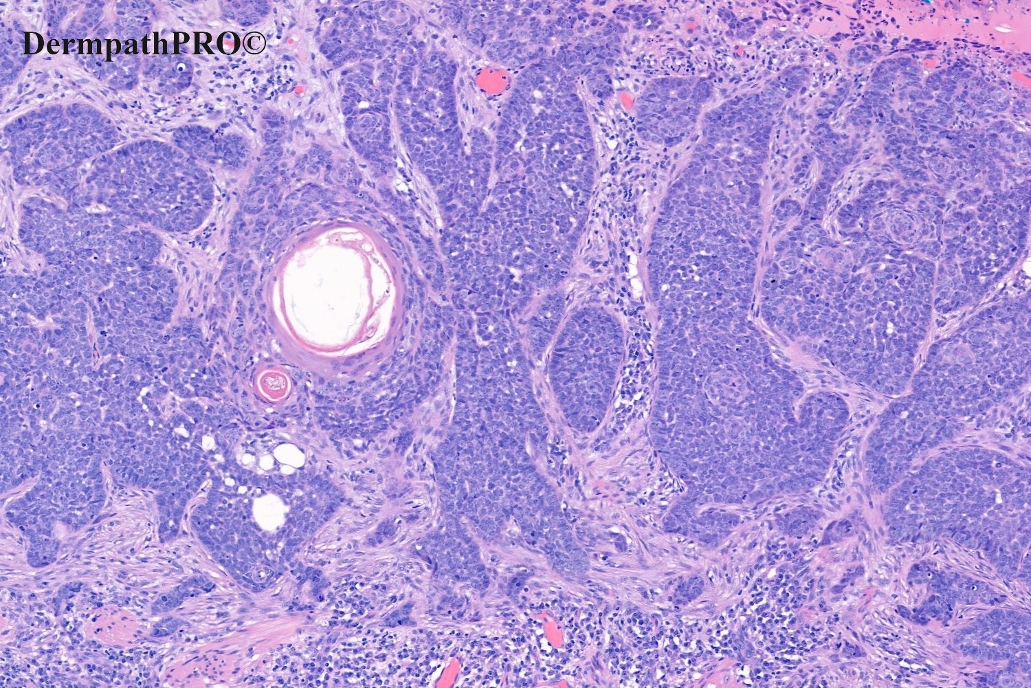
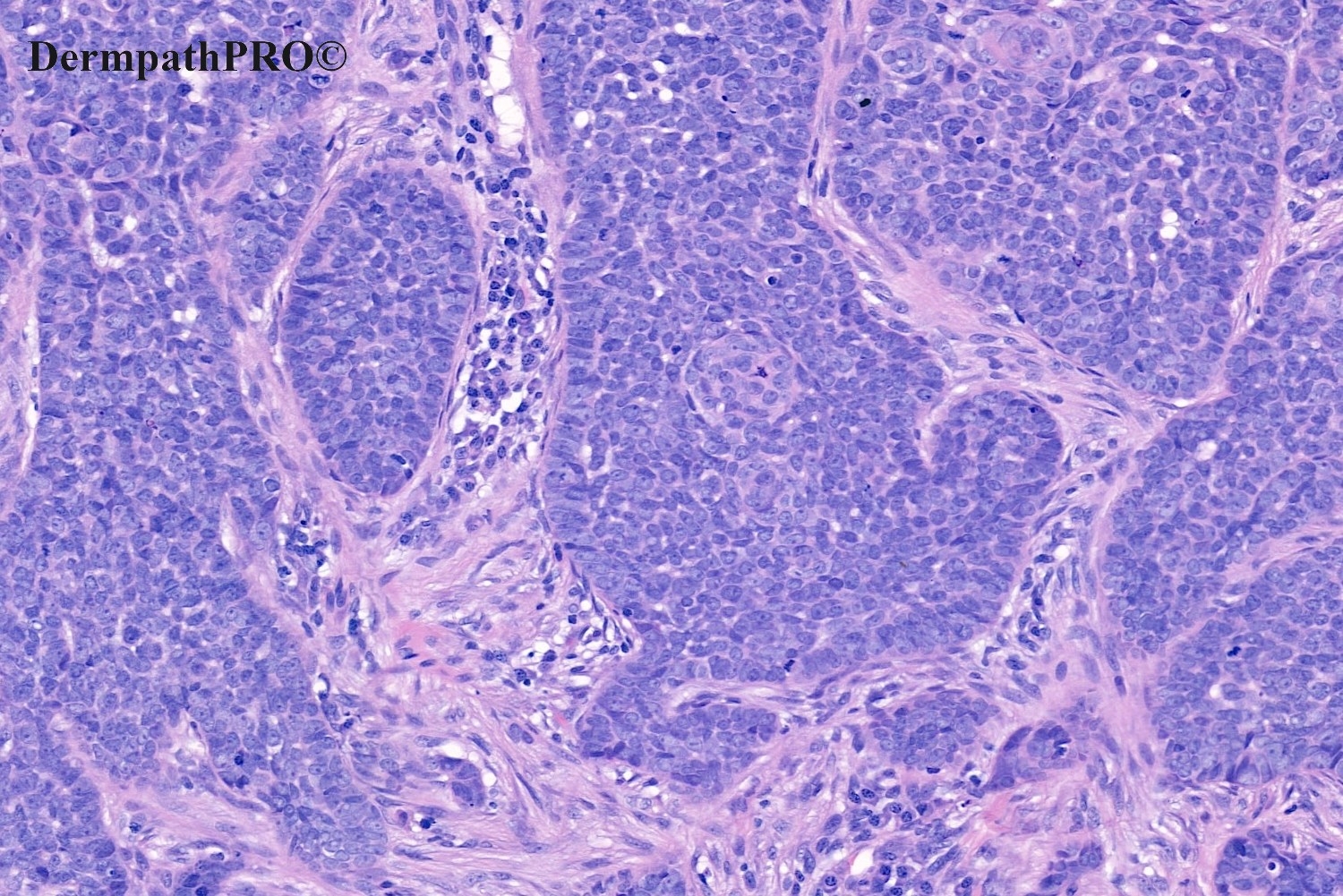
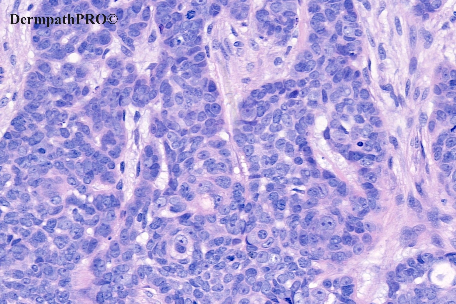
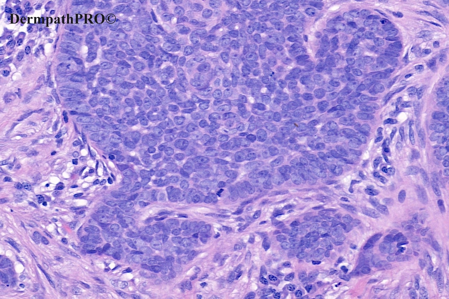
91M, Excision right cheek.


  • Like 1
  Report Record

User Feedback


Richard Logan

Posted

The low power appearance suggests an ulcerated basal cell carcinoma.  However, palisading is not well seen and there is no retraction artefact.  Some cells show bubbly cytoplasm.  I suspect this is a BCC mimic such as sebaceous carcinoma or basaloid SCC.  IH should help.

The other thing is there is a pronounced lymphocytic infiltrate as far down as the fat.  Perhaps there are two pathologies going on here such as CLL?

Share this comment


Link to comment
share_externally

3 hours ago, Richard Logan said:

The low power appearance suggests an ulcerated basal cell carcinoma.  However, palisading is not well seen and there is no retraction artefact.  Some cells show bubbly cytoplasm.  I suspect this is a BCC mimic such as sebaceous carcinoma or basaloid SCC.  IH should help.

The other thing is there is a pronounced lymphocytic infiltrate as far down as the fat.  Perhaps there are two pathologies going on here such as CLL?

Agree!

There are some palisades, but not in infiltrating solid aggregates. Instead I see a lobular, in situ, intra-sebaceous growth in superficial dermis, as well as it does the ocular sebaceous carcinoma. But this isn’t an ocular-periocular case( isn’t it?). Difficult, but favor a poorly differentiated sebaceous carcinoma. And agree with the CLL guesswork, also. 

Share this comment


Link to comment
share_externally

Alex-Ventura-Leon

Posted (edited)

BCC vs Basaloid SCC. I favor the first but IHC would be usefull (EMA, BerEP4)

Edited by Alex-Ventura-Leon
Typo

Share this comment


Link to comment
share_externally

Victor Delgado

Posted

it's a little bit weird, however it looks with neuroendocrine differentiation to me.

Share this comment


Link to comment
share_externally

Meenakshi Batrani

Posted

I would also keep the differential of sebaceous carcinoma, BCC and basaloid SCC.

Share this comment


Link to comment
share_externally

Krishnakumar subramanian

Posted

I would also keep the differential of sebaceous carcinoma,  basaloid SCC but thinking of CLL from the lymphocytes i dont know, i will get a CBC done

Share this comment


Link to comment
share_externally

Richard Logan

Posted

So it's BCC with neuroendocrine differentiation.  Is the degree of lymphocytic infitration significant, or just reactive to the tumour?

Share this comment


Link to comment
share_externally

Saleem Taibjee

Posted

Yes, well done, this is a BCC with neuroendocrine differentiation. These can be spotted at low power as basaloid tumours that show a BCC architecture, but with cells showing these larger 'high grade' nuclei. I suspect they are not uncommon, but get readily missed during busy routine sign-out.

As far as I'm aware, there are no reported cases of metastasis (?), and hence this may be a rather academic observation, but probably still worthwhile mentioning within the histology report but clearly indicating the distinction from Merkel cell carcinoma, etc.

After the comments above, I did go back and double-check the accompanying infiltrate on the glass slide. This comprises mostly mature plasma cells with normal morphology, with admixed lymophocytes. Hence I believe it is reactive. The patient has a normal ful blood count.

Share this comment


Link to comment
share_externally

Krishnakumar subramanian

Posted

Thanks a lot Dr for this interesting case

Share this comment


Link to comment
share_externally

On 15/09/2020 at 14:56, Saleem Taibjee said:

Yes, well done, this is a BCC with neuroendocrine differentiation. These can be spotted at low power as basaloid tumours that show a BCC architecture, but with cells showing these larger 'high grade' nuclei. I suspect they are not uncommon, but get readily missed during busy routine sign-out.

As far as I'm aware, there are no reported cases of metastasis (?), and hence this may be a rather academic observation, but probably still worthwhile mentioning within the histology report but clearly indicating the distinction from Merkel cell carcinoma, etc.

After the comments above, I did go back and double-check the accompanying infiltrate on the glass slide. This comprises mostly mature plasma cells with normal morphology, with admixed lymophocytes. Hence I believe it is reactive. The patient has a normal ful blood count.

What made you do chromogrannin?


Share this comment


Link to comment
share_externally


Join the conversation

You can post now and register later. If you have an account, sign in now to post with your account.

Guest
Add a comment...

×   Pasted as rich text.   Paste as plain text instead

  Only 75 emoji are allowed.

×   Your link has been automatically embedded.   Display as a link instead

×   Your previous content has been restored.   Clear editor

×   You cannot paste images directly. Upload or insert images from URL.

Loading...

×
×
  • Create New...