Jump to content
In this section we have spot diagnoses posted on a daily basis since June 2010, now over 4000! You can review the archived cases and read the suggested diagnoses by users and the final comment by the contributors.
Case are uploaded each week day by 10 am UK time with the correct diagnosis will generally be posted at 8 pm UK time. Why not view the most recent spot diagnosis and proffer a diagnosis?

Case Number : Case 2666 - 24 September 2020 Posted By: Saleem Taibjee

Please read the clinical history and view the images by clicking on them before you proffer your diagnosis.
Submitted Date :
   (0 reviews)

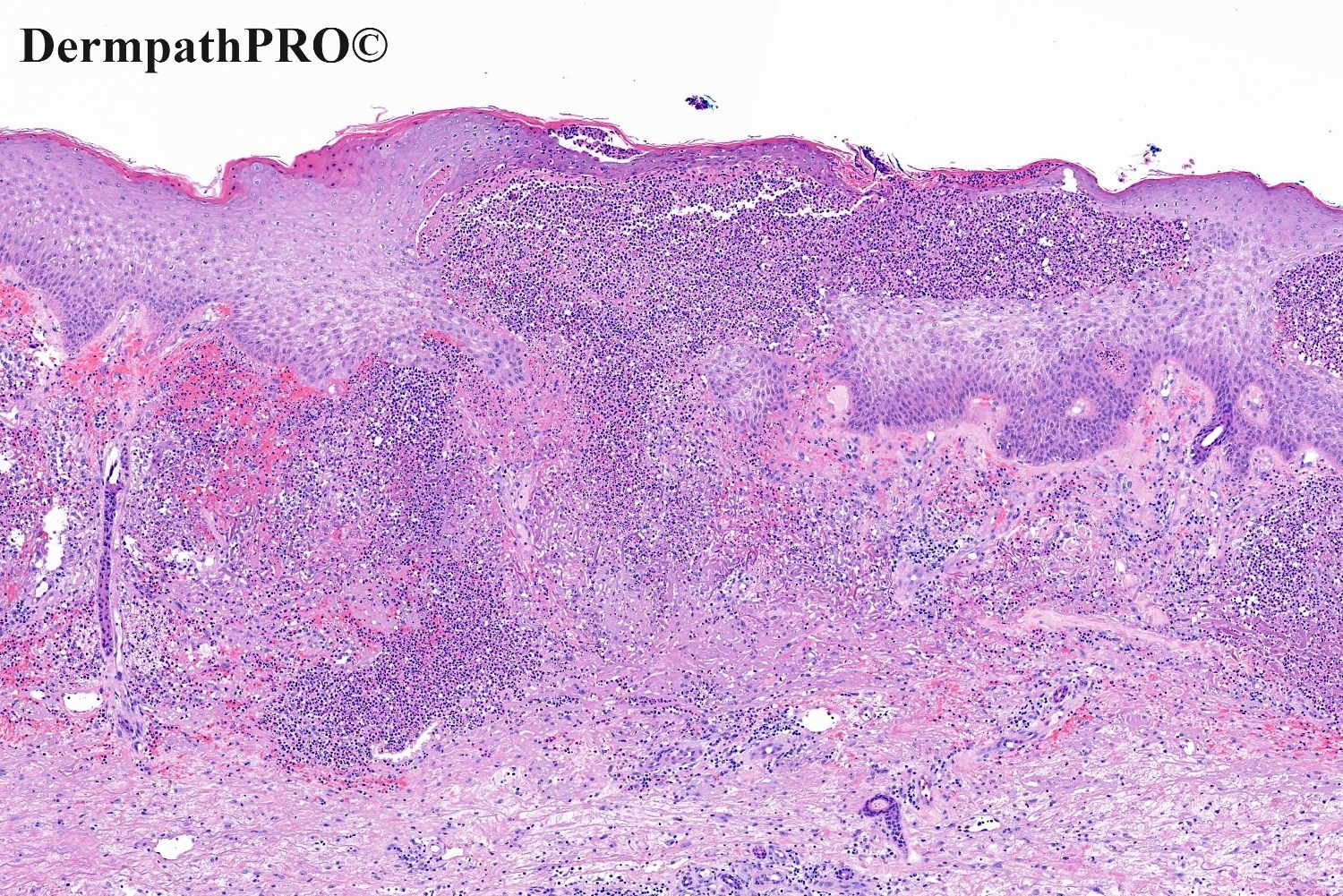
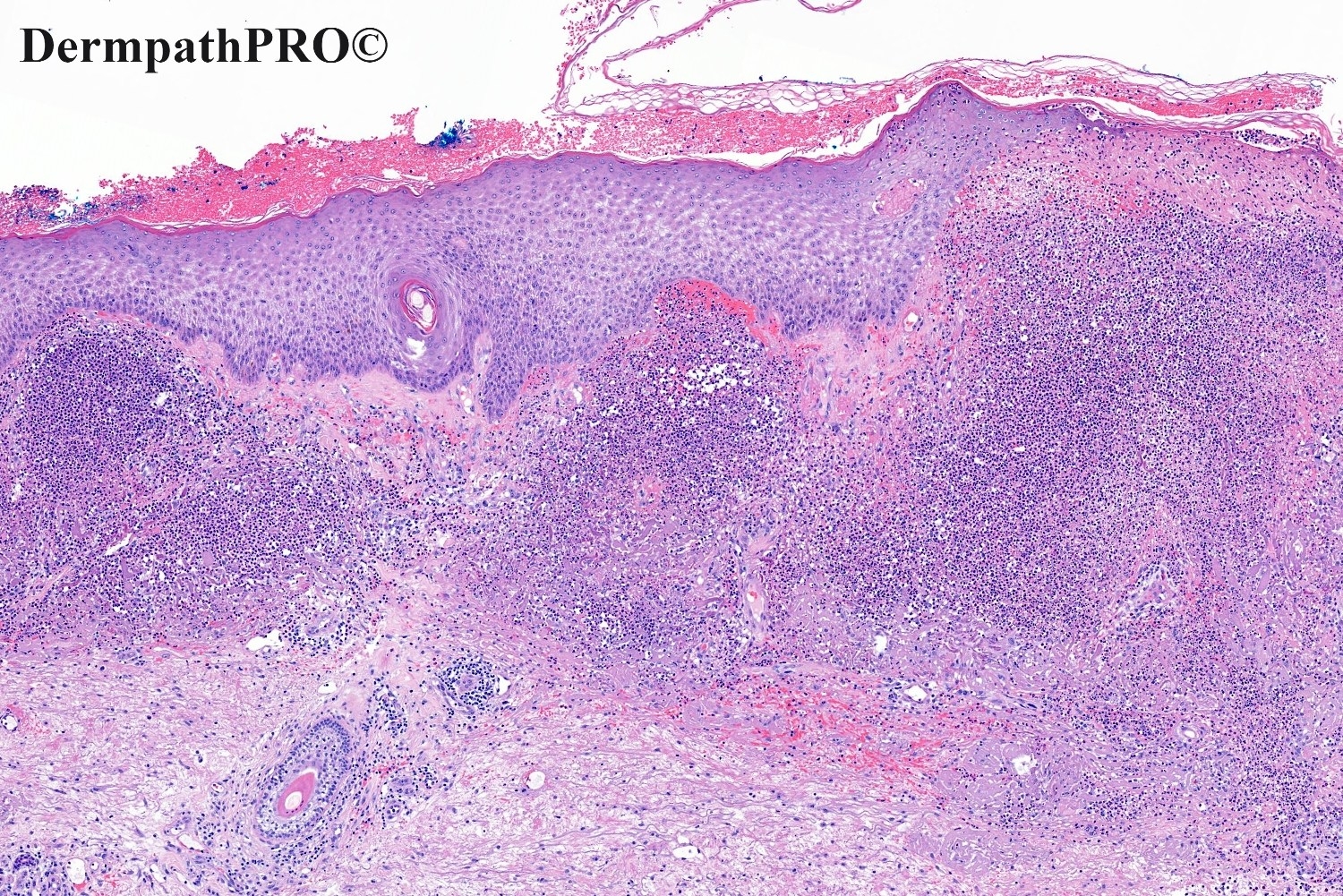
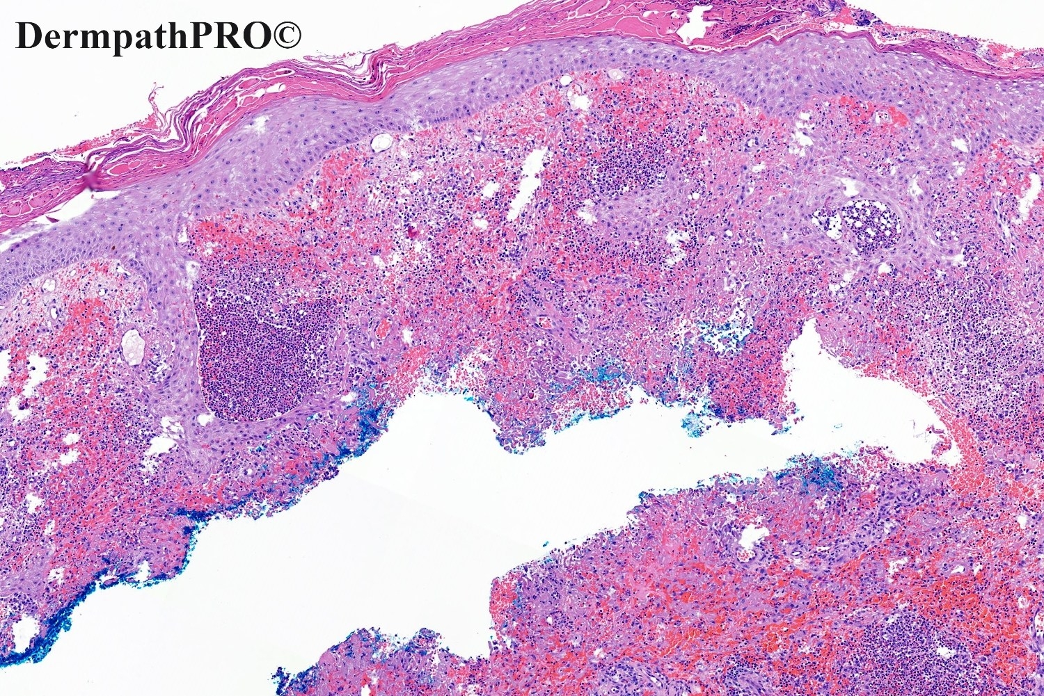
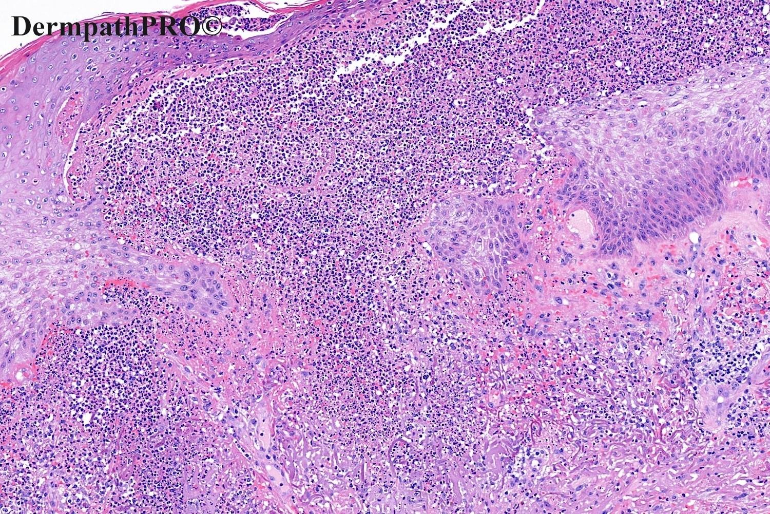
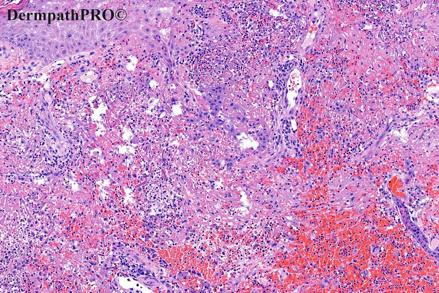
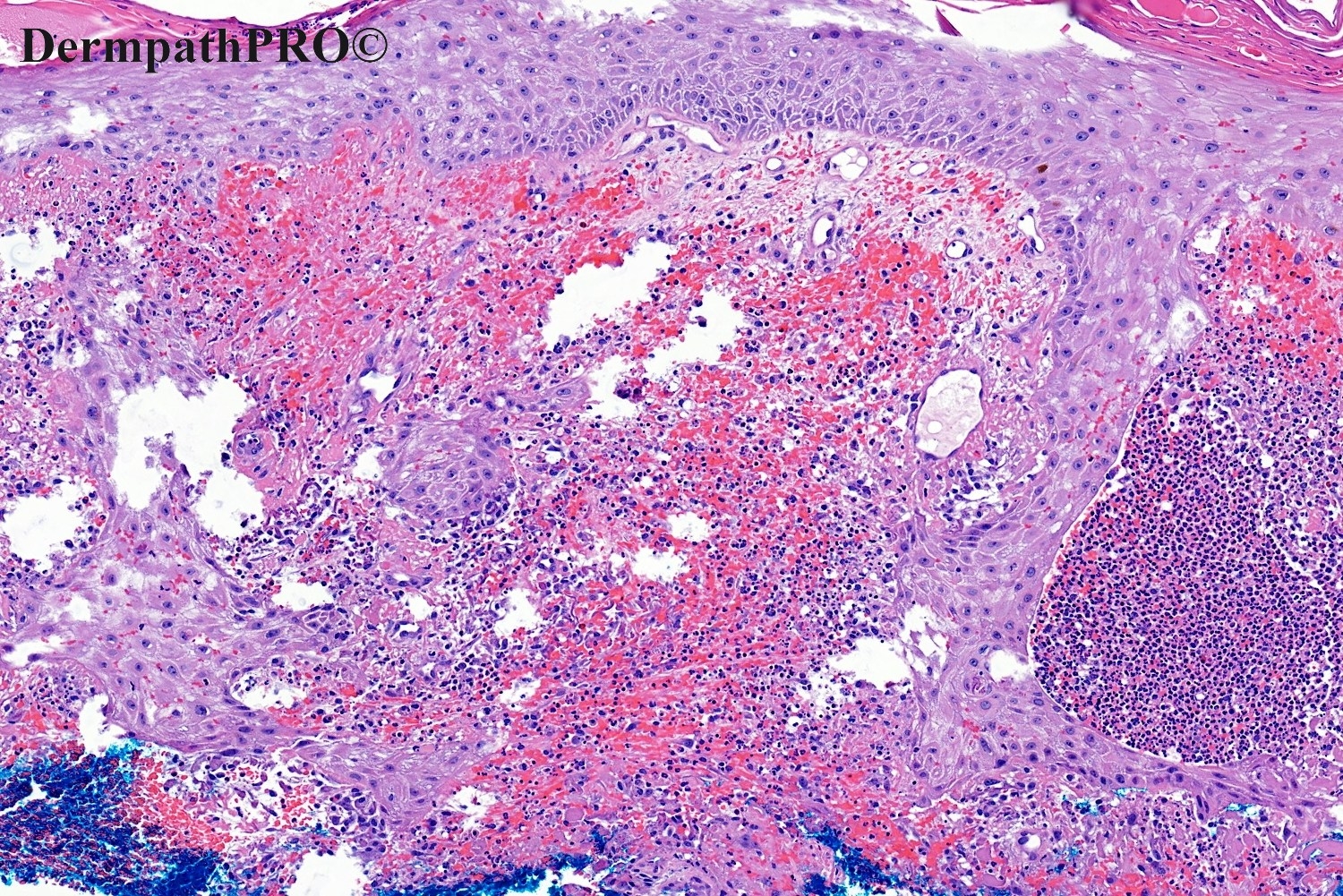
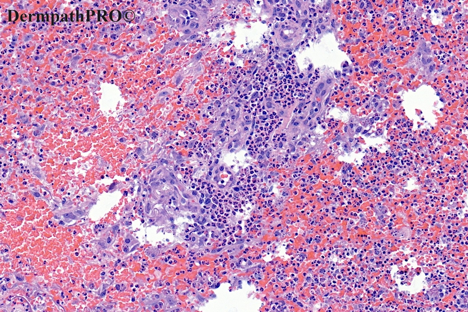
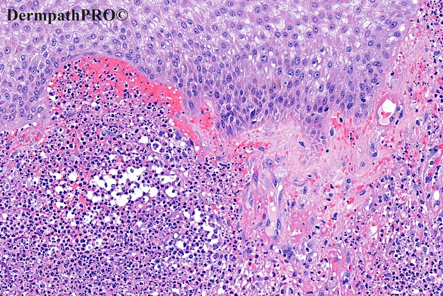
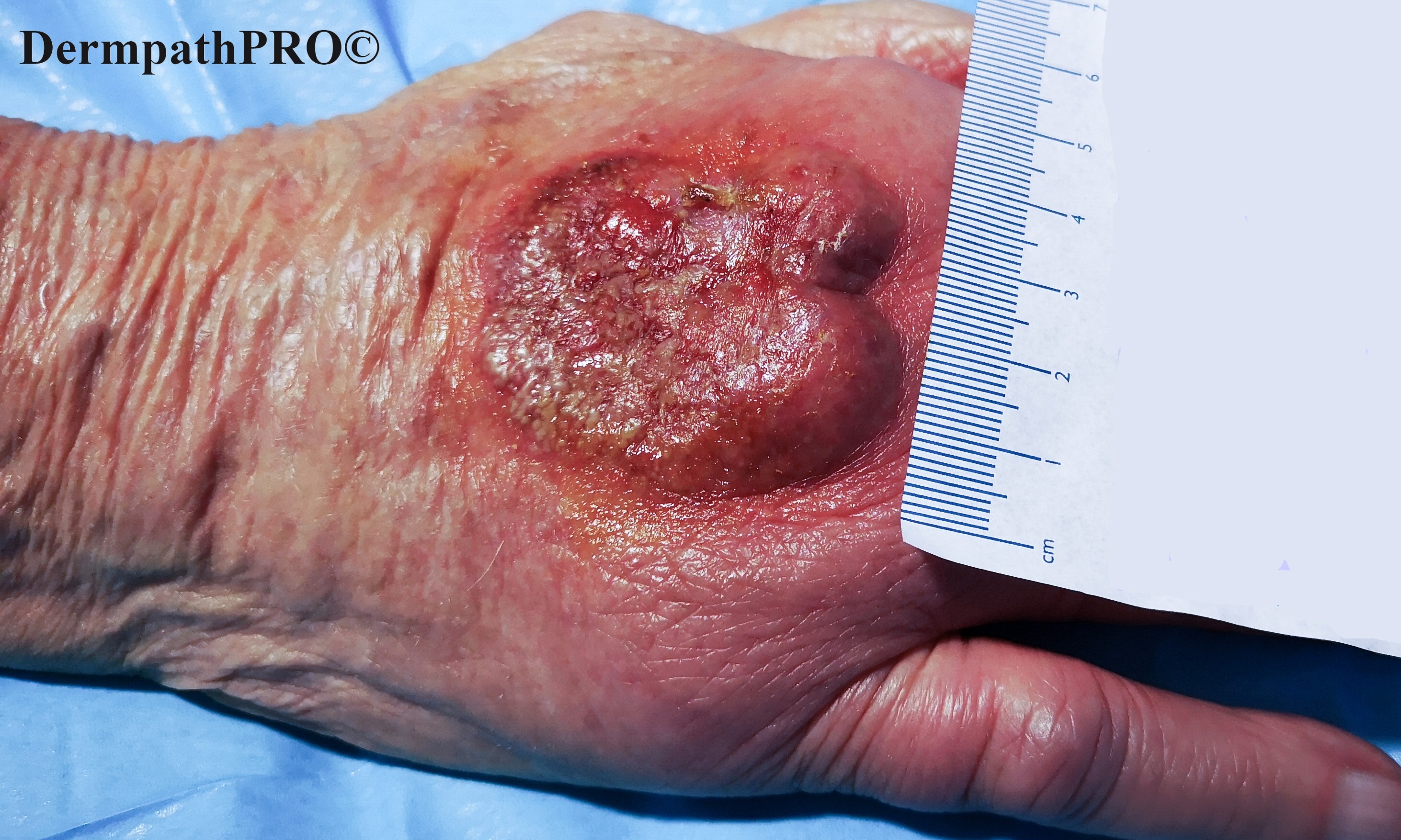
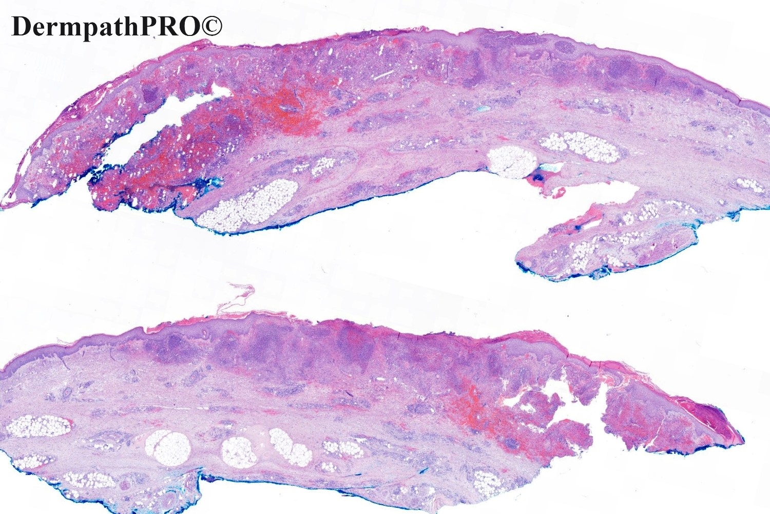
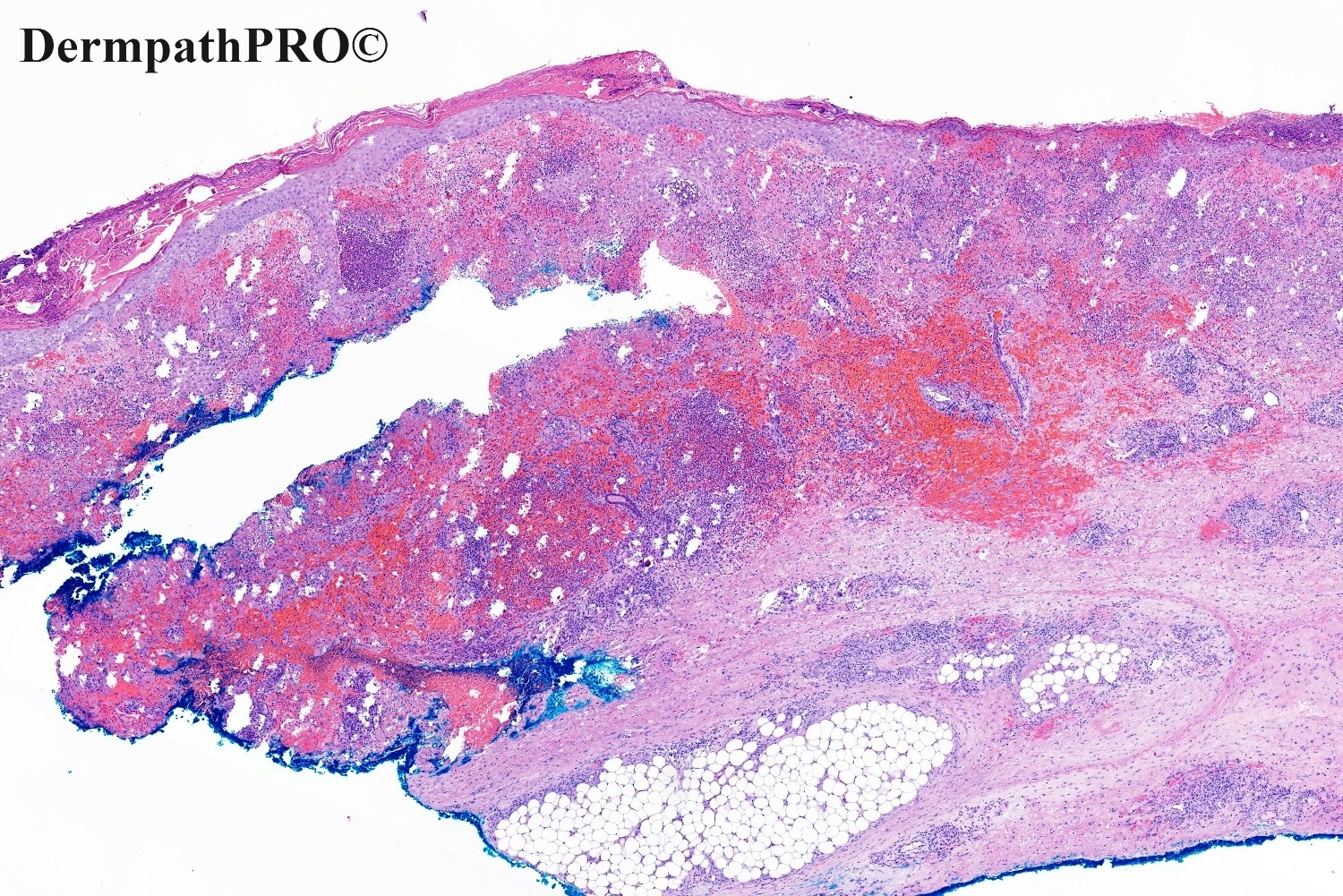
83F, Rapidly enlarging painful plaque dorsum of right hand. Recent hospital admission for iron deficiency anemia and pulmonary embolus.

  • Like 1
  Report Record

User Feedback


Richard Logan

Posted

I agree that there is much about this that suggests Sweet's syndrome.  The history of iron deficiency anaemia and recent pulmonary embolus suggests that there might be a gastro-intestinal neoplasm with which the Sweet's is associated. However, there are one or two other features giving me pause for thought. 

The clinical photograph has a nodularity to it which is rather more than might be expected for Sweet's and suggests local neoplasia, especially given the rapid growth, pain and presumably, single nature of the lesion.  There are cells in the superficial dermis which look epithelioid (images 8 and 9).  There is obviously an element of transepidermal elimination seen on other views (images 3 and 6) so perhaps these cells are also part of that process rather than representing some form of coincidental SCC.  I am sure some immunohistochemistry will throw light on this.

Share this comment


Link to comment
share_externally

Gabriel Tabosa

Posted

It made me think of Pyoderma gangrenosusm, as well.

Share this comment


Link to comment
share_externally

Richard Logan

Posted

A single lesion of pyoderma gangrenosum on the hand would be very usual.

Share this comment


Link to comment
share_externally

PG is also associated with an underlying disease, most commonly inflammatory bowel disease, rheumatic or haematological malignancy. So agree with Gabriel. Histologically PG fits better than SS...but, sincerely, I don’t have enough experience about this. 

Share this comment


Link to comment
share_externally

Richard Logan

Posted

I am still struck by the raised, nodular nature of this lesion which doesn't really fit clinically with pyoderma gangrenosum.  Obviously we can't palpate it which would give a clue as to whether the swelling is inflammatory or possibly at least partially neoplastic.  I think there is more than one pathology going on here.

Share this comment


Link to comment
share_externally

Saman Fatah

Posted

Once the sterile nature of the neutrophilic infiltrate is established via appropriate stains and tissue cultures then neutrophilic dermatosis of (dorsal) hands is most likely given the appearance of the a plaque that has a nodular edge one one end and tightly packed studded pustules on the other edge.
The transepidermal elimination of the dermal neutrophilic infiltrate is impressive and probably this biopsy is taken/included part of pustular edge.

Clinical correlation is essential especially any pyrexia and/or acute phase responses, though they may not be necessarily high in some cases such as MDS. 

Share this comment


Link to comment
share_externally

Pustular vasculitis of dorsal hands ( neutrophilic dermatosis of dorsal hands). There is evidence of vasculitis. I am worrying about am infection process. Need stains, or tissue culture.

Share this comment


Link to comment
share_externally

Krishnakumar subramanian

Posted

Neutrophilic dermatosis of dorsal hands, there is leukocytoclastic vasculitis with numerous neutrophils

considering the age get hematology consult to exclude myelodysplasia/PNH

Share this comment


Link to comment
share_externally

Meenakshi Batrani

Posted

I will also go with neutrophilic dermatosis of the dorsum of the hand. 

Share this comment


Link to comment
share_externally

A plaque studded with pustules and showing plenty of neutrophils histologically may be a lesion of pyoderma gangrenosum before ulceration. It could be a manifestation of pathergy if venepuncture was done at that site during hospitalization.

Share this comment


Link to comment
share_externally

Admin_Dermpath

Posted

Dear All,

Please see below the comment from Dr. Saleem Taibjee;

“This is a patient that I am looking after at the moment. It highlights the challenges of remote working and providing a dermatology opinion based on photos because as Richard indicates above, it was difficult to know whether this was inflammatory or neoplastic. It was only after examining the patient in the flesh and being able to palpate the lesion that I was able to determine a spongy rather than firm/hard consistency, suggesting inflammation rather than tumour.

I favored pyoderma gangrenosum based on the extensive neutrophilic infiltrate which undermines the epidermis, and after correlating with the clinical features, although I must admit that this is the first such case which I have seen on the hand. Neutrophilic dermatosis of the hands is a very reasonable differential diagnosis, but in my limited experience is usually bilateral (although I came across a case report of unilateral involvement) and with multiple lesions which may blister. But, of course, both conditions are likely to lie on a spectrum and confident distinction is not always possible.

As pointed out in the comments, it is important to rule out an underlying systemic disease (e.g. inflammatory arthritis, connective tissue disease, inflammatory bowel disease, hematological disorder, malignancy), although nothing has manifested thus far with this patient after various investigations.

Special stains for organisms were negative, and there was no improvement after antibiotic treatment. However, after 10 days of prednisolone, this is the impressive improvement shown below.”


Thanks

Saleem

23.09.20.jpg

Share this comment


Link to comment
share_externally

Krishnakumar subramanian

Posted

sir was the patient screened for paroxysmal nocturnal hemoglobinuria because of embolism and anemia. there is intravascular hemolysis

Share this comment


Link to comment
share_externally

Saleem Taibjee

Posted

I do not think this has been screened for, thanks for the suggestion. Presumably this would be rare to present at an advanced age?

Share this comment


Link to comment
share_externally


Join the conversation

You can post now and register later. If you have an account, sign in now to post with your account.

Guest
Add a comment...

×   Pasted as rich text.   Paste as plain text instead

  Only 75 emoji are allowed.

×   Your link has been automatically embedded.   Display as a link instead

×   Your previous content has been restored.   Clear editor

×   You cannot paste images directly. Upload or insert images from URL.

Loading...

×
×
  • Create New...