Jump to content
In this section we have spot diagnoses posted on a daily basis since June 2010, now over 4000! You can review the archived cases and read the suggested diagnoses by users and the final comment by the contributors.
Case are uploaded each week day by 10 am UK time with the correct diagnosis will generally be posted at 8 pm UK time. Why not view the most recent spot diagnosis and proffer a diagnosis?

Case Number : Case 2667 - 25 September 2020 Posted By: Dr. Richard Carr

Please read the clinical history and view the images by clicking on them before you proffer your diagnosis.
Submitted Date :
   (0 reviews)

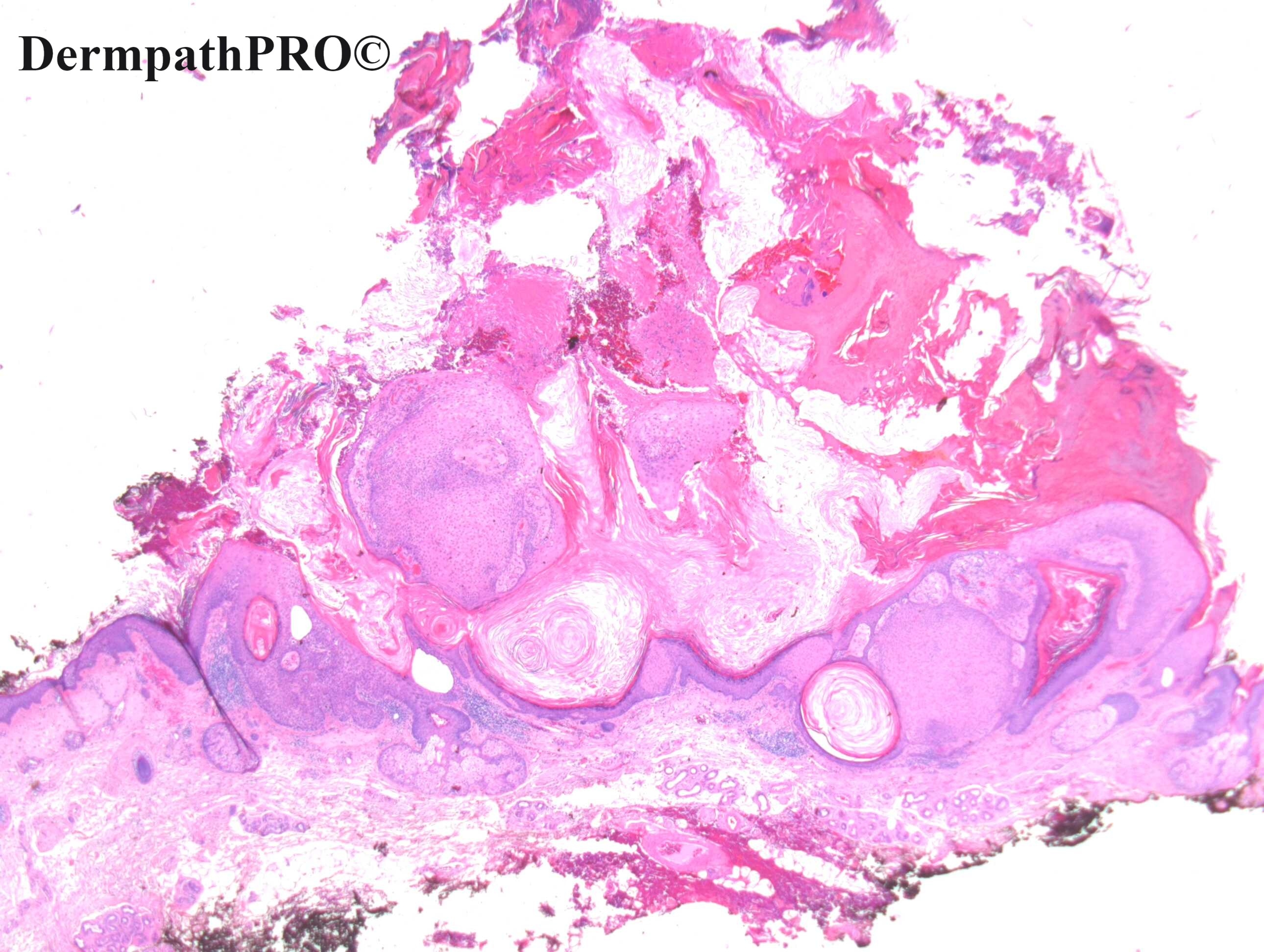
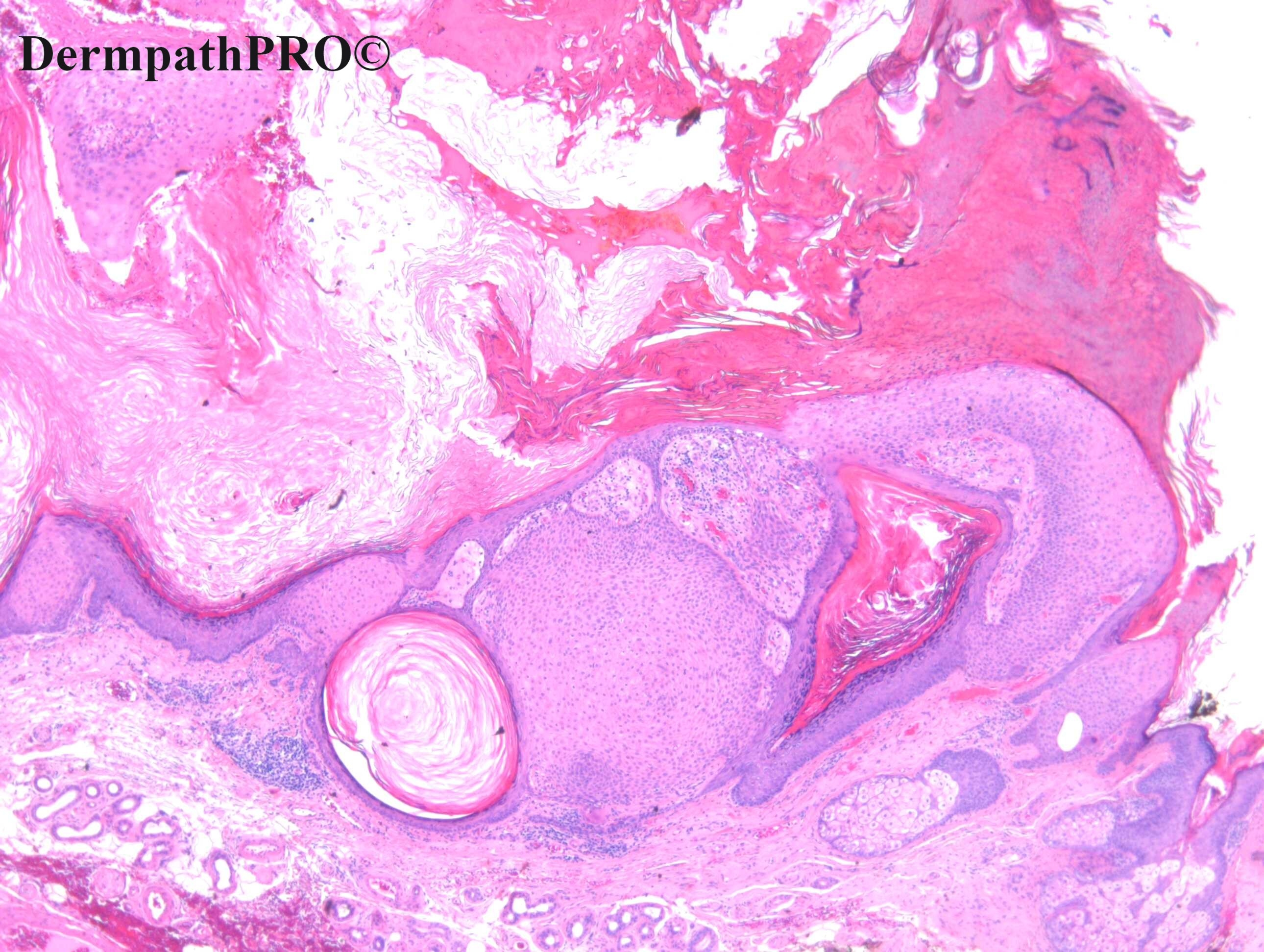
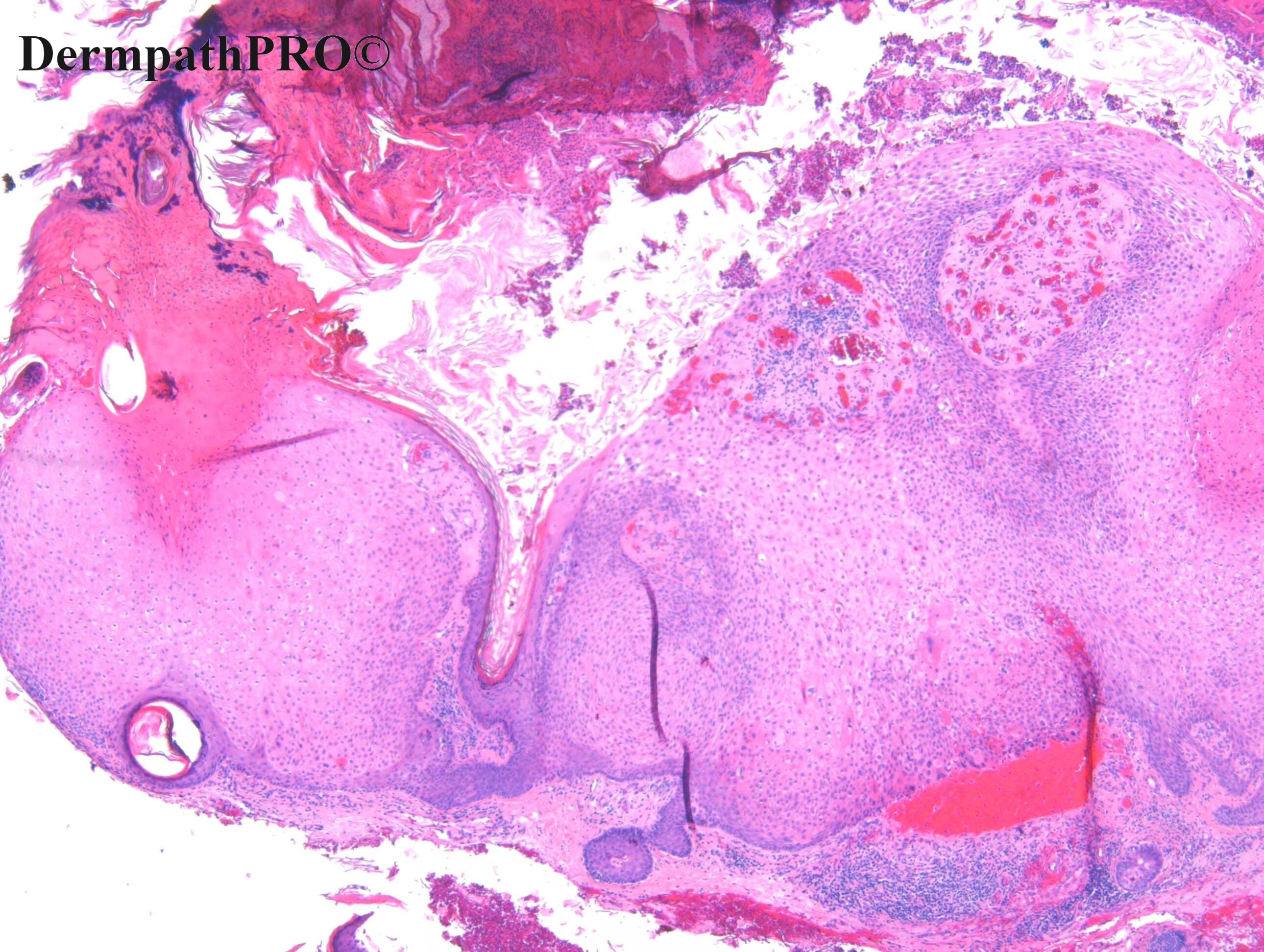
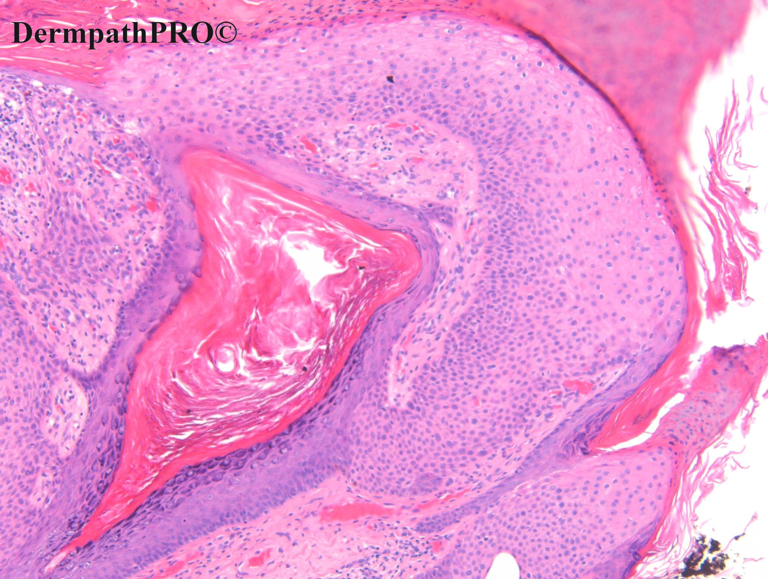
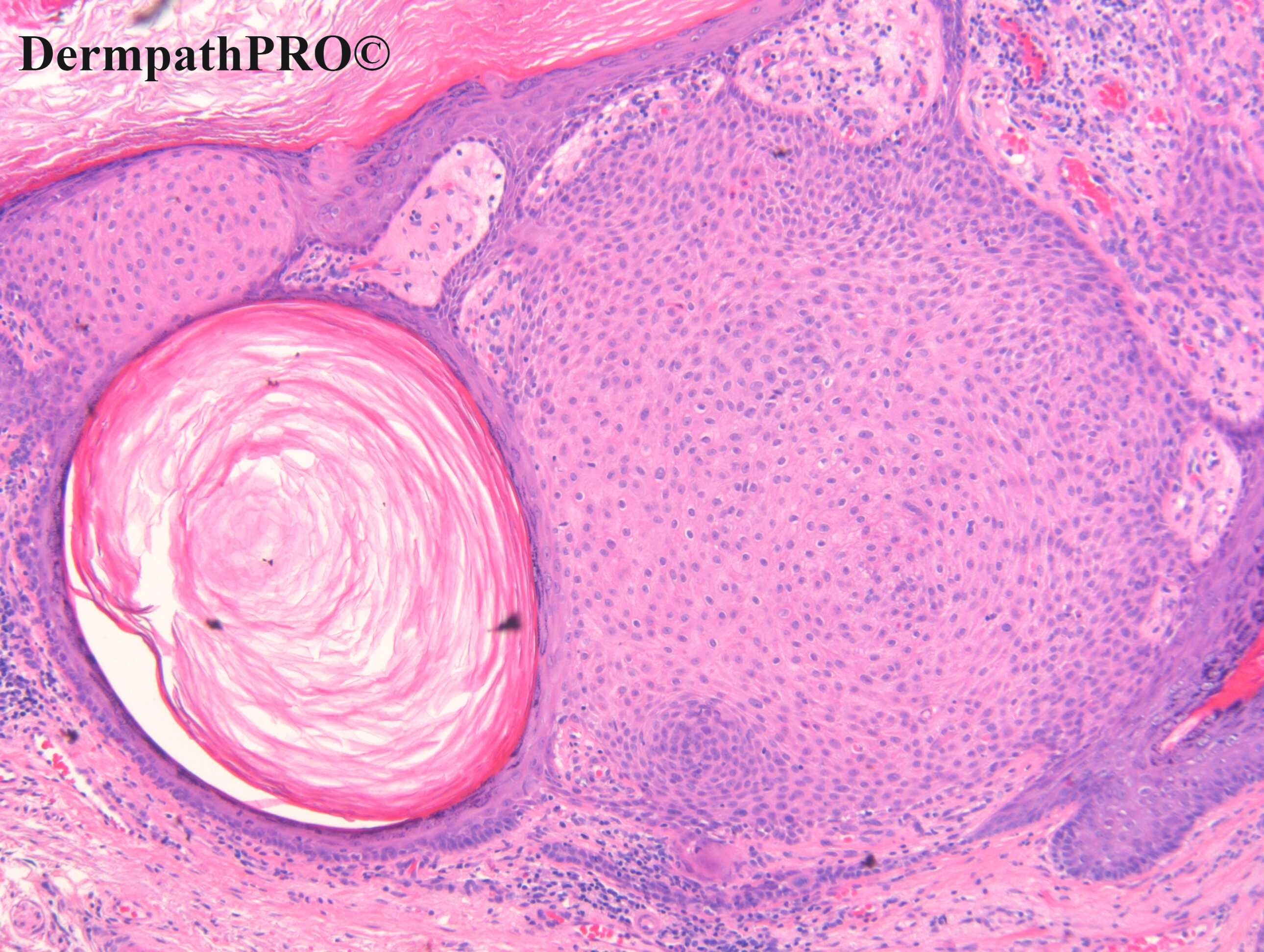
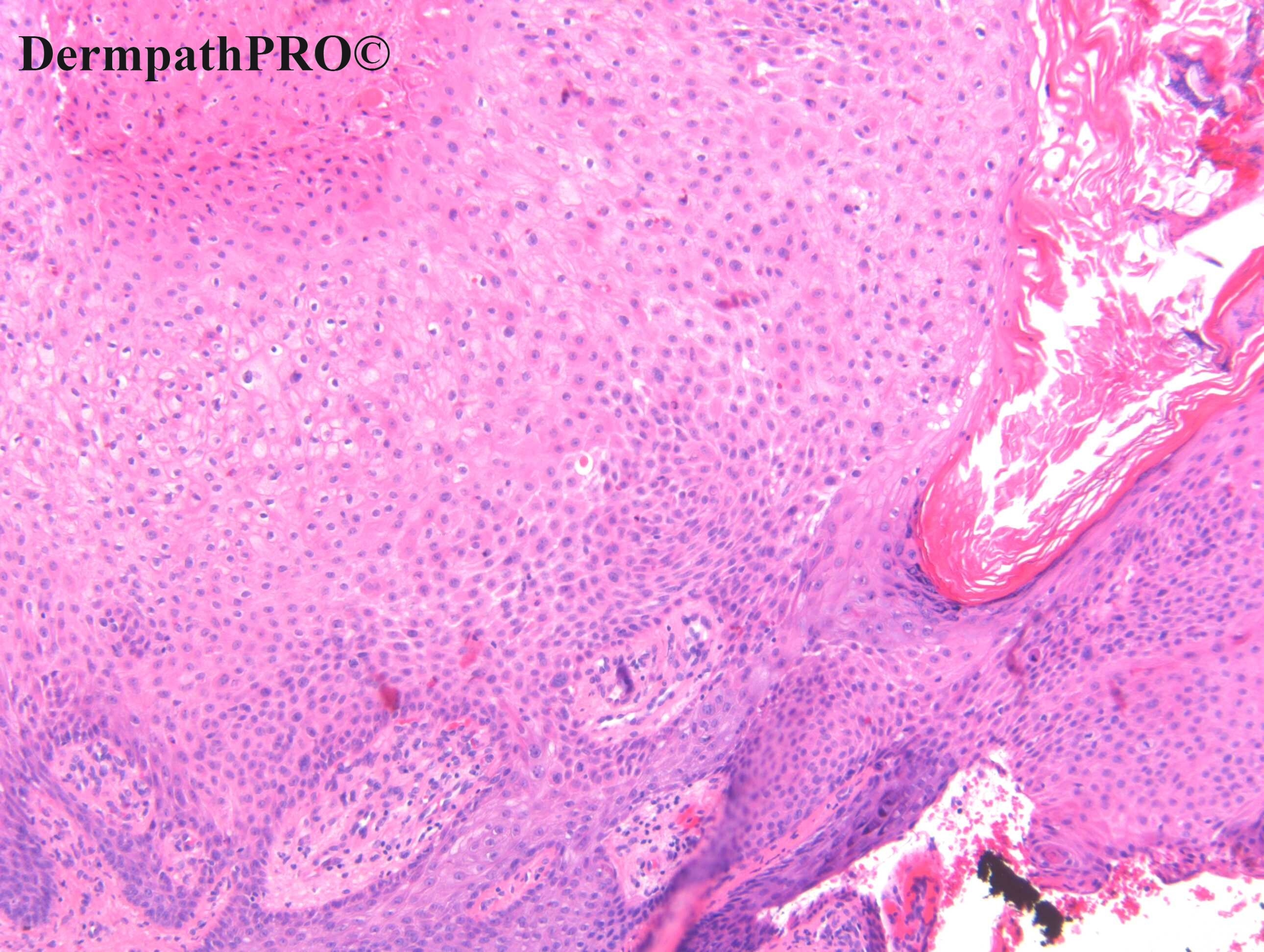
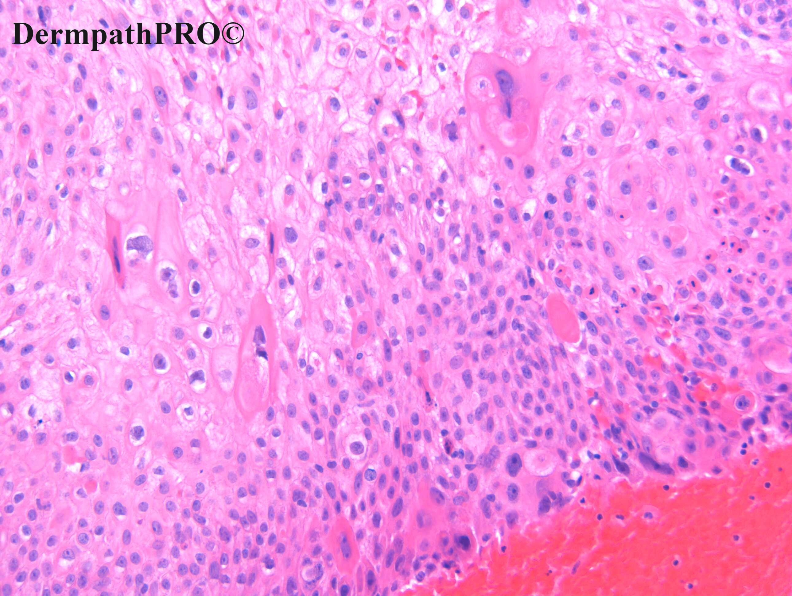
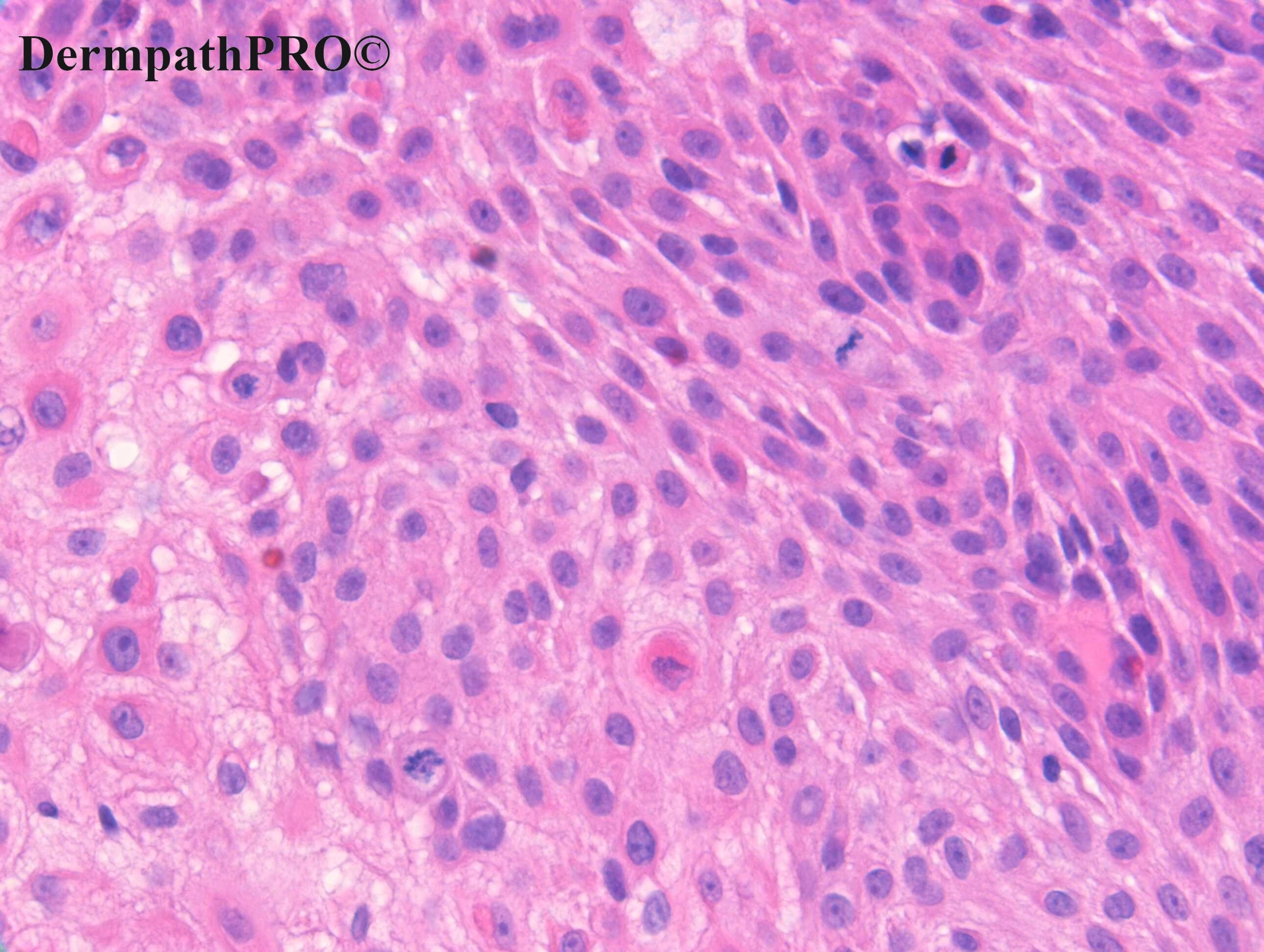
F80. Left cheek below medial left eye. ?BAK ?SCC

Edited by Admin_Dermpath


  Report Record

User Feedback


Krishnakumar subramanian

Posted

borst jadasson phenomenon in seb keratosis

Share this comment


Link to comment
share_externally

Meenakshi Batrani

Posted

Clonal SK. There seems to be some degree of dysplasia in last two figures but it is not full thickness for Bowen's disease. There may be varying degree of dysplasia from mild to carcinoma in-situ, developing in SK 

Share this comment


Link to comment
share_externally

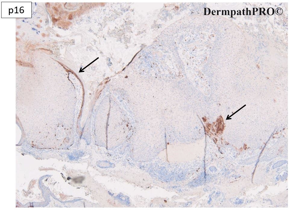
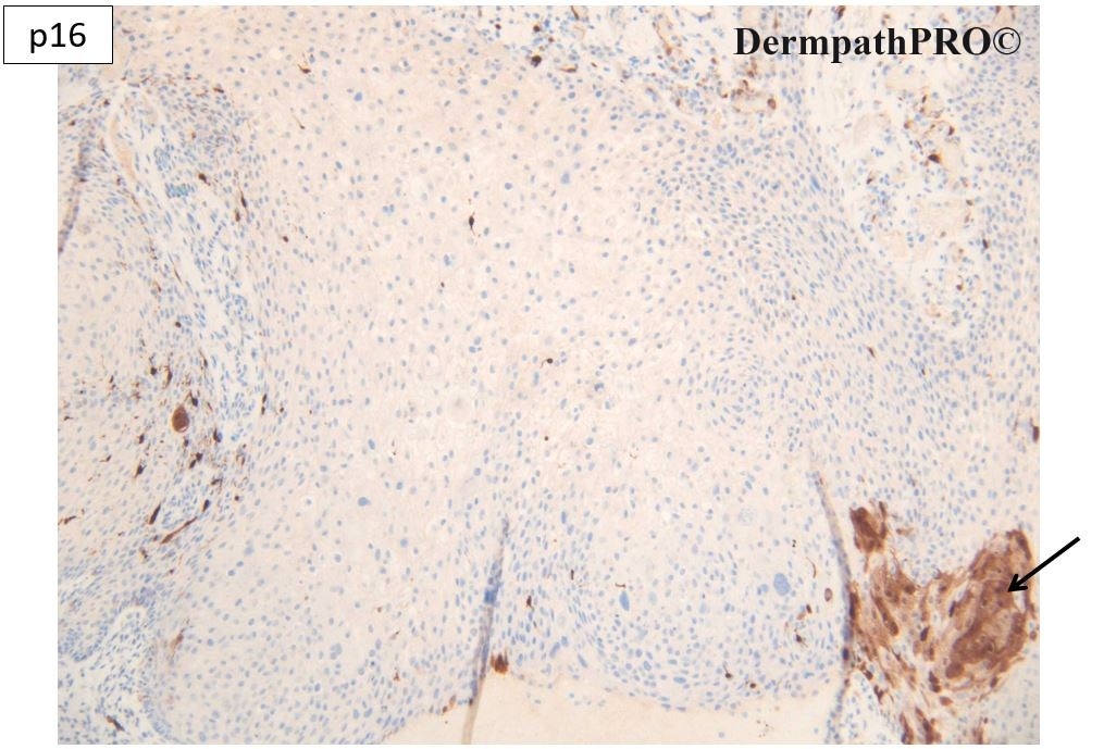
Pattern of p16 staining is the opposite of as expected in Bowen ( strong staining of all abnormal cells with sparing of basal cells ). Here I see strong staining of basal cells only ( as expected in actinic damage pattern ). What do you think about? 

Share this comment


Link to comment
share_externally

Krishnakumar subramanian

Posted

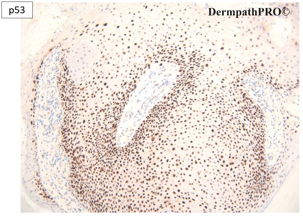
p53 is positive not only in basal cells but also in most of the above layer

suggest carcinoma in situ

Share this comment


Link to comment
share_externally

Dr. Richard Carr

Posted

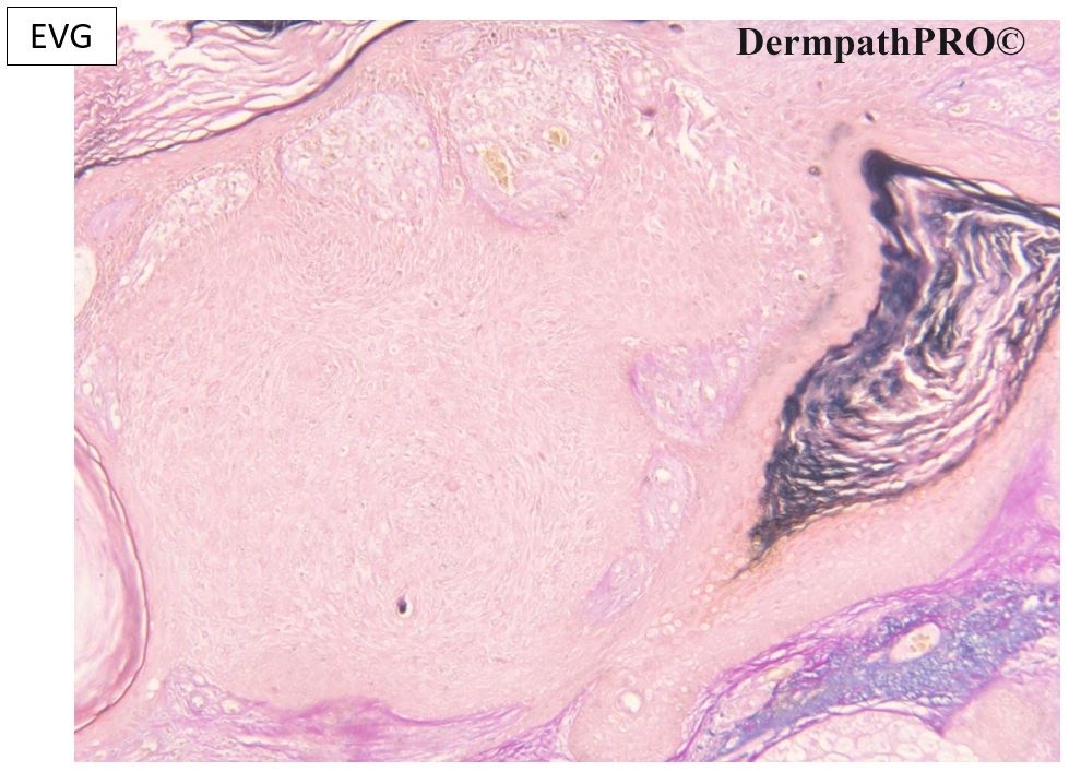
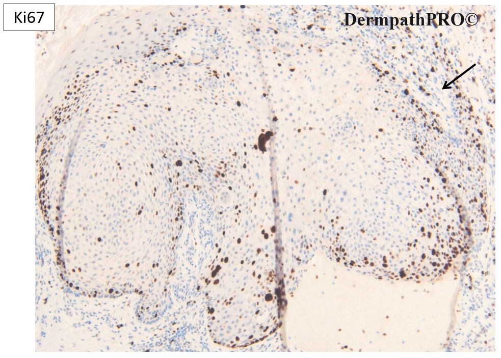
Okay this is another case that is not reading the textbooks. Agree totally this looks like a seborrhoeic keratosis / IFK clonal type. However there is focal dysplasia suggested by dyskeratosis, moderate cellular atypia, but also rather confluent parakeratosis and note the parakeratosis comes from both dips (folliclar pattern) and focally the surface fold. For me the p16 is completely null throughout the lesion (excluding melanocytes that are dendritic) which to me is highly suggestive of a genetic aberration. In sun-exposed skin in the elderly p16 would be expected to show patchy wild (mosaic) pattern staining at least in SEBK / IFK. p53 if regarded as aberrantly high wild type staining for SEBK i.e. >50% moderate & strong staining or moderate to strong staining in the most superficial (or inner) layers of a squamoproliferative lesion although not showing complete strong staining (inter-mixed weak and some negative nuclei) . Ki67 is less useful because you may see moderate & suprabasal staining in SEBK which is a proliferation of basal type cells. Based on the over all findings I prefer a designation of a follicular actinic keratosis with clonal SEBK-like architecture. Molecular studies will be required to confirm the specificity of null p16 in such lesions. p16 can be constitutively low in non-sun exposed skin and banal lesions but for me the null pattern here suggests malignant potential (this lesion clearly in situ) one point to note is he very abrupt transition from mosaic wild type in the adjacent epidermis and the null in the lesion. I will illustrate later follicular SCC that are characterised by striking follicular pseudoeptiheliomatous hyperplasia (fPEH) one of the reasons they may closely mimic KA but the fPEH shows mosaic p16.

Share this comment


Link to comment
share_externally


Join the conversation

You can post now and register later. If you have an account, sign in now to post with your account.

Guest
Add a comment...

×   Pasted as rich text.   Paste as plain text instead

  Only 75 emoji are allowed.

×   Your link has been automatically embedded.   Display as a link instead

×   Your previous content has been restored.   Clear editor

×   You cannot paste images directly. Upload or insert images from URL.

Loading...

×
×
  • Create New...