Jump to content
In this section we have spot diagnoses posted on a daily basis since June 2010, now over 4000! You can review the archived cases and read the suggested diagnoses by users and the final comment by the contributors.
Case are uploaded each week day by 10 am UK time with the correct diagnosis will generally be posted at 8 pm UK time. Why not view the most recent spot diagnosis and proffer a diagnosis?

Case Number : Case 2668 - 28 September 2020 Posted By: Iskander H. Chaudhry

Please read the clinical history and view the images by clicking on them before you proffer your diagnosis.
Submitted Date :
   (0 reviews)

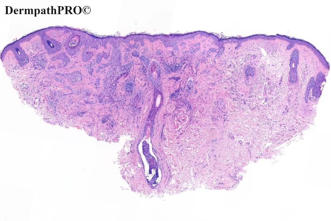
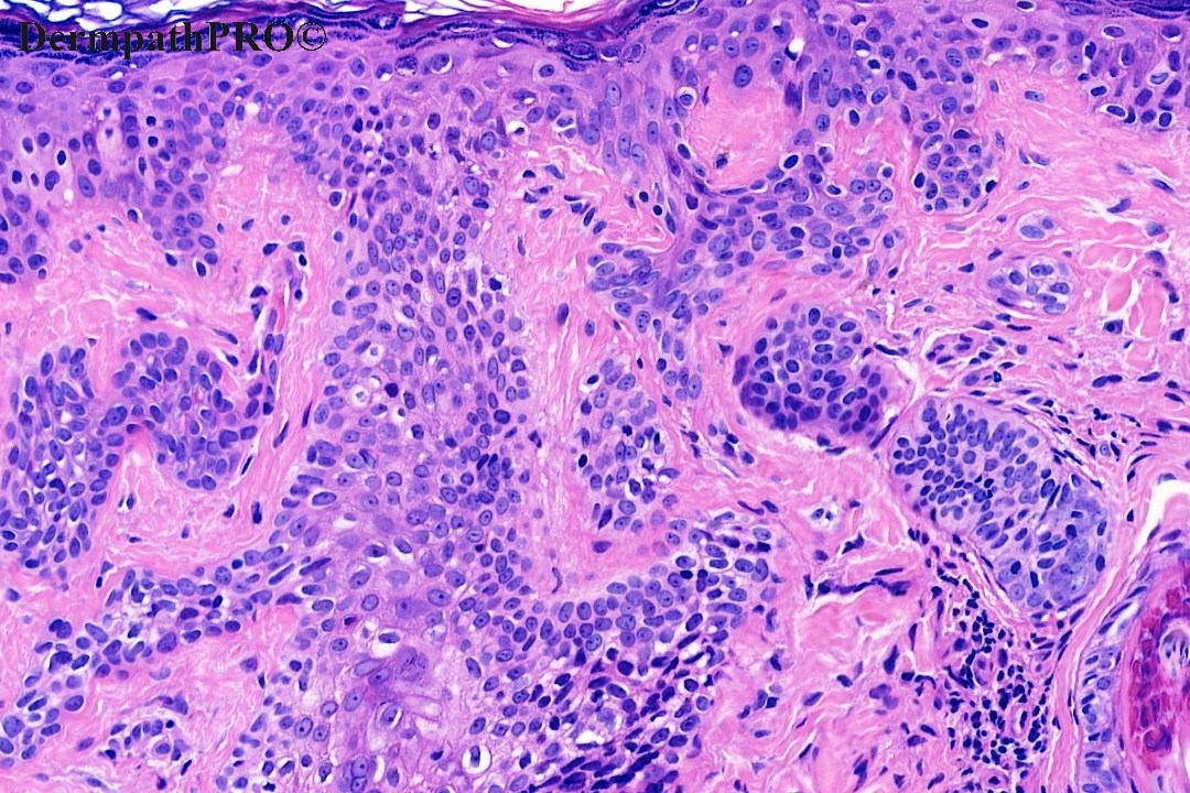
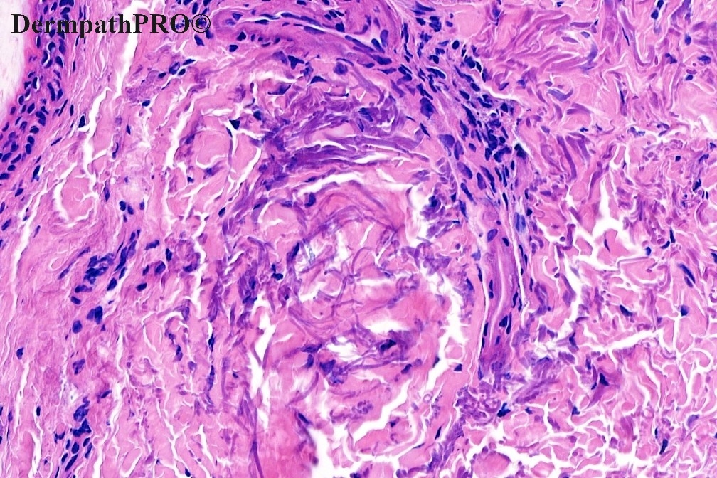
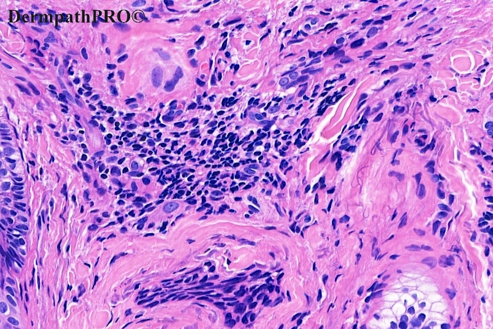
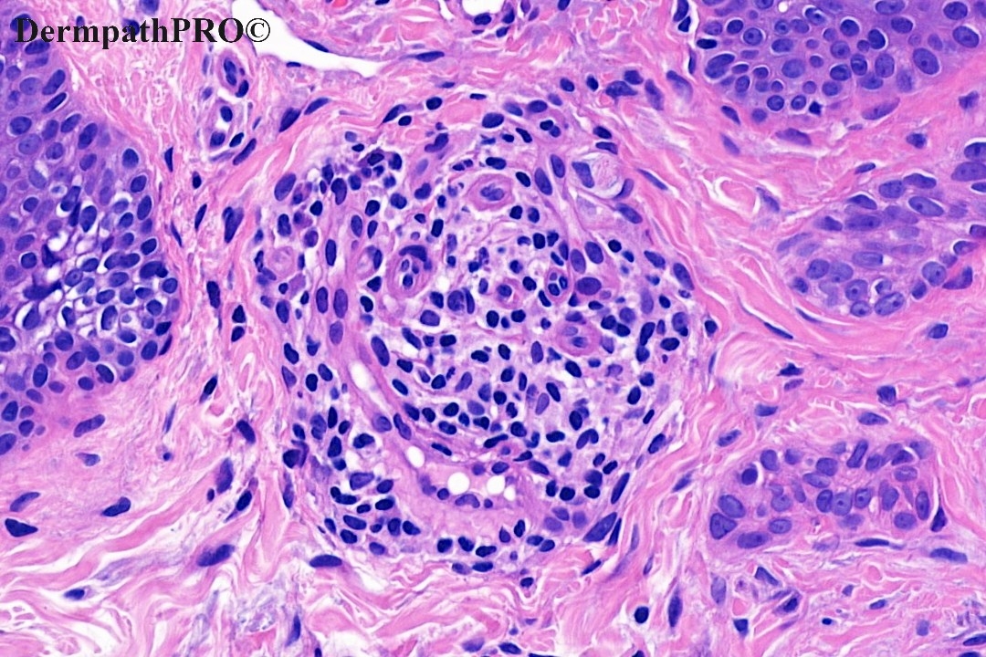
50M, 3-4mm indurated lesion right cheek , Punch biopsy. ?BCC


  Report Record

User Feedback


Richard Logan

Posted

I don't know what this is, and it seems no-one else is sufficiently confident about the diagnosis to venture a comment, so I'll start the ball rolling.  This lesion shows a sclerotic stromal reaction around islands of basophilic cells, some of which are originating from the epidermis.  In places there are cells which look trichilemmal, suggesting a desmoplastic trichilemmoma.  However, I see also areas where the cytology looks more poroma-like.  I'm not sure what to make of the elastic tissue alteration which is highlighted in figures 4,6 and 8.  I look forward to seeing what others make of this.

Share this comment


Link to comment
share_externally

solid carcinoma / solid variant of microcystic adnexal carcinoma.

Share this comment


Link to comment
share_externally

Meenakshi Batrani

Posted (edited)

?Desmoplastic trichoepithelioma

Edited by Meenakshi Batrani

Share this comment


Link to comment
share_externally

Please Dr Iskander: what’s the 2658 and 2664 dx?

Agree with ?DTE. 

Share this comment


Link to comment
share_externally

Eman El-Nabarawy

Posted

Solid variant of microcystic adnexal carcinoma.

Share this comment


Link to comment
share_externally

Admin_Dermpath

Posted

Thank you all - on this biopsy with the crust artefact the two main differentials are desmoplastic trichoepithelioma versus a micronodular basal cell carcinoma. We gave the differential but requested a complete excision. Microcystic adnexal carcinoma would typically show the BM thickening and cribriform architecture and is very rare in the skin. 

Iskander H. Chaudhry 

Share this comment


Link to comment
share_externally

Admin_Dermpath

Posted

On 30/09/2020 at 18:46, vincenzo said:

Please Dr Iskander: what’s the 2658 and 2664 dx?

Agree with ?DTE. 

These now have comments attached thanks 

Share this comment


Link to comment
share_externally

Dr. Richard Carr

Posted

IHC can be quite useful for this sort of thing. I was wondering about a low grade carcinoma like MAC (not ducts in the images). It looks low grade and indolent. 

Share this comment


Link to comment
share_externally


Join the conversation

You can post now and register later. If you have an account, sign in now to post with your account.

Guest
Add a comment...

×   Pasted as rich text.   Paste as plain text instead

  Only 75 emoji are allowed.

×   Your link has been automatically embedded.   Display as a link instead

×   Your previous content has been restored.   Clear editor

×   You cannot paste images directly. Upload or insert images from URL.

Loading...

×
×
  • Create New...