Jump to content
In this section we have spot diagnoses posted on a daily basis since June 2010, now over 4000! You can review the archived cases and read the suggested diagnoses by users and the final comment by the contributors.
Case are uploaded each week day by 10 am UK time with the correct diagnosis will generally be posted at 8 pm UK time. Why not view the most recent spot diagnosis and proffer a diagnosis?

Case Number : Case 2806- 8 April 2021 Posted By: Saleem Taibjee

Please read the clinical history and view the images by clicking on them before you proffer your diagnosis.
Submitted Date :
   (0 reviews)

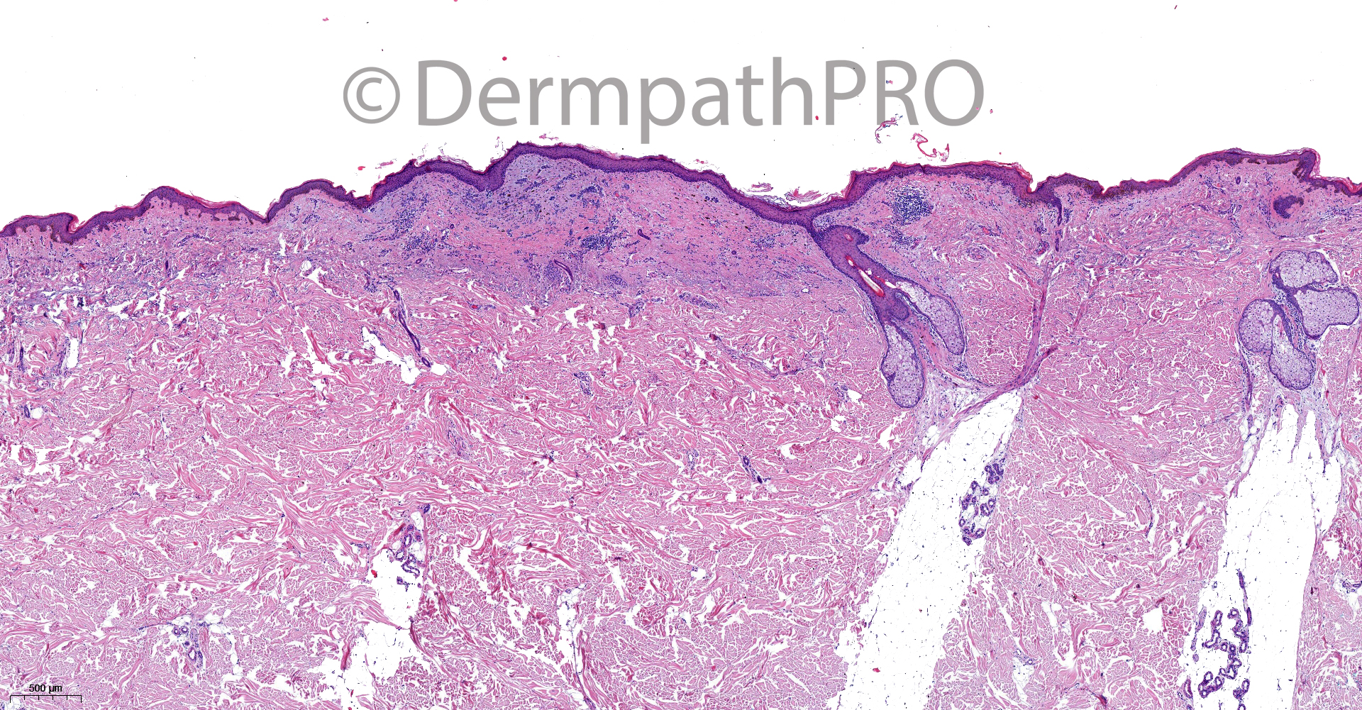
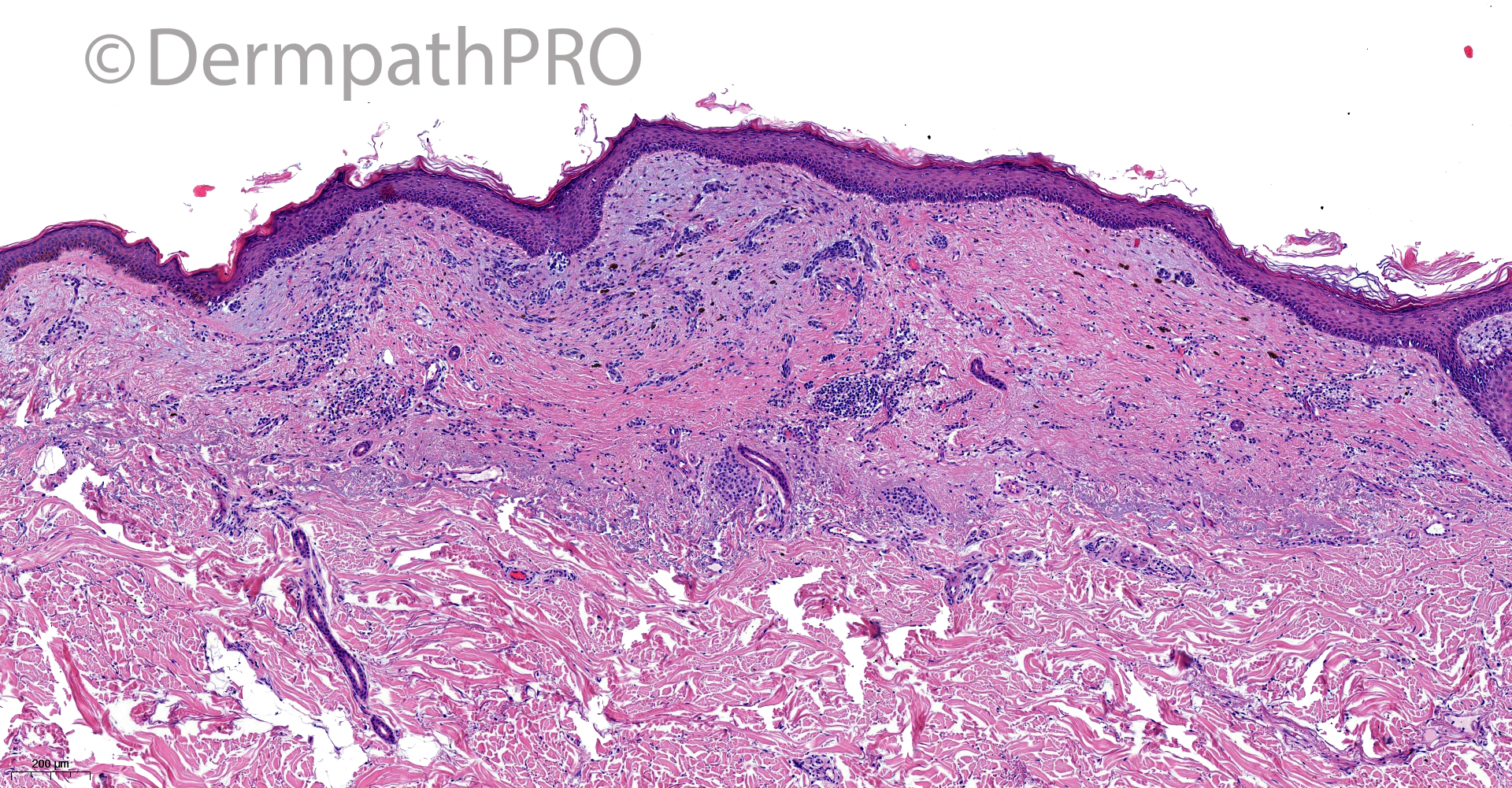
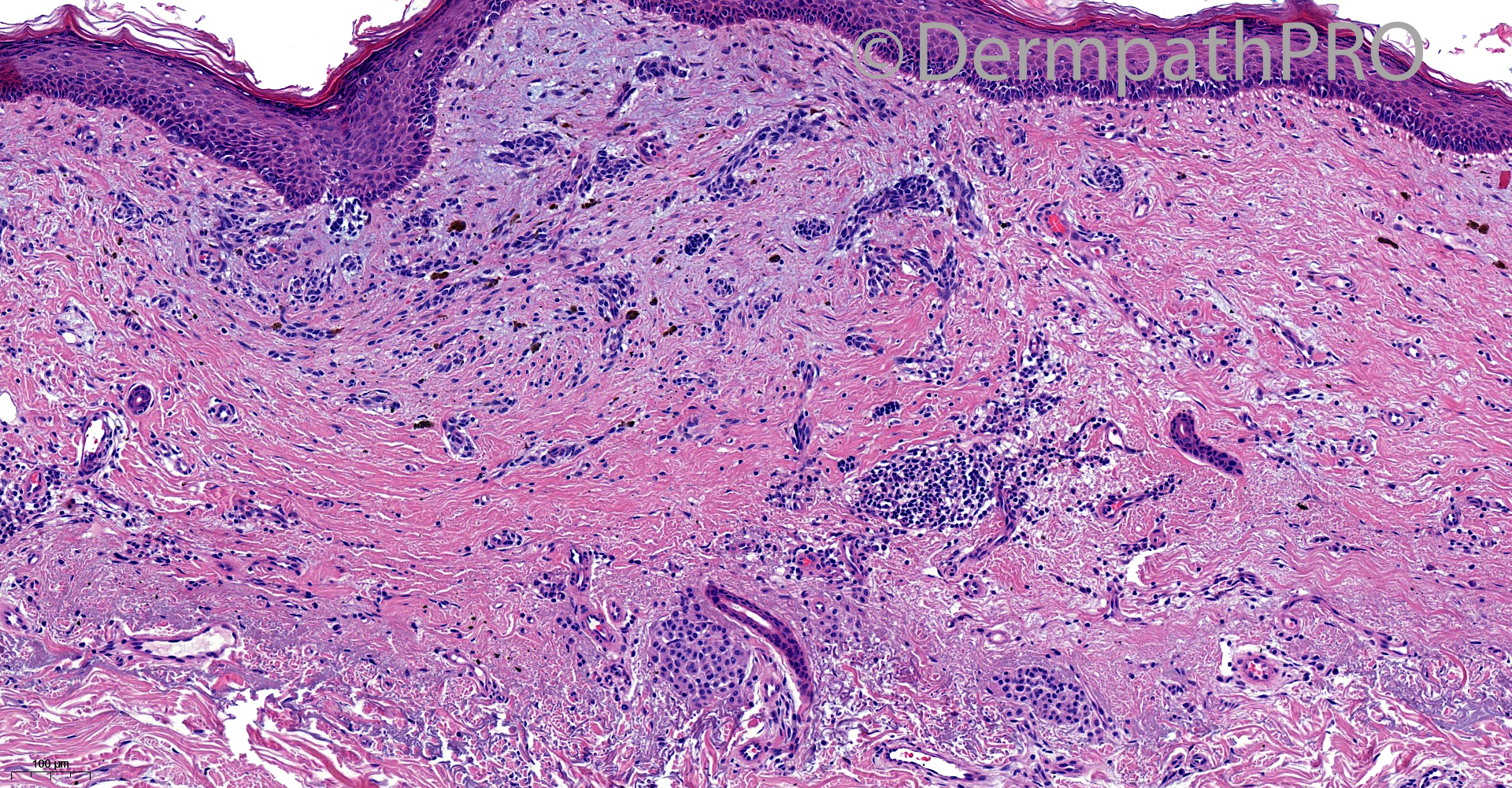
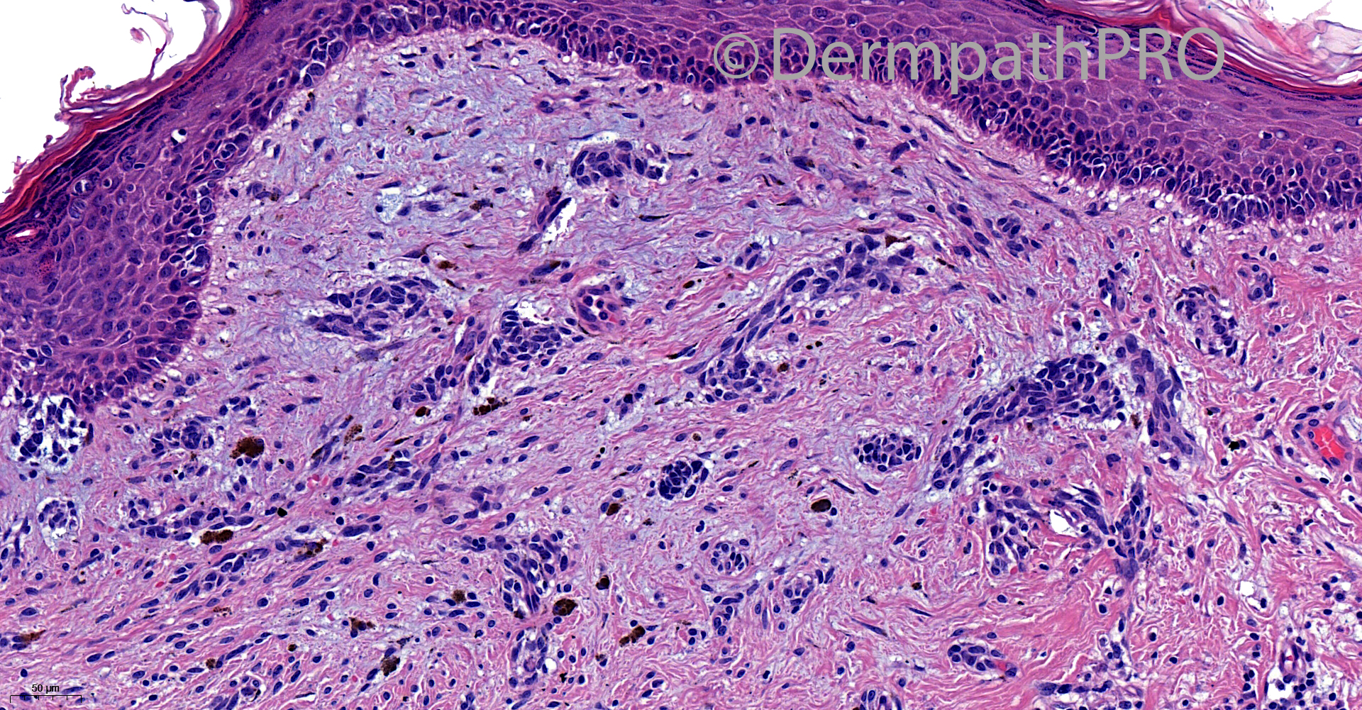
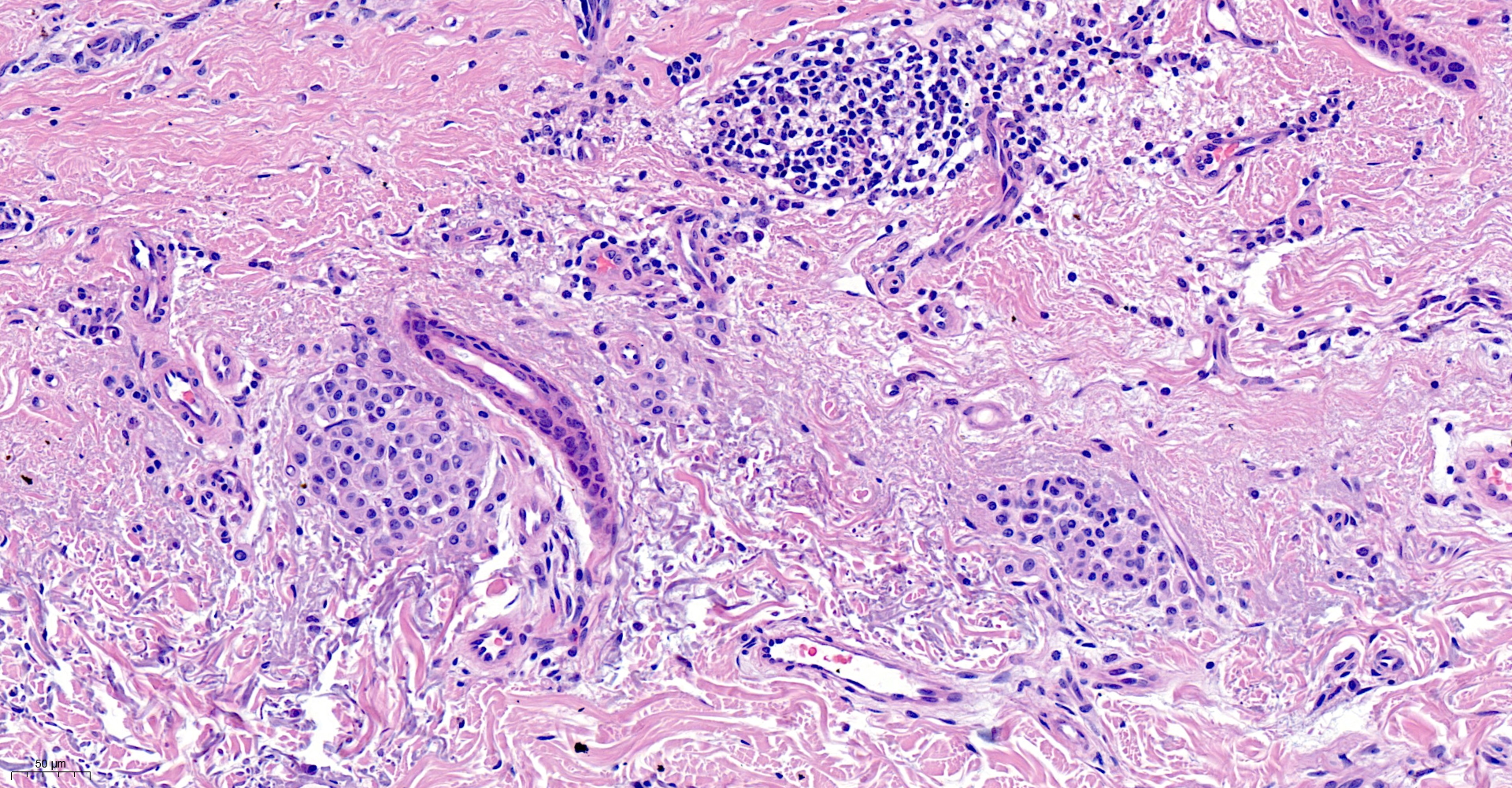
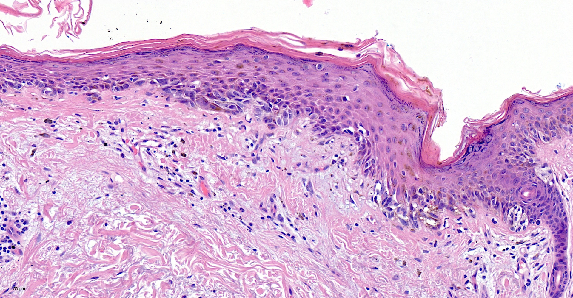
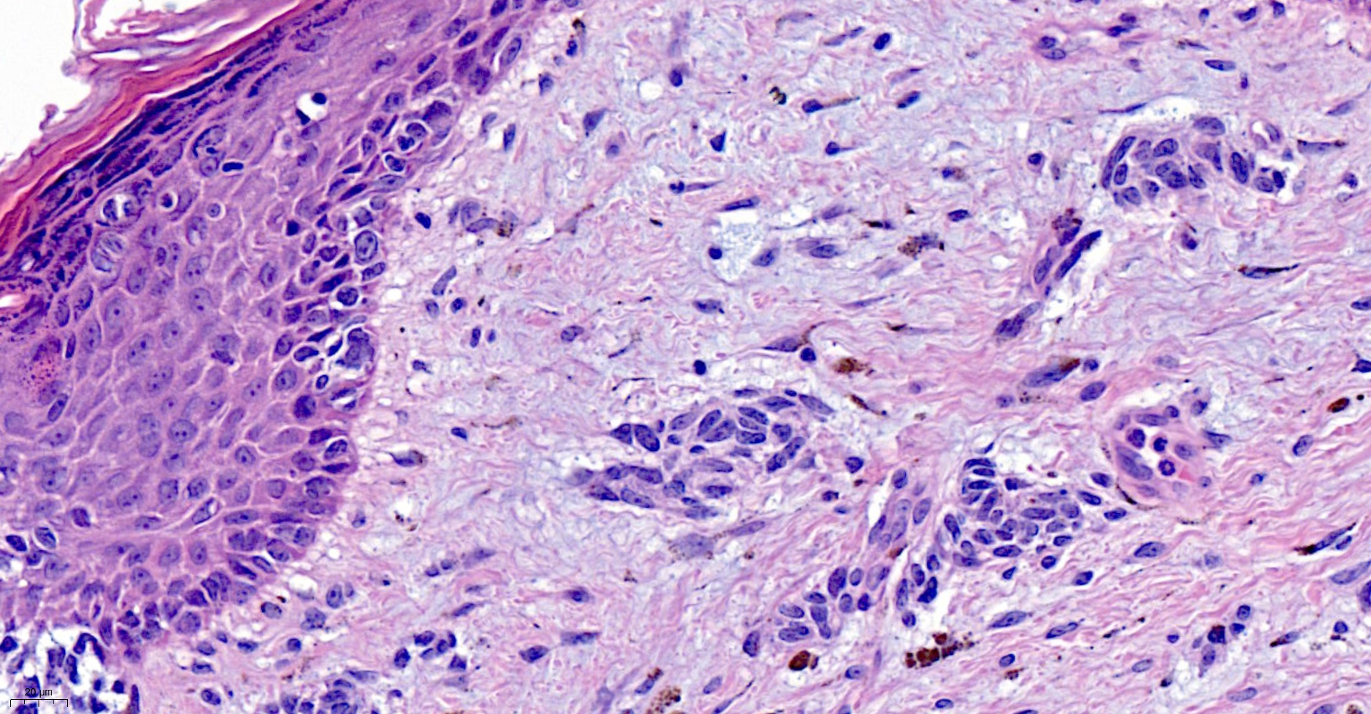
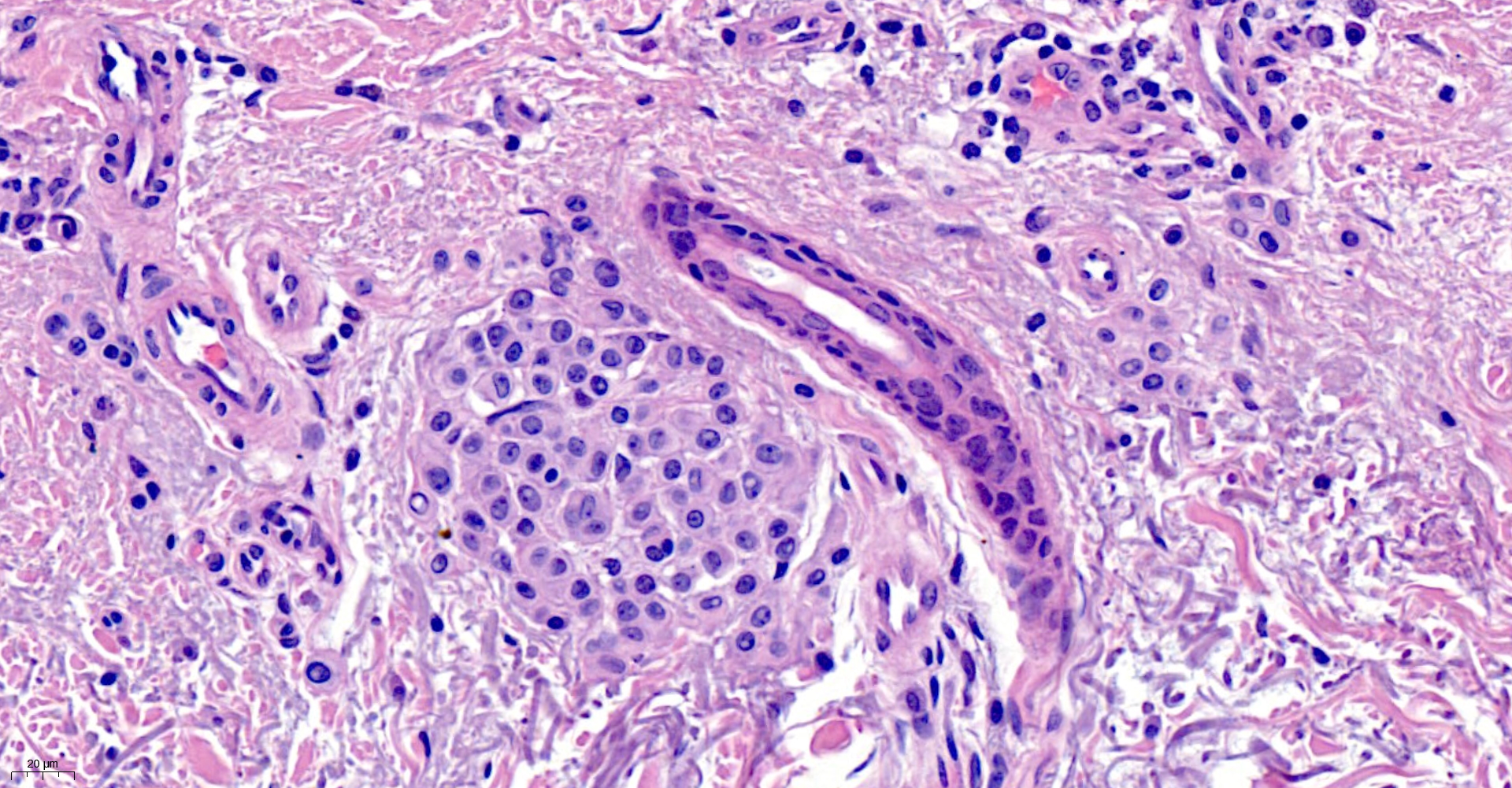
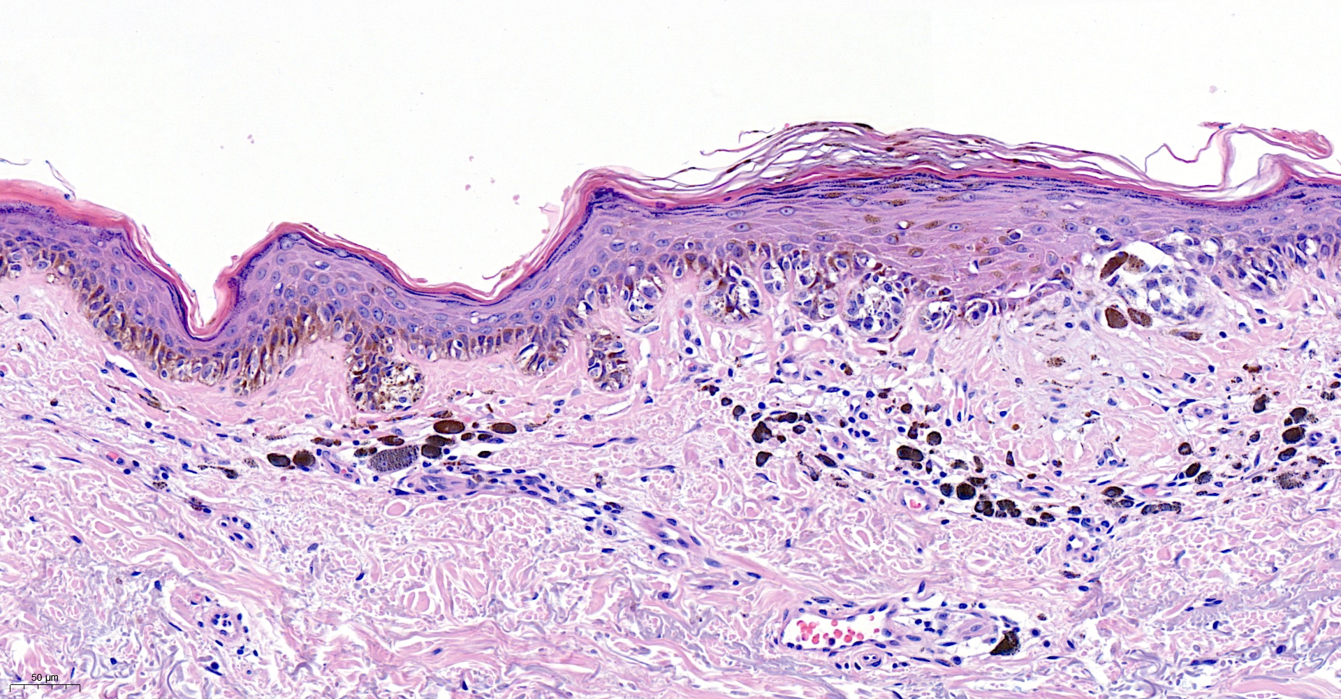
83M pigmented lesion upper back

  • Like 1
  • Thanks 1
  Report Record

User Feedback


A three layers organized growth, with deep residual intradermal nevus, intermediate scar tissue, and intraepidermal predominantly basal atypical melanocytic infiltration. This could be a recurrent Nevus...but would like to see the right lesionale shoulder  magnification, also.  

Share this comment


Link to comment
share_externally

Richard Logan

Posted

I agree with Vincenzo's assessment.

Share this comment


Link to comment
share_externally

Saleem Taibjee

Posted

Unfortunately once again not all of the images have been posted for this case. I will contact the admin team.

Share this comment


Link to comment
share_externally

Saleem Taibjee

Posted

I can see that the additional histology images have now been posted for this case above.

The clinical images and other histology images have also now been posted for the case 2801 1st April.

Share this comment


Link to comment
share_externally

This could be a perfect recurrent nevus, were it not for images 6 and 7, which point to an asymmetrical growth, which exceeds the limits of the scar. How about a lentigo maligna contiguous to a recurrent nevus?

Share this comment


Link to comment
share_externally

Saleem Taibjee

Posted

This is a tricky case. In the end I favoured melanoma (with partial regression) arising in a pre-existing naevus. I was concerned by the rather extensive lateral component, which does extend beyond the confines of the scarring. Furthermore, I was trying to illustrate in the images that the dermal component varies in morphology, with subtle cytological atypia and resemblance to the overlying junctional component e.g. images 2 and 6 above, in comparison to unequivocal benign naevus with a different morphology e.g. image 7 (last image).

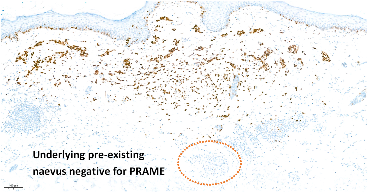
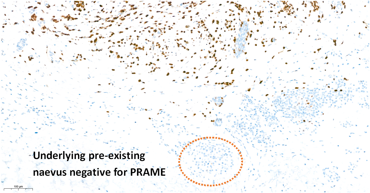
I have been using PRAME IHC in such cases, and it does seem to be helpful in distinguishing invasive melanoma (naevoid) from pre-existing naevus in such cases.

I am unable to attach images to my posts at the moment, so I will ask the admin team if they can upload the PRAME images which indicate positive staining in the presumed melanoma component, in contrast to negative within the deeper benign naevus foci.

BW

Saleem

Share this comment


Link to comment
share_externally


Join the conversation

You can post now and register later. If you have an account, sign in now to post with your account.

Guest
Add a comment...

×   Pasted as rich text.   Paste as plain text instead

  Only 75 emoji are allowed.

×   Your link has been automatically embedded.   Display as a link instead

×   Your previous content has been restored.   Clear editor

×   You cannot paste images directly. Upload or insert images from URL.

Loading...

×
×
  • Create New...