Jump to content
In this section we have spot diagnoses posted on a daily basis since June 2010, now over 4000! You can review the archived cases and read the suggested diagnoses by users and the final comment by the contributors.
Case are uploaded each week day by 10 am UK time with the correct diagnosis will generally be posted at 8 pm UK time. Why not view the most recent spot diagnosis and proffer a diagnosis?

Case Number : Case 2807- 9 April 2021 Posted By: Dr. Richard Carr

Please read the clinical history and view the images by clicking on them before you proffer your diagnosis.
Submitted Date :
   (0 reviews)

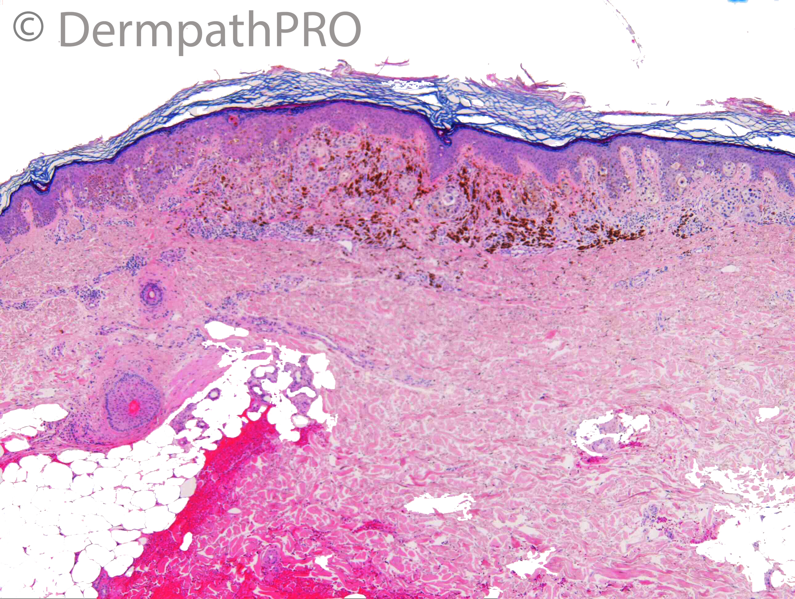
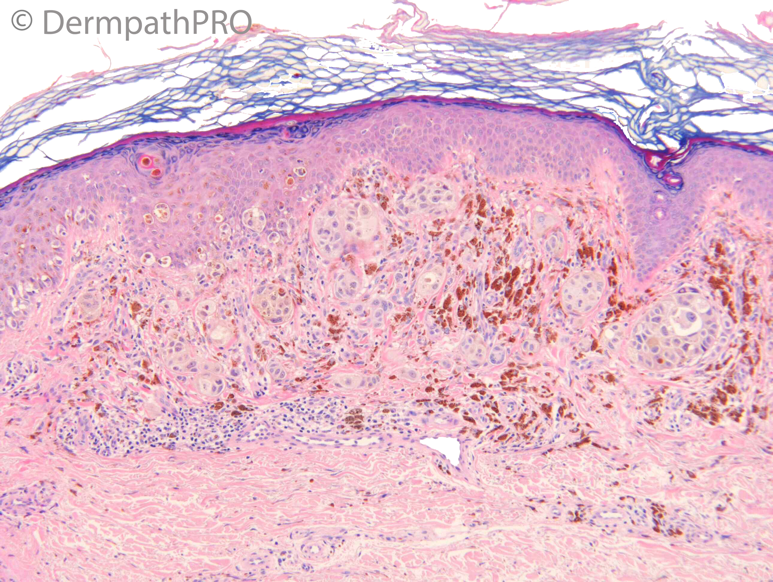
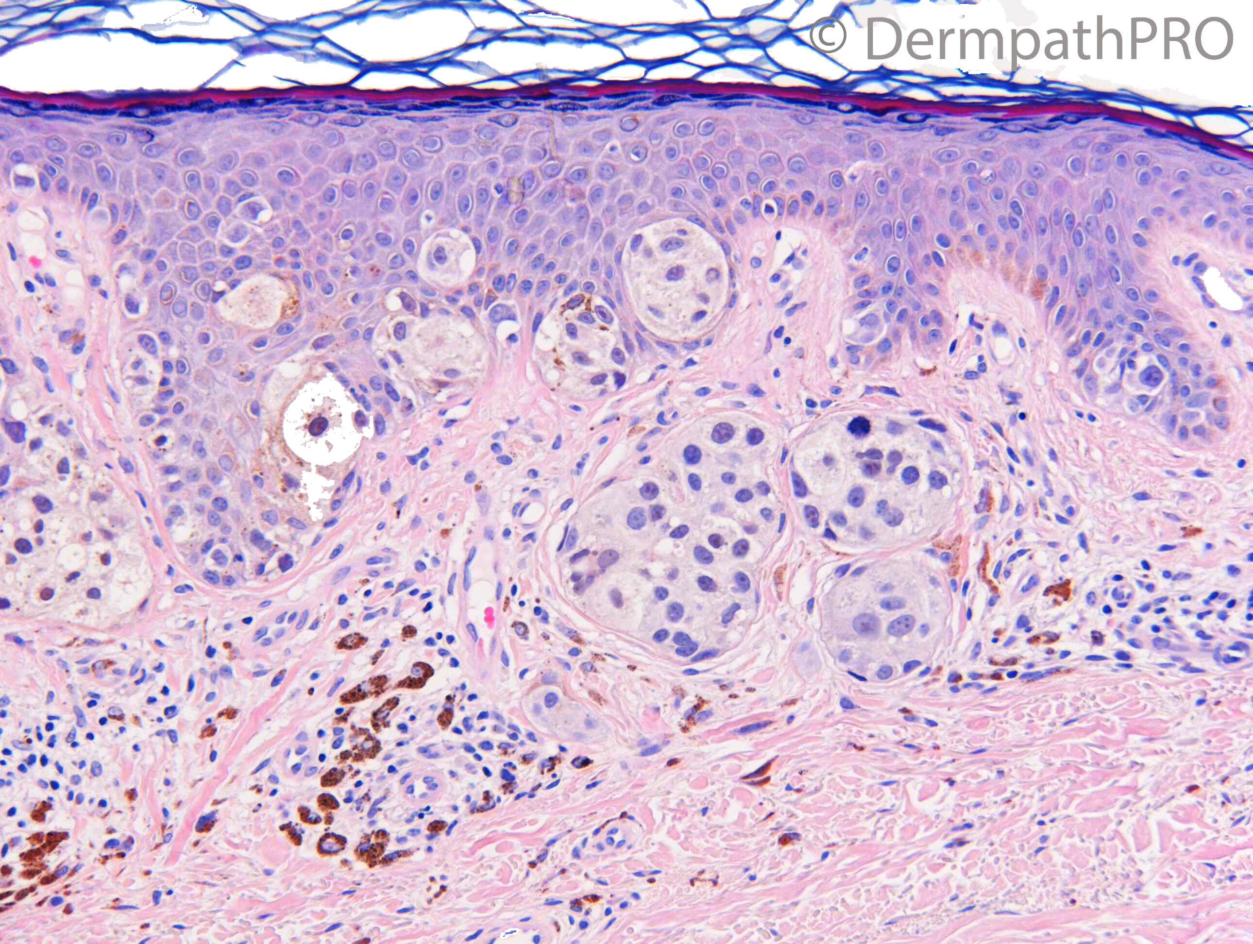
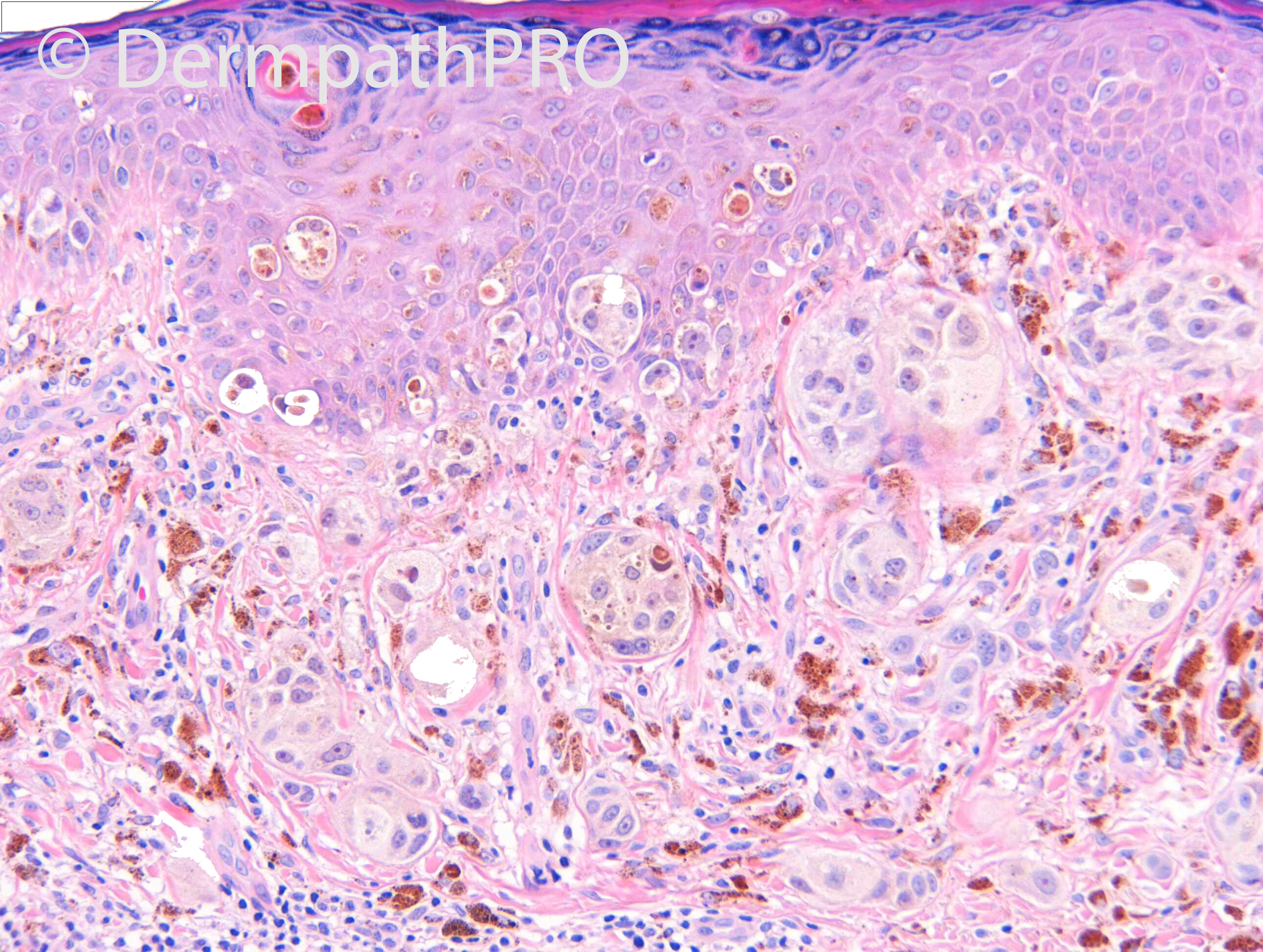
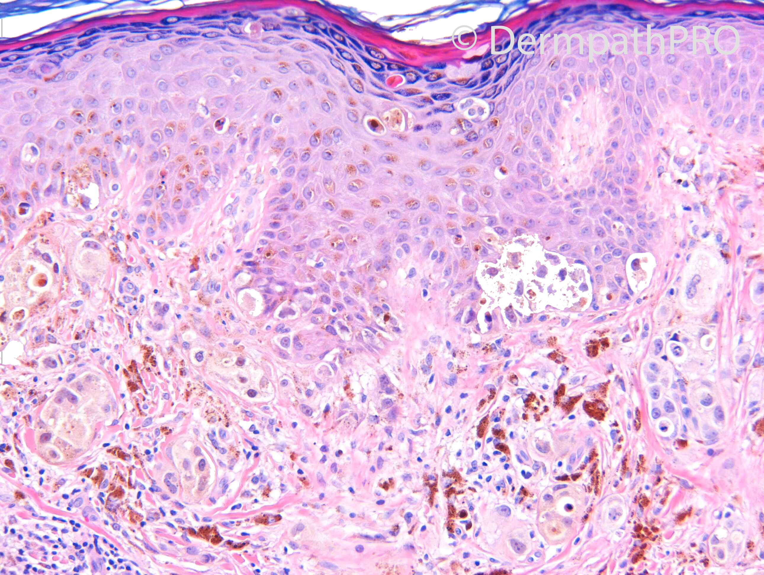
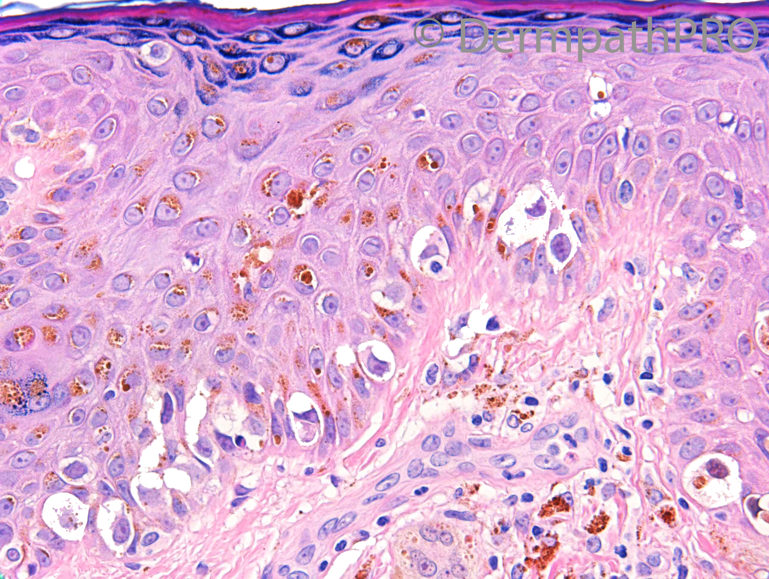
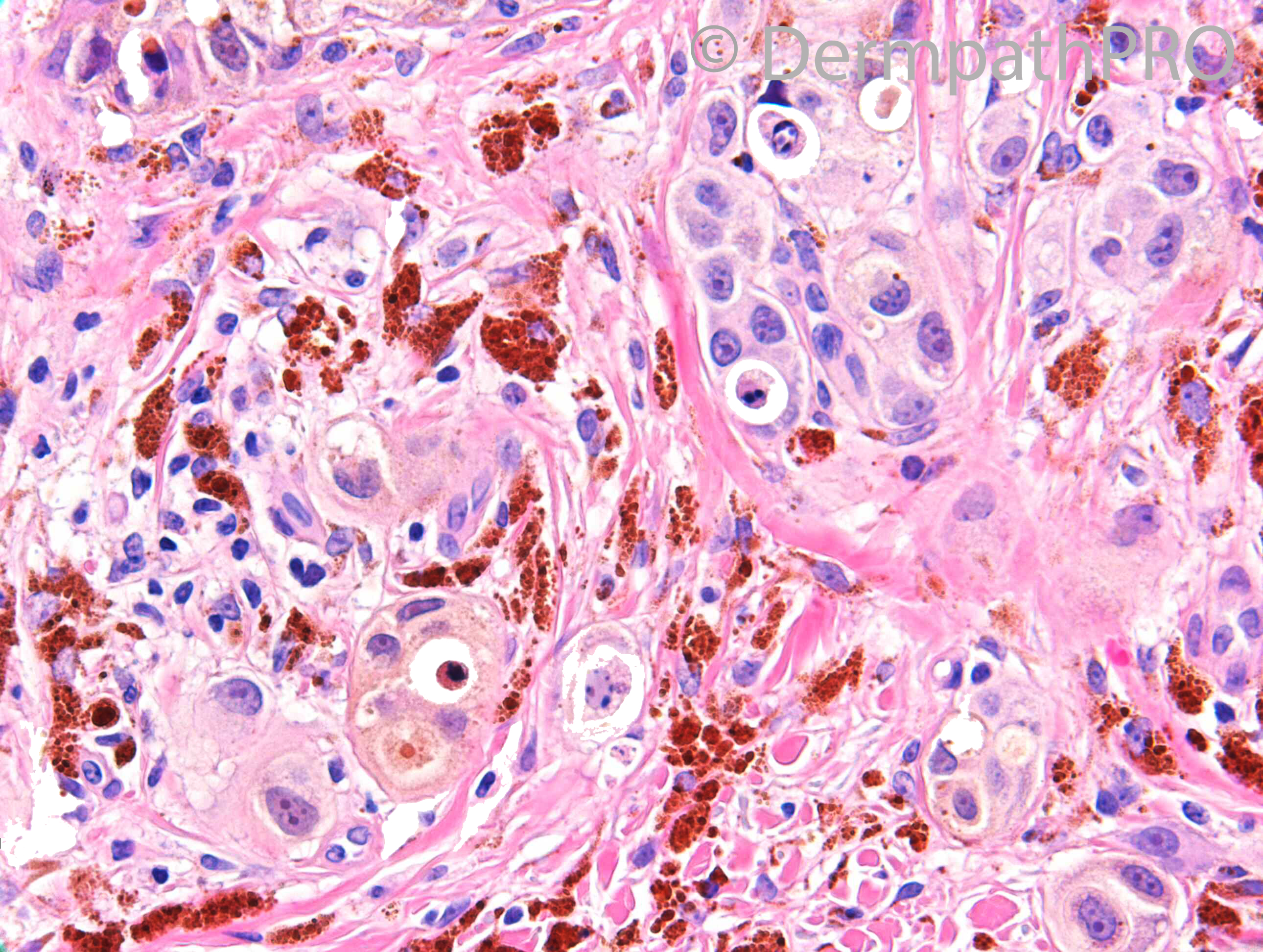
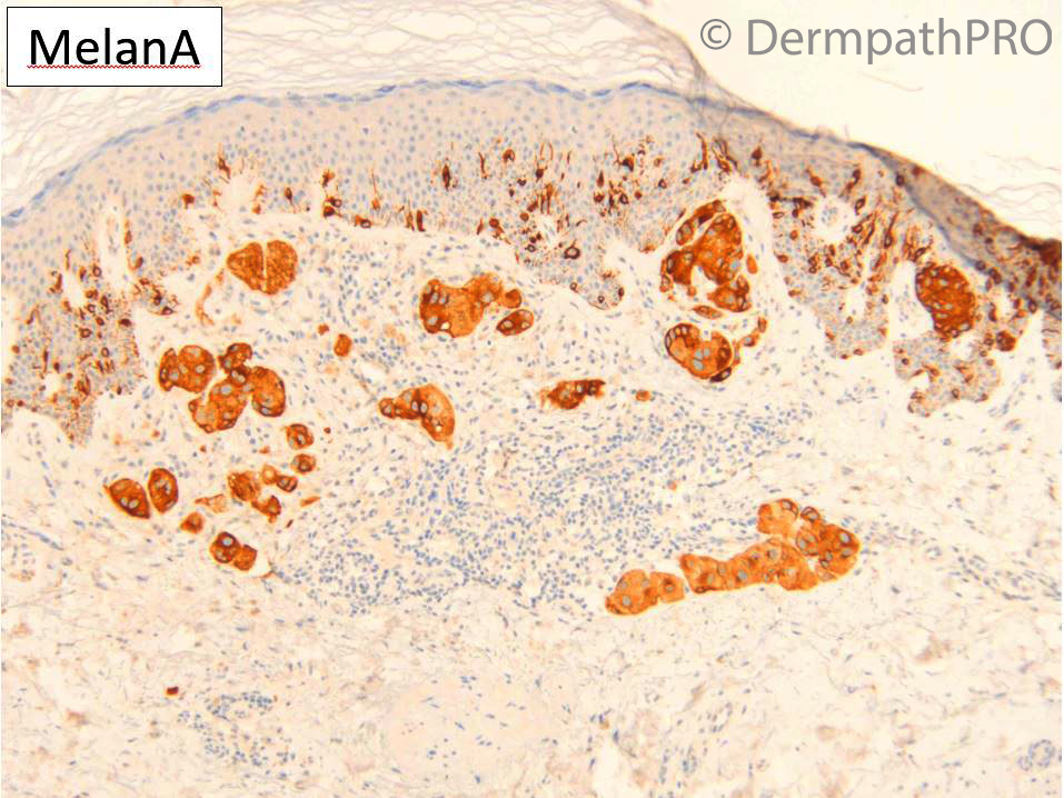
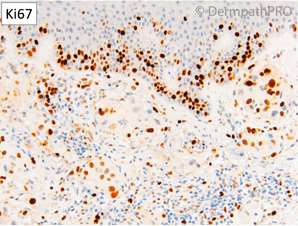
F60. Right lateral thigh. 1 year history of a darkly pigmented cutaneous nodule. ?MM, ?Dermatofibroma.
H&Ex8; IHCx5 at 6pm please

  Report Record

User Feedback


Richard Logan

Posted

Melanocytic lesions are not my strong suit but I would go for this being a superfically invasive malignant melanoma.

Share this comment


Link to comment
share_externally

On 09/04/2021 at 15:08, Richard Logan said:

Melanocytic lesions are not my strong suit but I would go for this being a superfically invasive malignant melanoma.

Yes. SSM. 

Share this comment


Link to comment
share_externally

Krishnakumar subramanian

Posted

Nodular melanoma

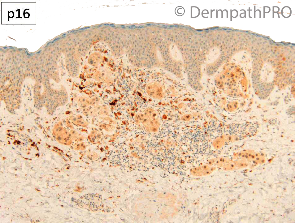
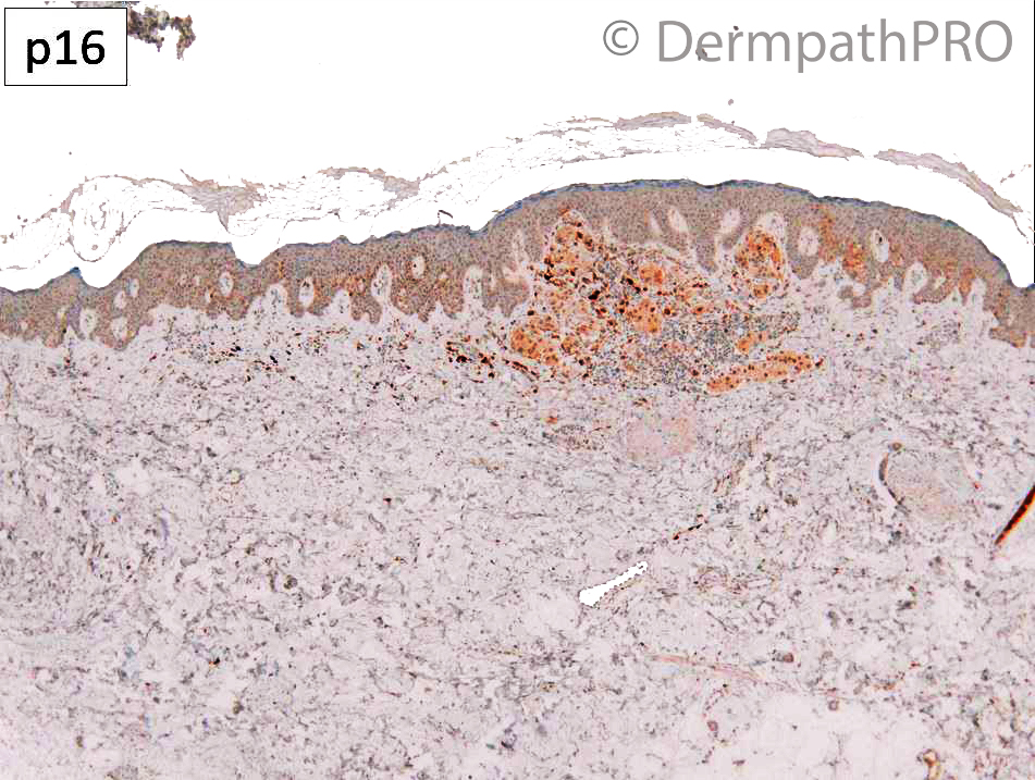
nuclear atypia and loss of 16 supports melanoma

Share this comment


Link to comment
share_externally

Meenakshi Batrani

Posted

Superficial spreading invasive melanoma

Share this comment


Link to comment
share_externally

Dr. Richard Carr

Posted

Okay thanks and I have to agree with you all. However this is a very small 0.6mm thick lesion with one mitosis in the dermal component. Given the risk for metastasis being so low I admitted uncertainty but advised management as for pT1a melanoma (in 2018) - currently that would be complete excision often up to 1cm and follow-up for 1 year. She developed ipsilateral LN dis. in 2020 BRAF+ve at which point I did the p16 and agree it's a knock-out pattern. If I'd done BRAF IHC and it was positive (excluding a Spitz MELTUMP) with a p16 knock-out I'd probably have given a firmer diagnosis of melanoma at the time. I suppose the question is about the approach to thin lesions we are suspicious for melanoma but not certain that have approximately ~1% risk of developing metastases but unquantifiable prospectively in an individual patient. I've tended to not over-diagnose melanoma because 99% will have no further problem and all 99% will likely suffer varying degrees of anxiety and be subjected to overtreatment often (excessive margins of excision when simple excision with 3 to 5mm clinical & 2 to 3mm histological margin and watchful waiting should suffice). So should we call everything we are worried about melanoma to let us sleep in our beds or should be be pragmatic and let 99 patients who won't come to harm from conservative complete removal and call it uncertain but expected low risk for metastasis?  Having been humbled and troubled by rare cases of thin lesions that I got wrong (with the benefit of hindsight and clinical outcome), and rarely 2 times in a 20+ year career, with devastating clinical outcomes, I have to remember the lesions were fully removed and they would not have had SLNBx or prolonged follow-up for pT1a lesions and remember those 100's of patients that were not over treated or made over anxious. This is the art of medicine there is no possibility of knowing what is the least harm to the many and Richard no matter how long you do it these lesions will always be challenging and subjective. I struggle with them nearly every day in my own and referral practice and often it comes down the the philosophical matter of trying to do least harm.

Share this comment


Link to comment
share_externally


Join the conversation

You can post now and register later. If you have an account, sign in now to post with your account.

Guest
Add a comment...

×   Pasted as rich text.   Paste as plain text instead

  Only 75 emoji are allowed.

×   Your link has been automatically embedded.   Display as a link instead

×   Your previous content has been restored.   Clear editor

×   You cannot paste images directly. Upload or insert images from URL.

Loading...

×
×
  • Create New...