Jump to content
In this section we have spot diagnoses posted on a daily basis since June 2010, now over 4000! You can review the archived cases and read the suggested diagnoses by users and the final comment by the contributors.
Case are uploaded each week day by 10 am UK time with the correct diagnosis will generally be posted at 8 pm UK time. Why not view the most recent spot diagnosis and proffer a diagnosis?

Case Number : Case 2812- 16 April 2021 Posted By: Dr. Richard Carr

Please read the clinical history and view the images by clicking on them before you proffer your diagnosis.
Submitted Date :
   (0 reviews)

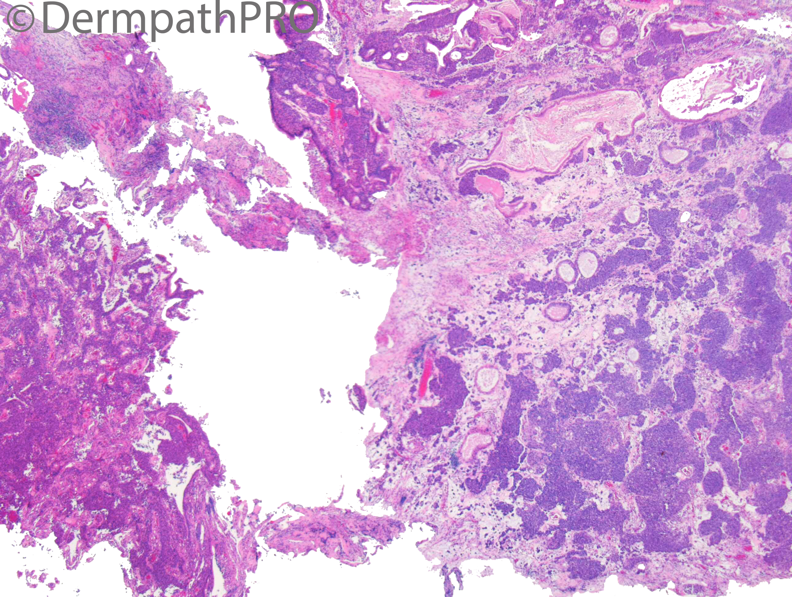
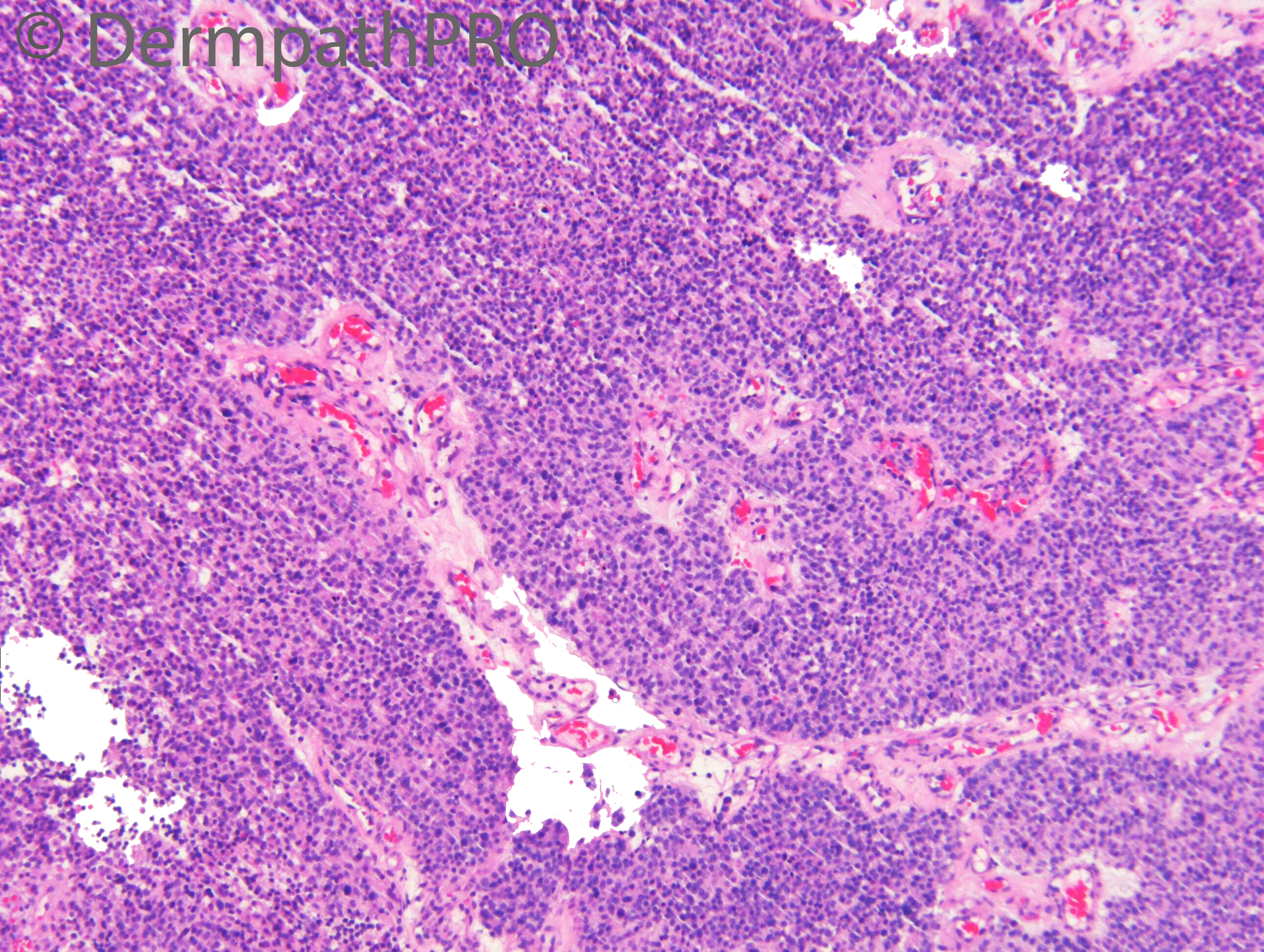
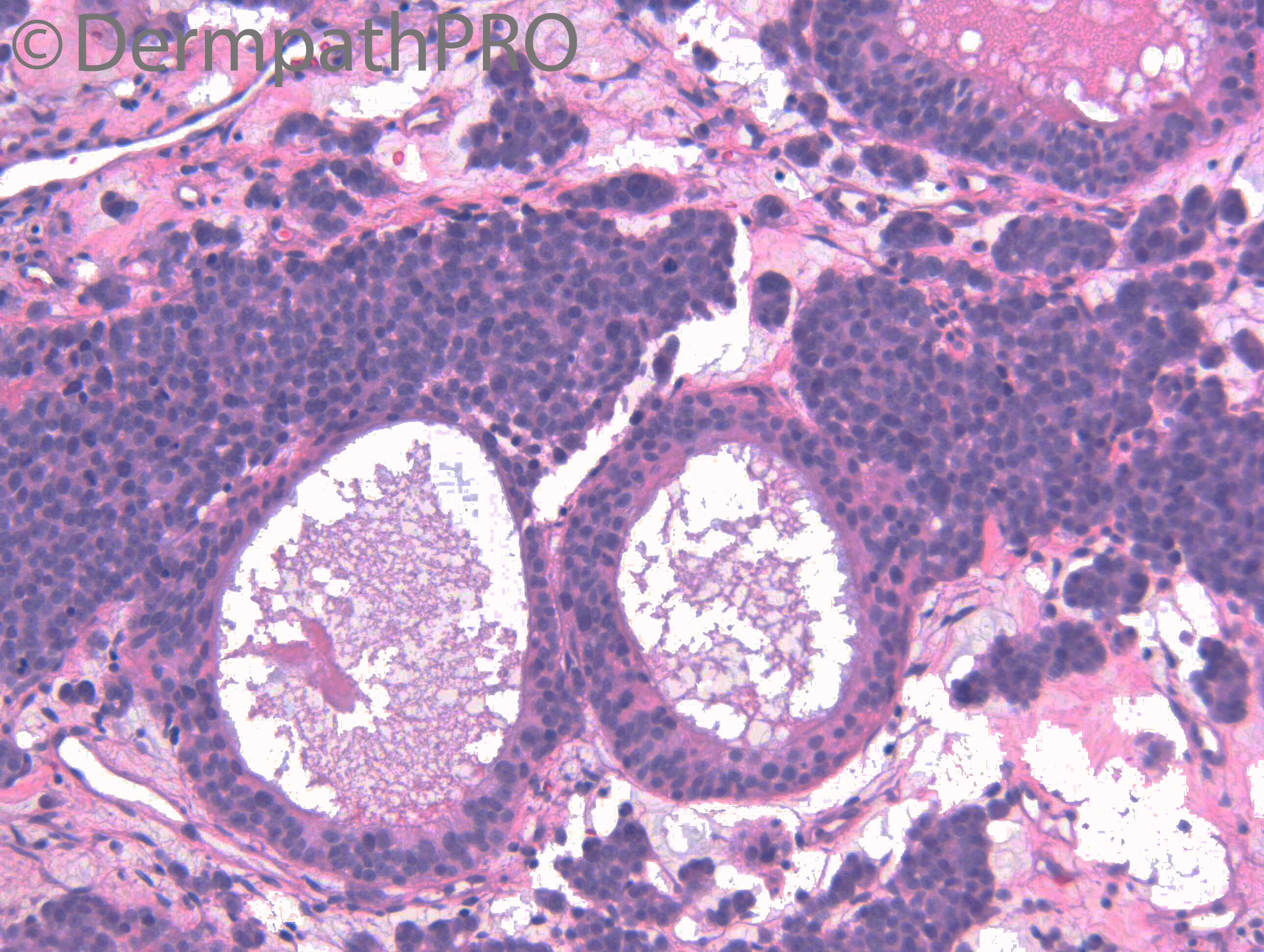
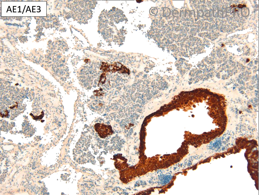
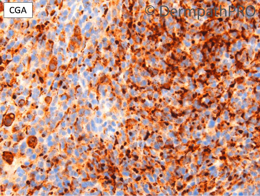
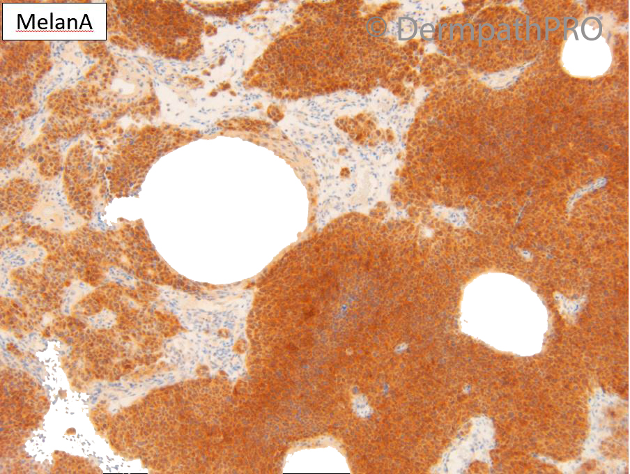
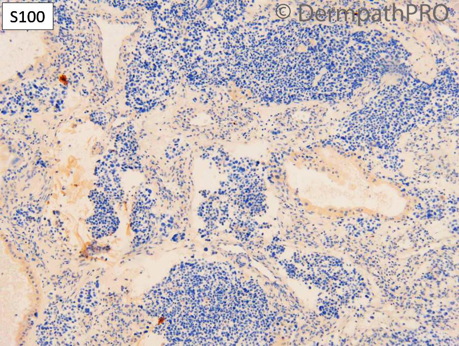
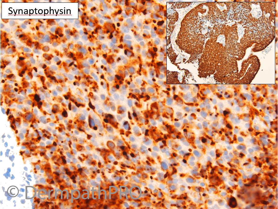
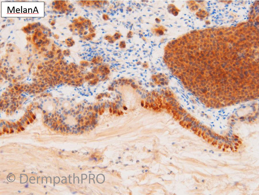
M75. h/o Ocular melanoma 2016. Developed nasal cavity mass.


  Report Record

User Feedback


Krishnakumar subramanian

Posted

rosette like structures are seen. But not much necrosis

go with olfactory neuroblastoma

Share this comment


Link to comment
share_externally

Krishnakumar subramanian

Posted

pigmentary Olfactory neuroblastoma

Share this comment


Link to comment
share_externally

Dr. Richard Carr

Posted

I was asked to review the case with the question can this be metastatic melanoma (hence the "dermatopathological" opinion from a colleague. The pan-keratin appeared negative, the S100 negative and the melanA looks funny but possible positive. I expanded the panel to include Sox10, HMB45 - negative and BerEP4 which was diffusely positive. Could not see rosettes actually they seem to be epithelial structures and very close scrutiny there was very focal dot-like positive staining for Cam5.2 and Pan-keratins. So final diagnosis awaits review by the ENT / neuro team at a local major centre but I concluded as follows: DDx Neuroendocrine carcinoma, olfactory neuroblastoma, NUT midline carcinoma, PNET. So far not managed to see the tumour from the eye (we could not yet trace the histopathology).

Share this comment


Link to comment
share_externally

Interesting case, important to think of non-dermatopathological differentials during skin consults!

Share this comment


Link to comment
share_externally

Dr. Richard Carr

Posted

I've now seen the opinion of the specialist ENT pathologist as follows:

Further calcitonin, prolactin, pit-1, p63 protein, Merkel cell marker, HMB45, desmin, CD99, NUT-1, TBX19, SN5 and NR5a studies are negative. Repeat melan A is also paradoxically negative. 
Overall, therefore, these appearances are most in keeping with part of an invasive, poorly differentiated non-small cell (large cell) neuroendocrine carcinoma. In the absence of any alternative origin these appearances are in keeping with a primary sino-nasal derivation. The possibility of a second, irradiation-induced malignancy cannot be excluded. The morphology and immunopheonotype militate against metastatic malignant melanoma, Merkel cell carcinoma, rhabdomyosarcoma, sino-nasal undifferentiated carcinoma (SNUC), undifferentiated carcinoma of nasopharyngeal-type (UCNT; lympho-epithelial carcinoma, small cell undifferentiated neuroendocrine carcinoma (SNUC), myo-pericytic malignancy and lymphoma. 

Share this comment


Link to comment
share_externally


Join the conversation

You can post now and register later. If you have an account, sign in now to post with your account.

Guest
Add a comment...

×   Pasted as rich text.   Paste as plain text instead

  Only 75 emoji are allowed.

×   Your link has been automatically embedded.   Display as a link instead

×   Your previous content has been restored.   Clear editor

×   You cannot paste images directly. Upload or insert images from URL.

Loading...

×
×
  • Create New...