Jump to content
In this section we have spot diagnoses posted on a daily basis since June 2010, now over 4000! You can review the archived cases and read the suggested diagnoses by users and the final comment by the contributors.
Case are uploaded each week day by 10 am UK time with the correct diagnosis will generally be posted at 8 pm UK time. Why not view the most recent spot diagnosis and proffer a diagnosis?

Case Number : Case 2821- 29 April 2021 Posted By: Saleem Taibjee

Please read the clinical history and view the images by clicking on them before you proffer your diagnosis.
Submitted Date :
   (0 reviews)

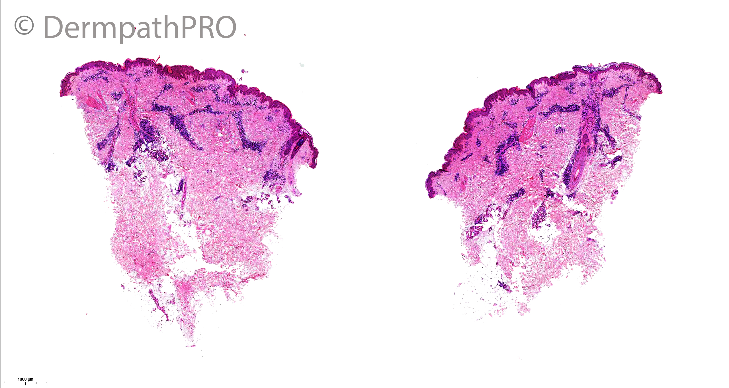
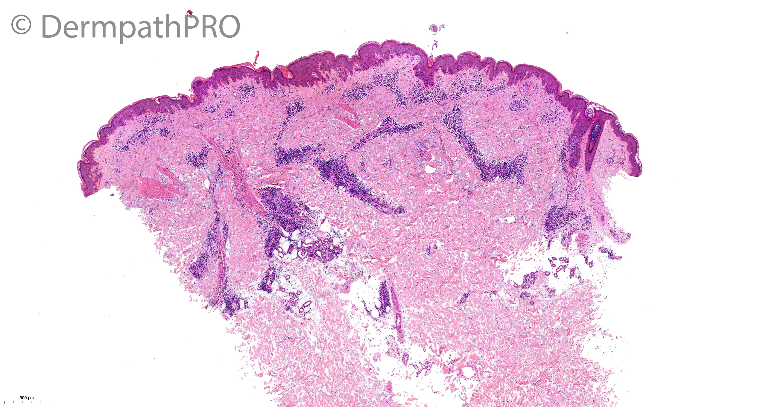
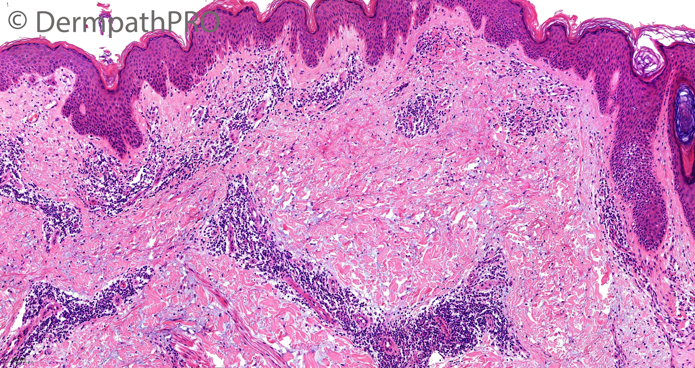
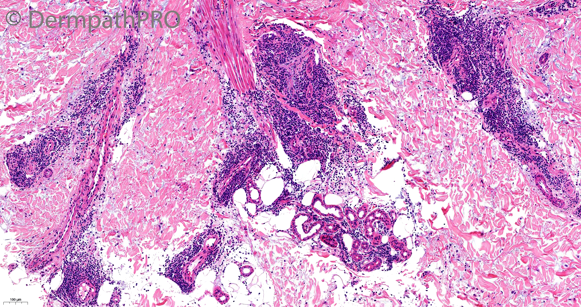
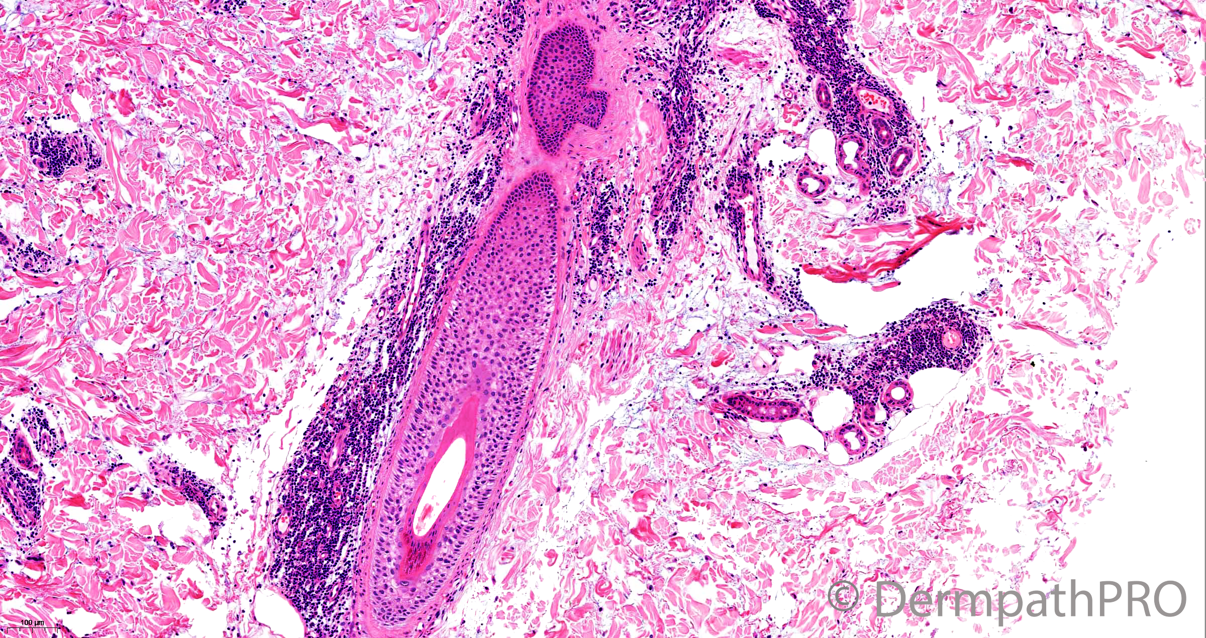
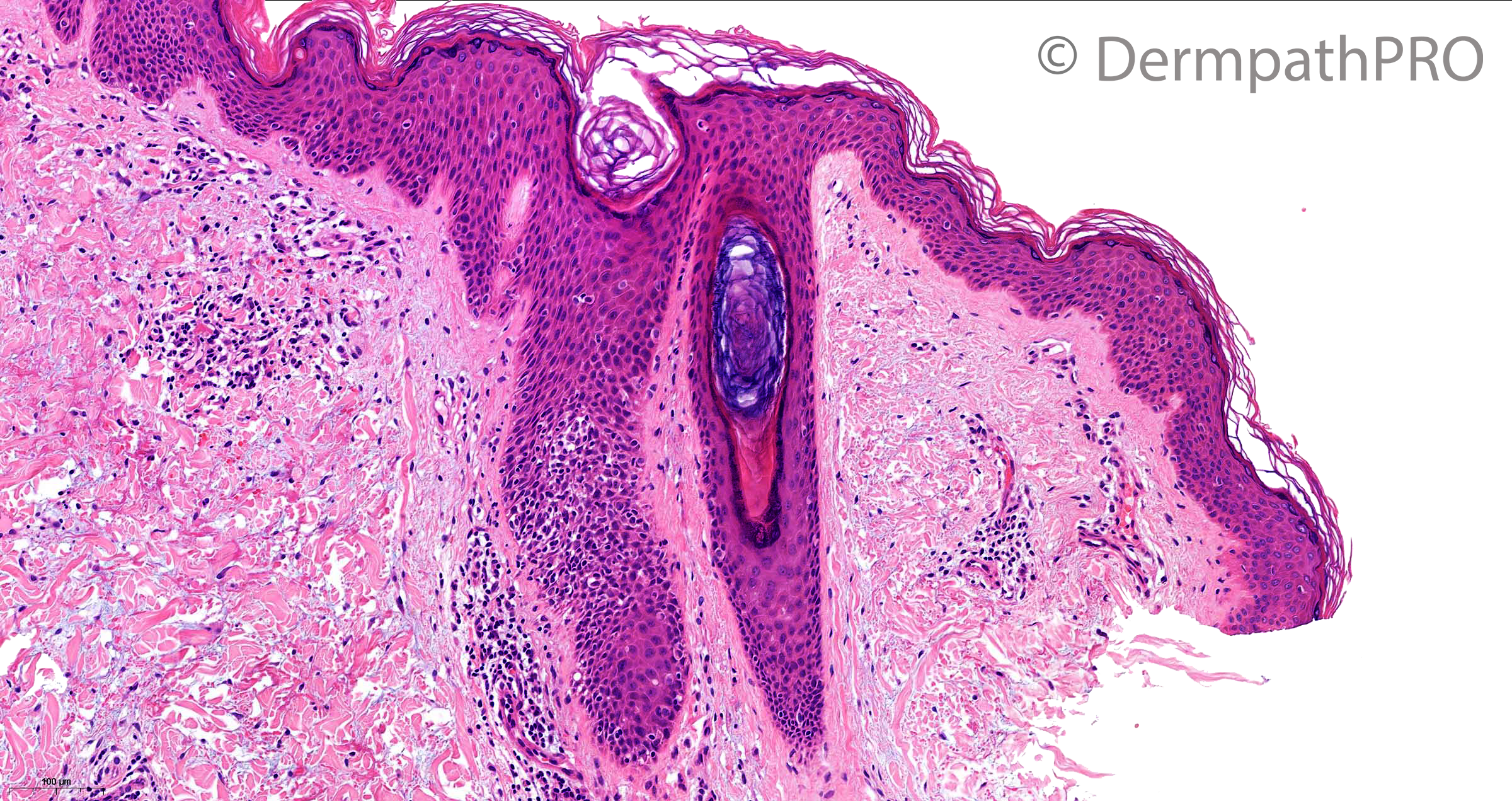
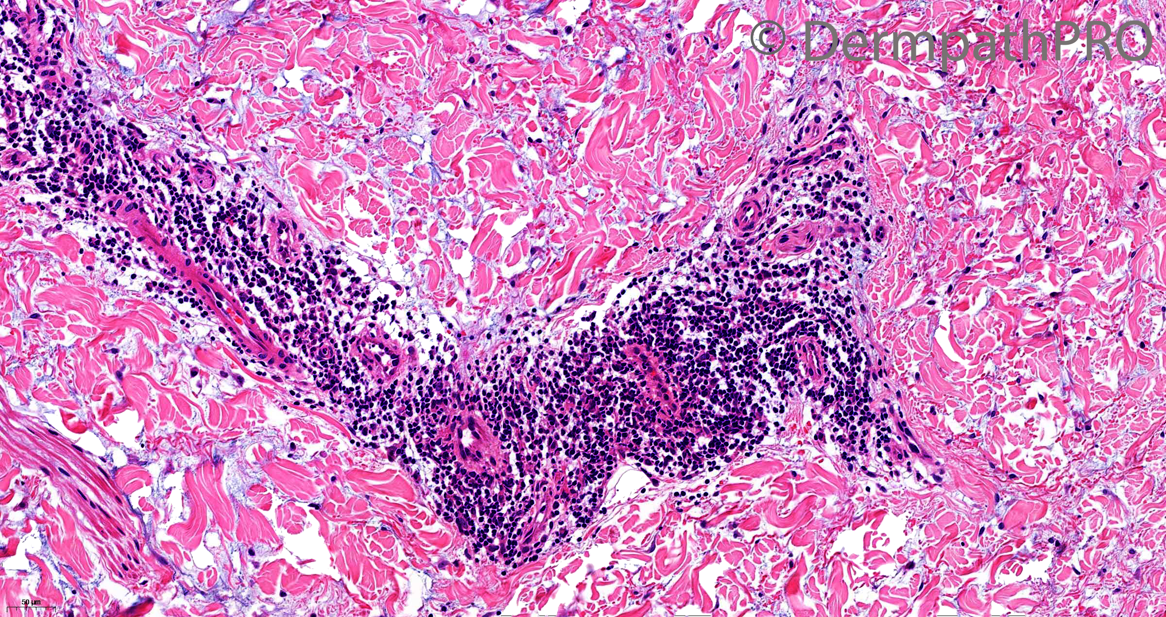
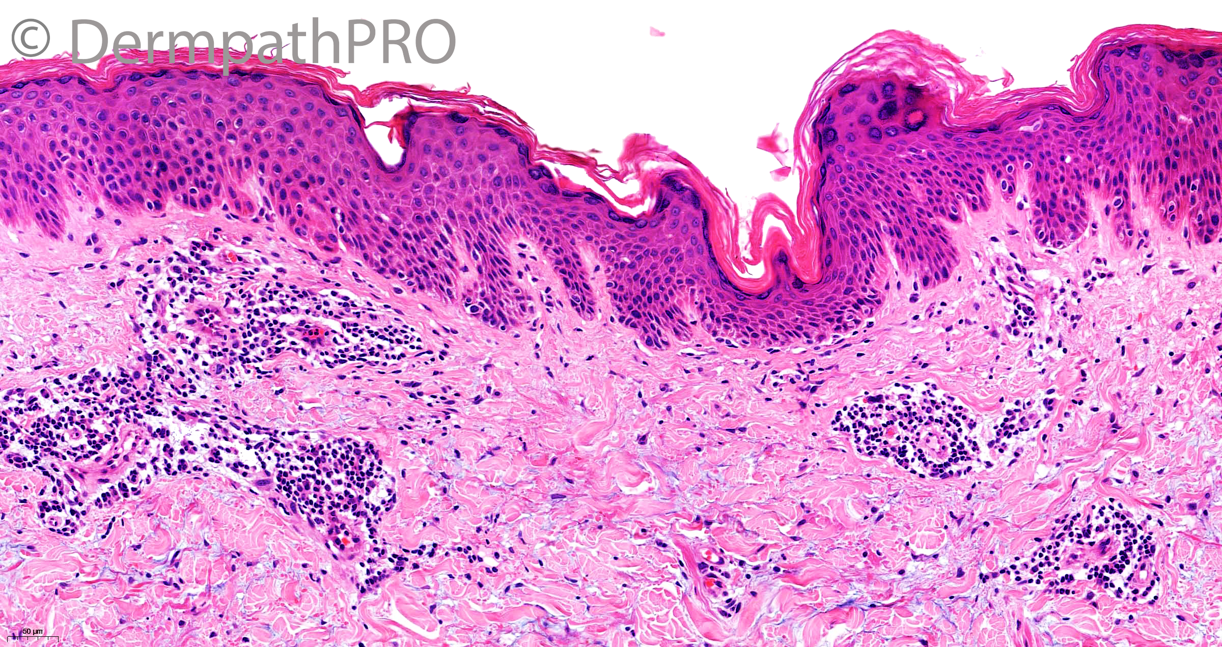
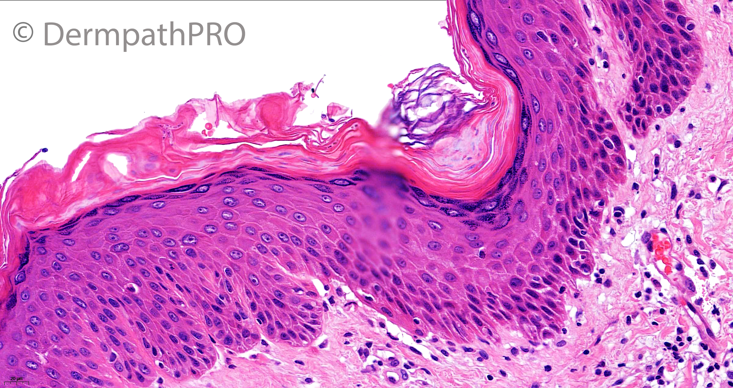
38F, itchy erythematous rash


  Report Record

User Feedback


daniellindsay

Posted

Superficial and deep perivascular and periadnexal lymphocytic infiltrate with focal interface component and follicular plugging. Would consider discoid lupus here?

Share this comment


Link to comment
share_externally

Richard Logan

Posted

There is a tight, peri-vascular and peri-adnexal lymphohistiocytic infiltrate showing some folliculotropism. Interface change is very mild.

I am struck by the dermal mucin deposition, and for that reason I think this is more likely to be reticular erythematous mucinosis, although lupus and Jessner's lymphocytic infiltrate are also possibilities.

Share this comment


Link to comment
share_externally

Mohamed khaled

Posted

I goes with cutaneous lupus and I am convinced that there are many names under the umbrella of  such disease, eg REM, Jessiner’s lymphocytic infiltrate, ....

Share this comment


Link to comment
share_externally

Meenakshi Batrani

Posted

Mucin and superficial and deep dermal infiltrate are the most significant findings with subtle interface. In the spectrum of lupus erythematosus- REM or some other form. 

Share this comment


Link to comment
share_externally

Some hyphae are seen in the stratum corneum.

Share this comment


Link to comment
share_externally

Agree with Lupus...with associated mycosis?

Share this comment


Link to comment
share_externally

Meenakshi Batrani

Posted

20 hours ago, cem said:

Some hyphae are seen in the stratum corneum.

Yes I also noticed some hyphae like structures, but then in view of other findings especially mucin, I thought I am imagining.

Share this comment


Link to comment
share_externally

Saman Fatah

Posted

This pattern of inflammation can be seen in a number of diseases, some are probably closely-related/on a spectrum while others may be same disease but given different names overtime by various authors. The density of the lymphoid infiltrate, dermal mucin deposition and interface changes can histologically favour one over the other (some of this is rather subjective) and close correlation with clinical appearance, serology and patient’s background is essential.

Tumid LE, Jessner’s Lymphocytic Infiltrate, REM and a very early plaque of DLE which has not matured yet to show the other classical surface features  can show such pattern.

REM is rare but has a rather distinctive appearance on central chest/inframammary or central back and it will be interesting to see if Saleem share photos of the eruption at a latter stage.

I still struggle to confidently differentiate between tumid LE and JLI clinically and they share more similarities than differences if you are in the lumpers camp (like me) rather than splitters.

Share this comment


Link to comment
share_externally

Saleem Taibjee

Posted

The PAS stain is shown below. As one or two of you correctly identified, there were numerous hyphae indicating tinea corporis.

This recent case was quite a surprise for me. Like yourselves, I went down the 'superfical and deep inflammation' differential (c.f. Weedon's 'L's list), but was starting to feel that I would not be able to make a specific diagnosis. Our private laboratory routinely cuts a PAS section in additional to H&E for all rashes. I had missed the hyphae on H&E, but casually looked at the PAS (I did not disclose that the clinician did include tinea in the differential) at which point my jaw dropped. Of course, this case also illustrates what a wonderful resource Weedon's textbook is, dermatophyte is listed in the differential of superficial and deep inflammation.

Here is the relevant excerpt from Weedon:

The often quoted mnemonic of diseases causing this pattern of inflammation is the eight ‘L’ diseases – light reactions, lymphoma (including pseudolymphomas), leprosy, lues (syphilis), lichen striatus, lupus erythematosus, lipoidica (includes necrobiosis lipoidica and incomplete forms of granuloma annulare), and lepidoptera (used incorrectly in the mnemonic to refer to arthropod bites and other parasitic infestations). To the eight ‘L diseases’ should be added ‘DRUGS’ – drug reactions, as well as dermatophyte infections, reticular erythematous mucinosis, urticaria (chronic urticaria and the urticarial stages of bullous pemphigoid and herpes gestationis), gyrate erythemas (deep type), and scleroderma (particularly the localized variants).

For some time I have been debating whether routinely cutting PAS for inflammatory skin cases within our lab is cost-effective, but this recent case has definitely made me back-track on that! There are similar debates to be had on other potential cost-savings/efficiencies. For example, we recently discussed in our lab whether a negative control is required/cost-effective for immunohistochemistry? How often does a negative control really affect our interpretation? With regards to punch biopsies, Richard Carr has recommended generally bisecting 4mm (or larger) punch biopsies. Other labs embed whole and take initial levels. Over the years, seeing both practices, I am sure that the bisecting option is the more efficient i.e. much lower rate of requiring to request further levels.

BW, Saleem

13372_40.0x PASc.jpg


13372_40.0x PASb.jpg

13372_40.0x PAS.jpg

Share this comment


Link to comment
share_externally

Richard Logan

Posted

A salutory tale - thanks Saleem.

Share this comment


Link to comment
share_externally

Saman Fatah

Posted

Thanks for sharing this case, was not aware dermatophyte can cause such pattern.  

Share this comment


Link to comment
share_externally

Richard Logan

Posted

Saleem, do you think that this inflammatory pattern implies that the fungus was involving the follicles?  Did the PAS show deeper follicular involvement?


Share this comment


Link to comment
share_externally

Saleem Taibjee

Posted

Good thought. I didn't see any deeper involvement. But perhaps this might require examination of multiple levels to find such a follicular focus.

Share this comment


Link to comment
share_externally


Join the conversation

You can post now and register later. If you have an account, sign in now to post with your account.

Guest
Add a comment...

×   Pasted as rich text.   Paste as plain text instead

  Only 75 emoji are allowed.

×   Your link has been automatically embedded.   Display as a link instead

×   Your previous content has been restored.   Clear editor

×   You cannot paste images directly. Upload or insert images from URL.

Loading...

×
×
  • Create New...