Jump to content
In this section we have spot diagnoses posted on a daily basis since June 2010, now over 4000! You can review the archived cases and read the suggested diagnoses by users and the final comment by the contributors.
Case are uploaded each week day by 10 am UK time with the correct diagnosis will generally be posted at 8 pm UK time. Why not view the most recent spot diagnosis and proffer a diagnosis?

Case Number : Case 2891 - 05 August 2021 Posted By: Saleem Taibjee

Please read the clinical history and view the images by clicking on them before you proffer your diagnosis.
Submitted Date :
   (0 reviews)

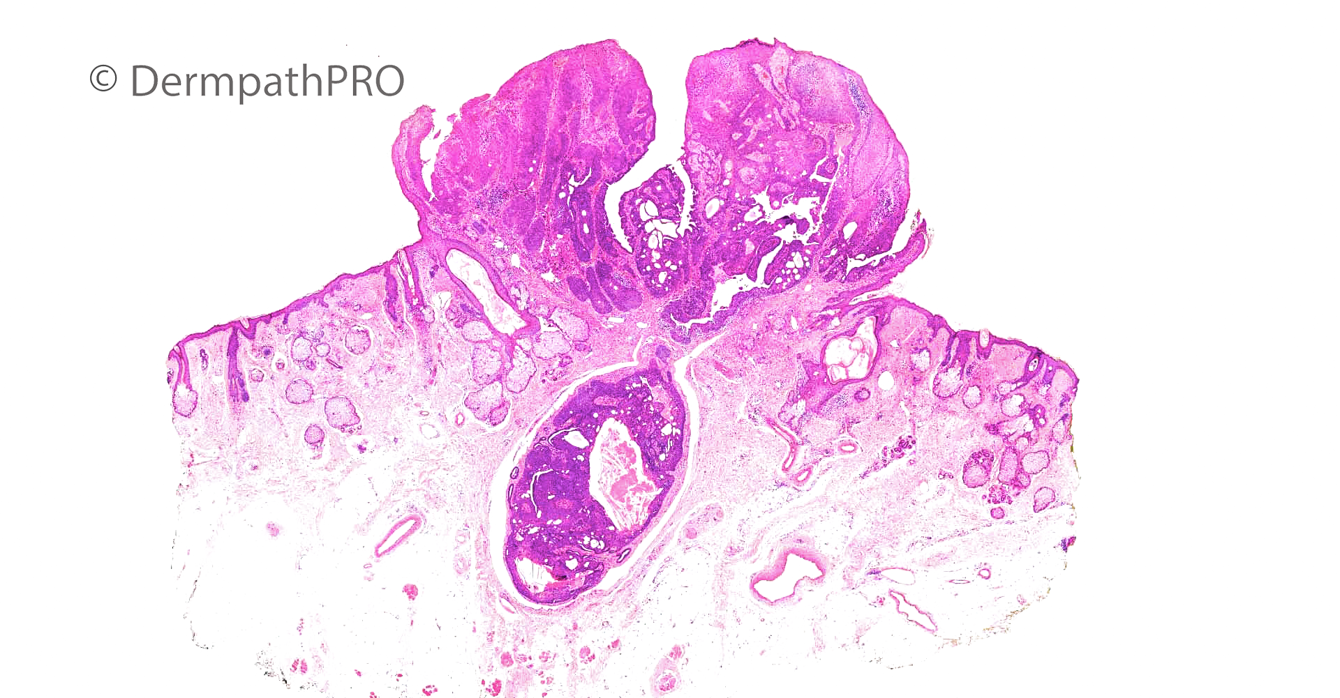
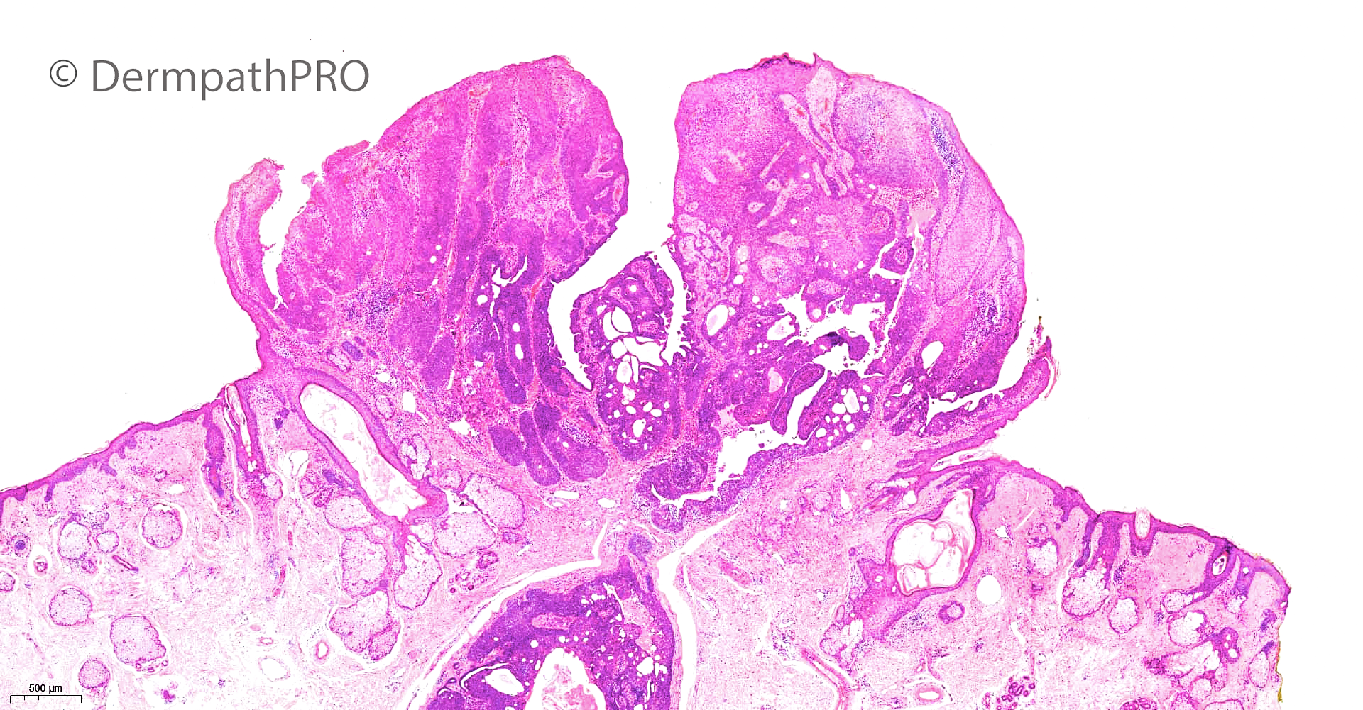
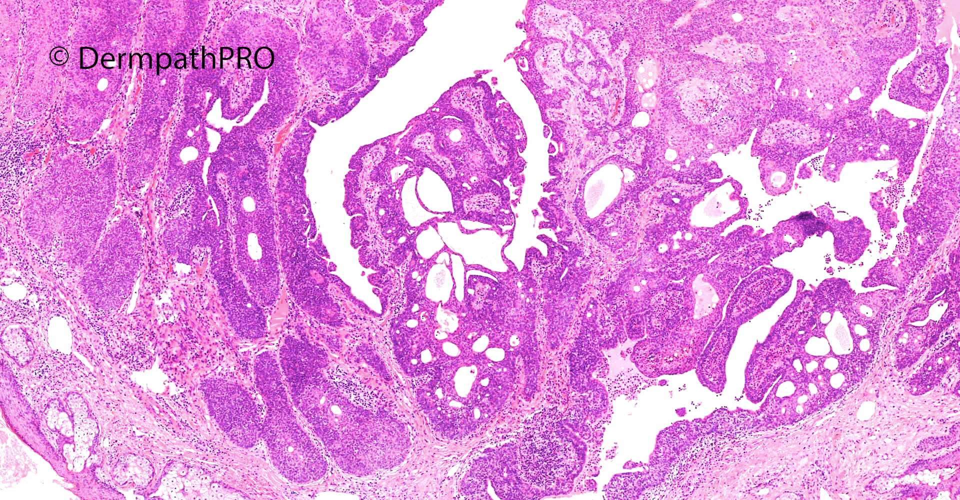
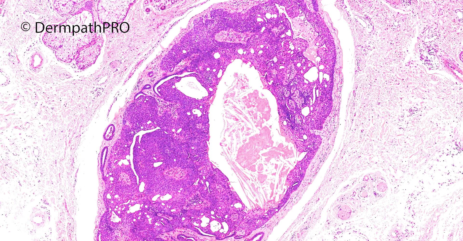
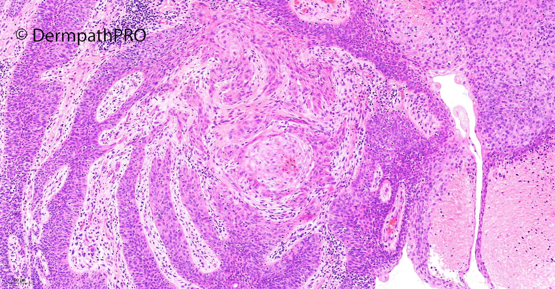
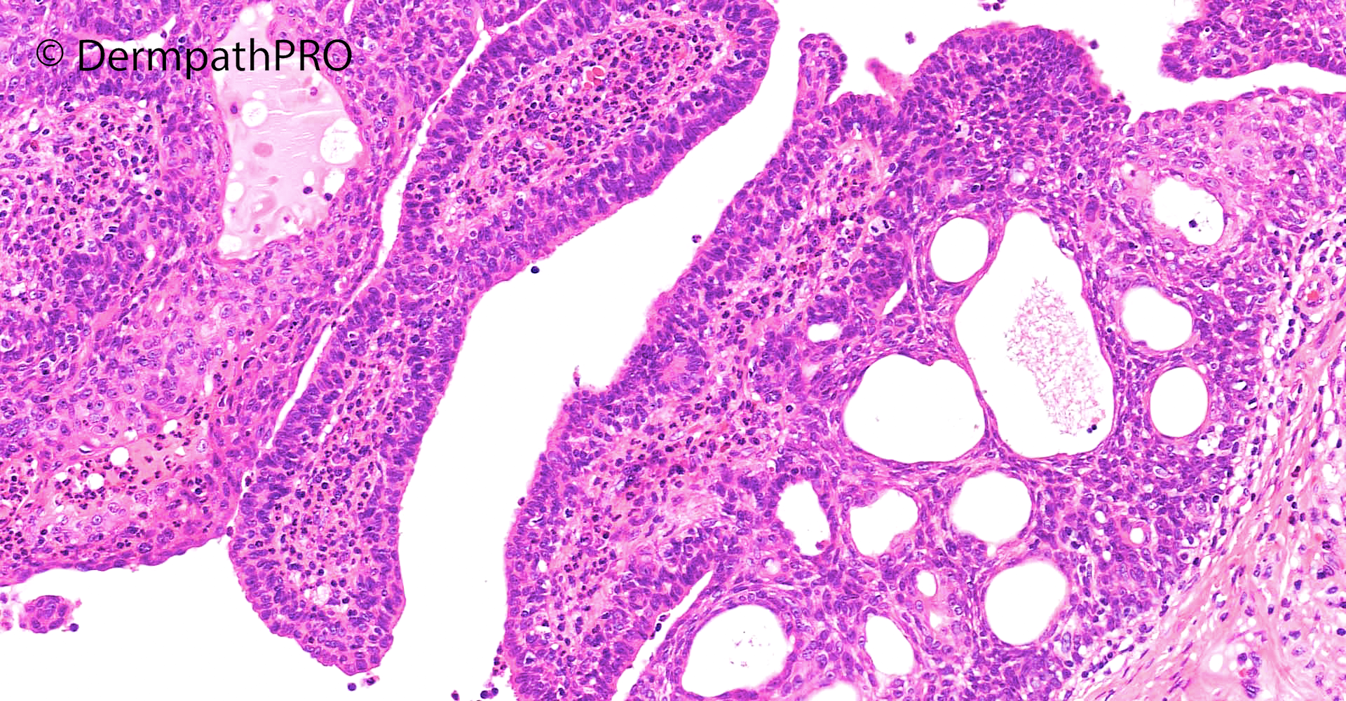
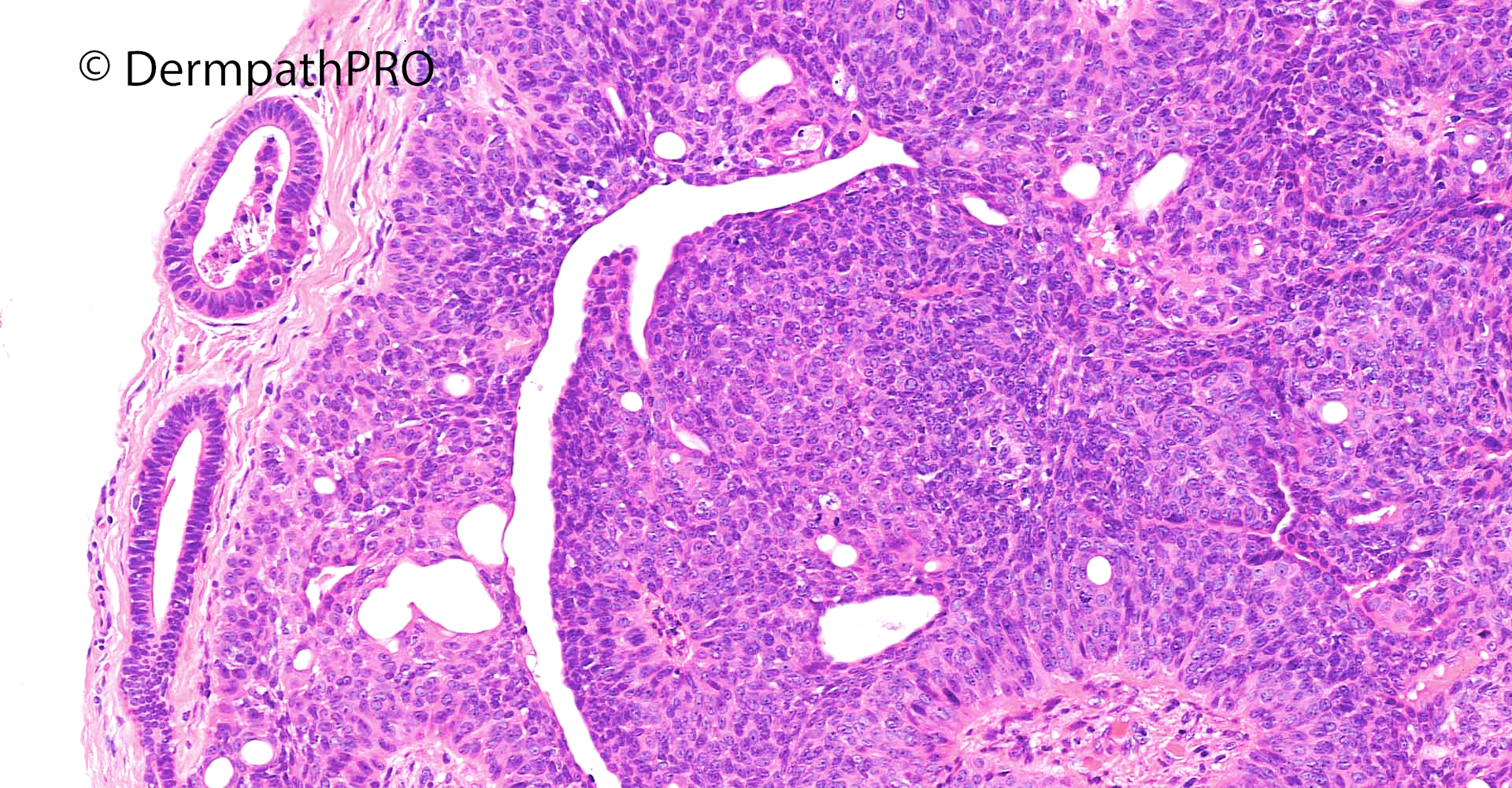
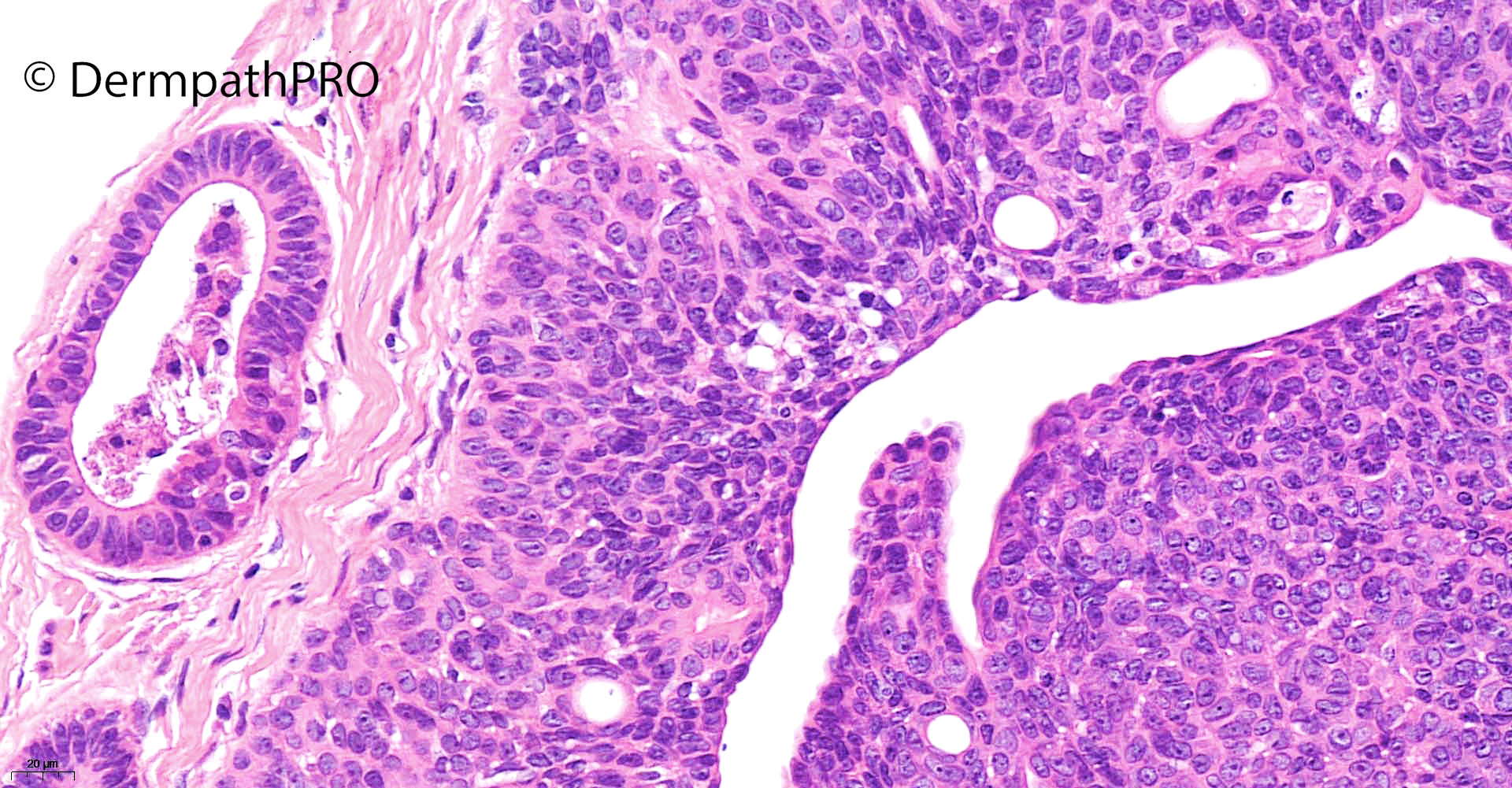
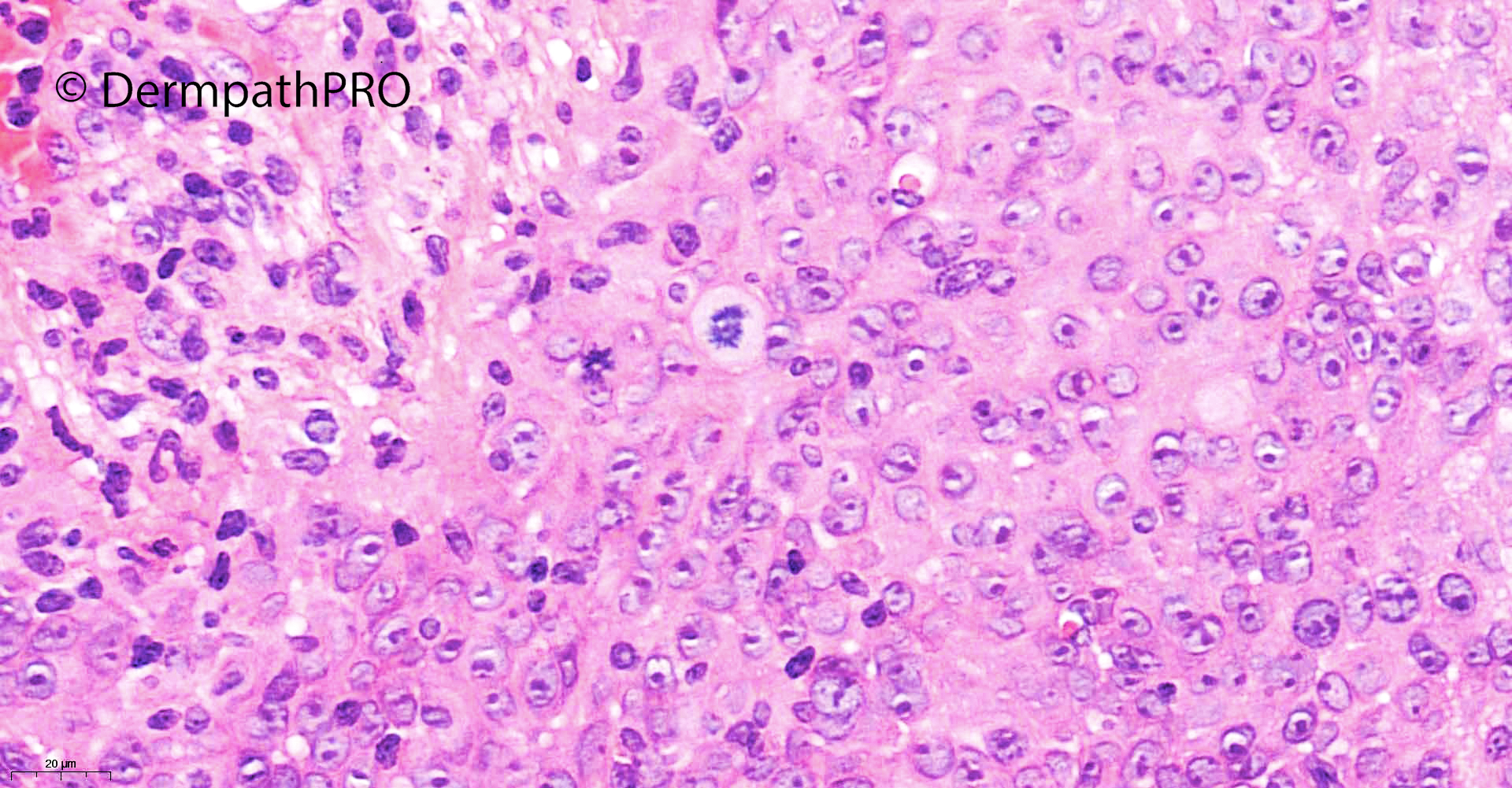
64M Longstanding telangiectatic papule on forehead ?BCC


  Report Record

User Feedback


Meenakshi Batrani

Posted

Overall silhouette appears to be of SCAP, however, cytology is atypical.  I would consider Syringocystadenocarcinoma papilliferum in situ. Even the lower dermal nodule appears well circumscribed, frank invasion is not evident.  

Share this comment


Link to comment
share_externally

Eman El-Nabarawy

Posted (edited)

Nevus sebaceous with syringocystadnoadenocarcinoma papilliferum in situ.

Edited by Eman El-Nabarawy

Share this comment


Link to comment
share_externally

Trichilemmoma + SCAP , both atypical, but I don’t think really carcinomatous. 

Share this comment


Link to comment
share_externally

Eman El-Nabarawy

Posted

4 hours ago, vincenzo said:

Trichilemmoma + SCAP , both atypical, but I don’t think really carcinomatous. 

I also thought of desmoplastic trichilemmoma but couldn't be sure!

Share this comment


Link to comment
share_externally

Krishnakumar subramanian

Posted

SCAP with malignant change

Share this comment


Link to comment
share_externally

Patient may have nevus sebaceus with syringocystadenoma papilliferum. Background epidermis may be interpreted as Bowen's disease with follicular extension. not really trichilemmoma to me.

Share this comment


Link to comment
share_externally

Saleem Taibjee

Posted

Thanks for the comments on this interesting recent case which I did not find straightforward.

I too felt that there were features of syringocystadenoma papilliferum. However, as is often the case with sweat gland tumours, these tumours can be difficult to firmly classify, with possible hybrid features of poroma / hidradenoma (deeper focus) in this case. As you know we can see hybrid lesion in the context of pre-existing naevus sebaceous, but I could not identify this in this case.

With regards to possible dysplasia / malignant change, I found that tricky in this case. I did display an area of more mitotically active / mildly pleomorphic change in the last image above to help stimulate discussion. This area corresponded to a more superficial and eroded aspect of the tumour. I think we not infrequently see such change in the context of poroma lesions, and hence I was not 100% convinced for dysplasia (but could not fully exclude). For interest, p53 is shown below. Although there is quite prominent staining, it does nonethless seem to be wild-type pattern. Following comments above of possible trichilemmoma-like change, I have since requested CD34, but this is negative (not shown).

BW, Saleem

32143_1.0x p53 labelled.jpg

32143_10.0x p53.jpg32143_20.0x p53b.jpg


Share this comment


Link to comment
share_externally


Join the conversation

You can post now and register later. If you have an account, sign in now to post with your account.

Guest
Add a comment...

×   Pasted as rich text.   Paste as plain text instead

  Only 75 emoji are allowed.

×   Your link has been automatically embedded.   Display as a link instead

×   Your previous content has been restored.   Clear editor

×   You cannot paste images directly. Upload or insert images from URL.

Loading...

×
×
  • Create New...