Jump to content
In this section we have spot diagnoses posted on a daily basis since June 2010, now over 4000! You can review the archived cases and read the suggested diagnoses by users and the final comment by the contributors.
Case are uploaded each week day by 10 am UK time with the correct diagnosis will generally be posted at 8 pm UK time. Why not view the most recent spot diagnosis and proffer a diagnosis?

Case Number : Case 2892 - 06 August 2021 Posted By: Dr. Richard Carr

Please read the clinical history and view the images by clicking on them before you proffer your diagnosis.
Submitted Date :
   (0 reviews)

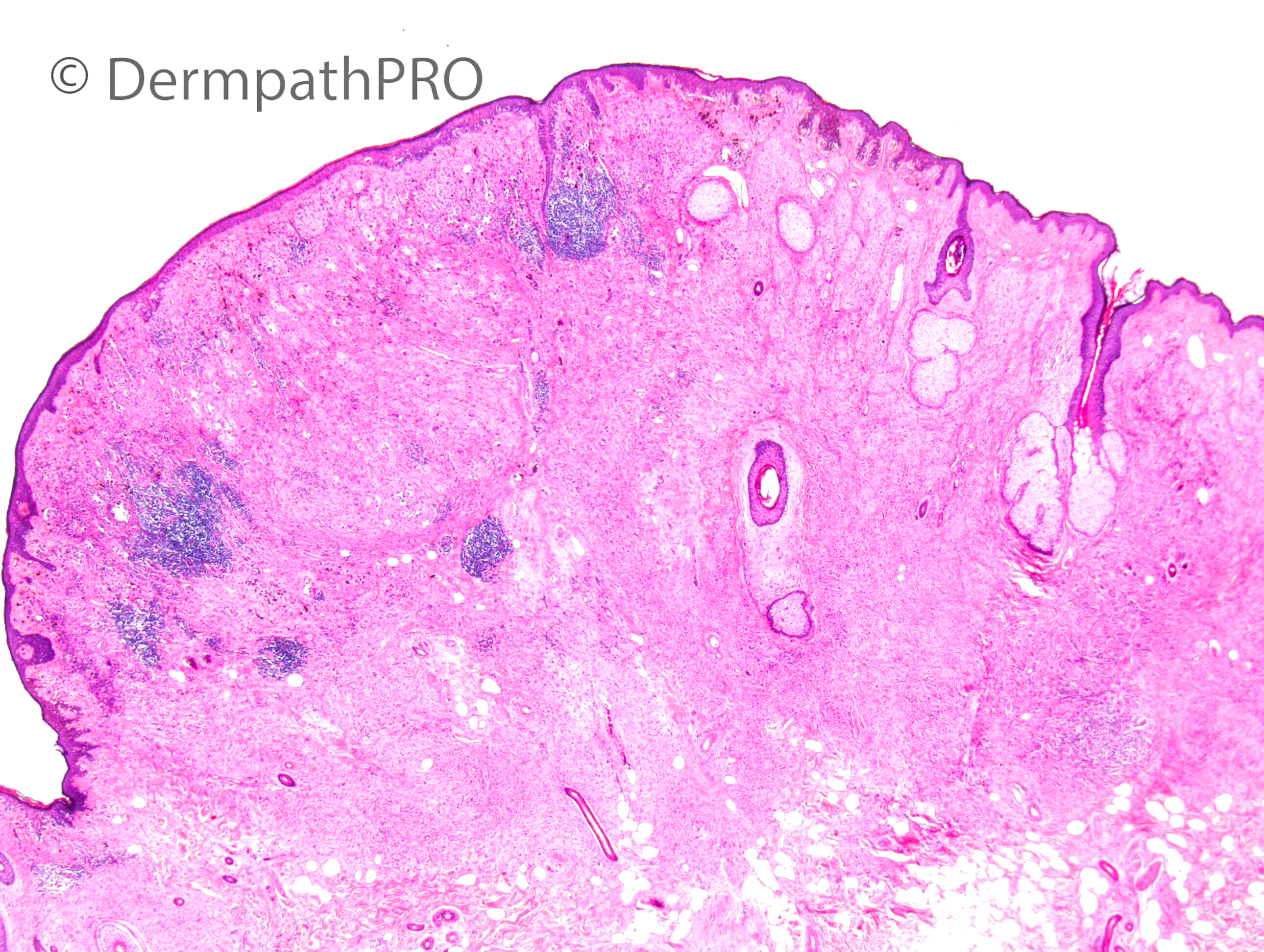
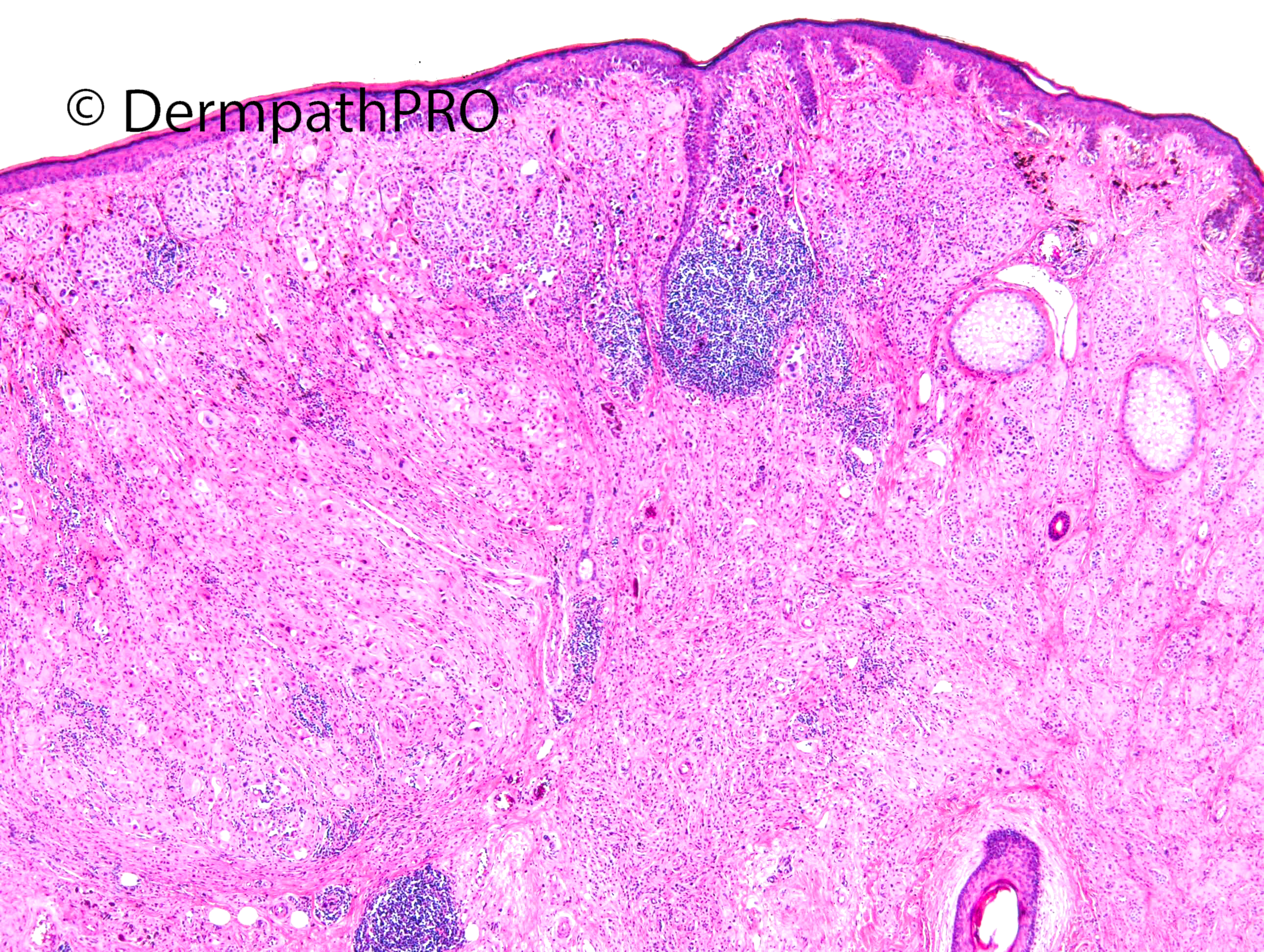
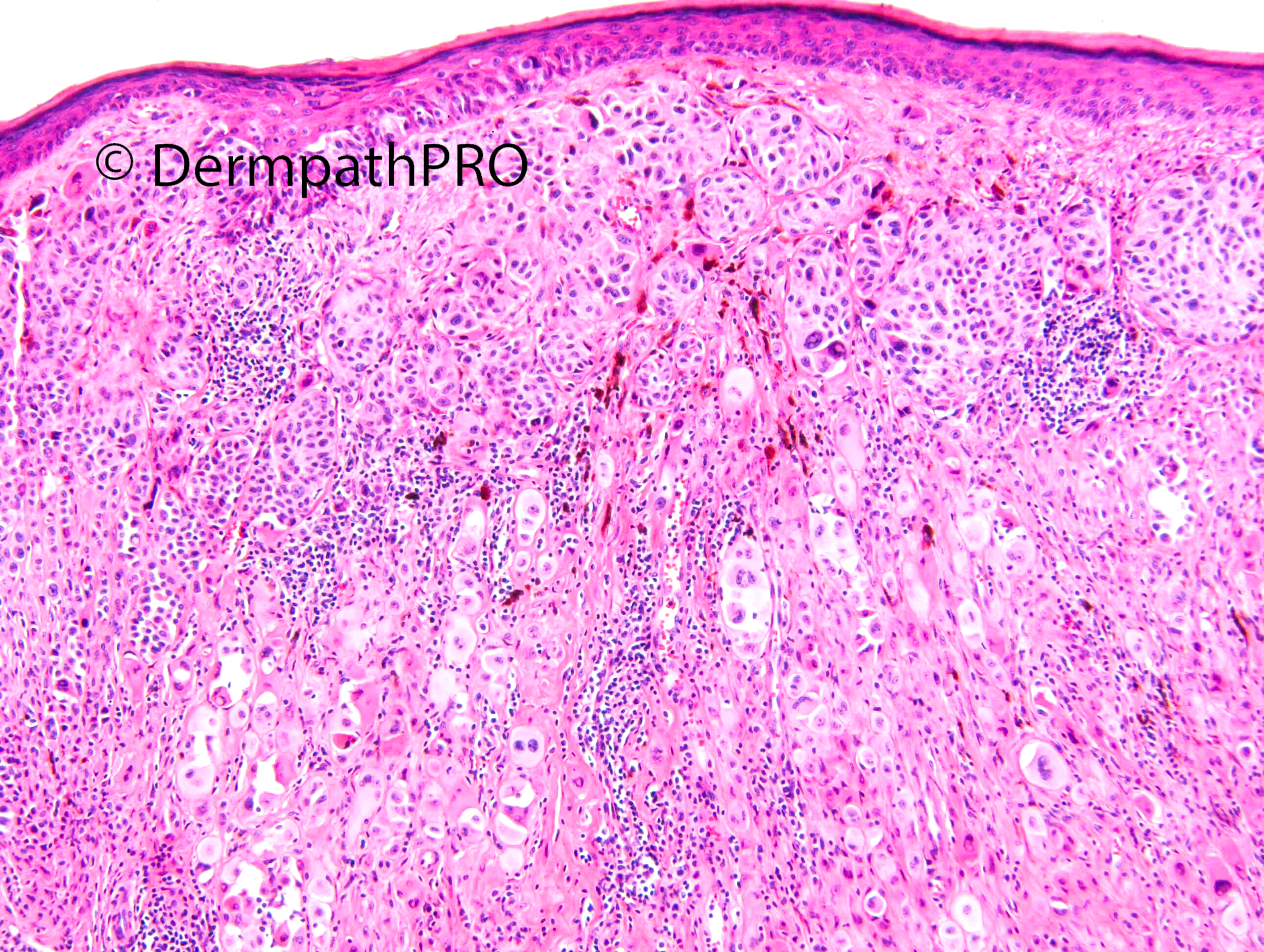
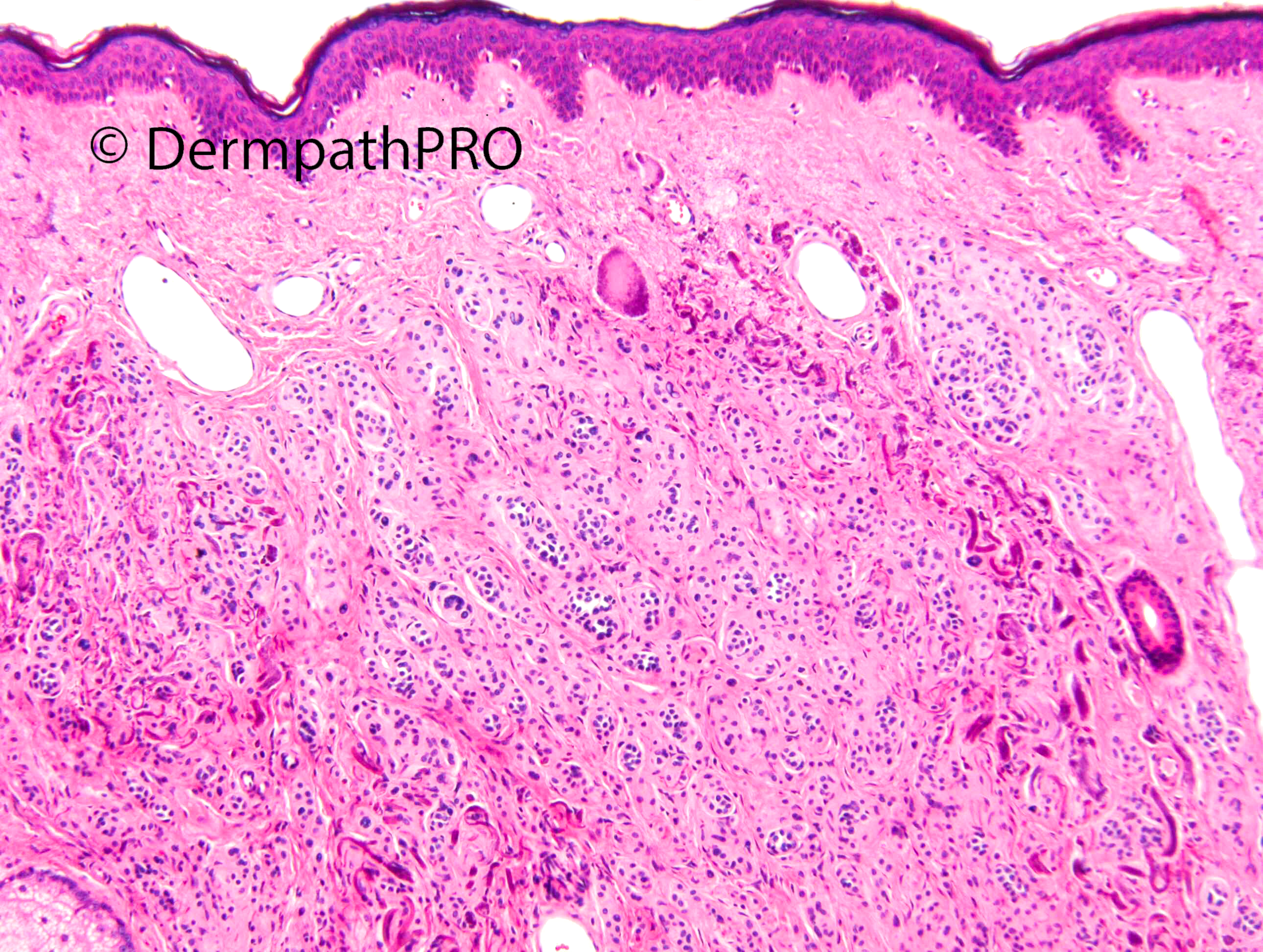
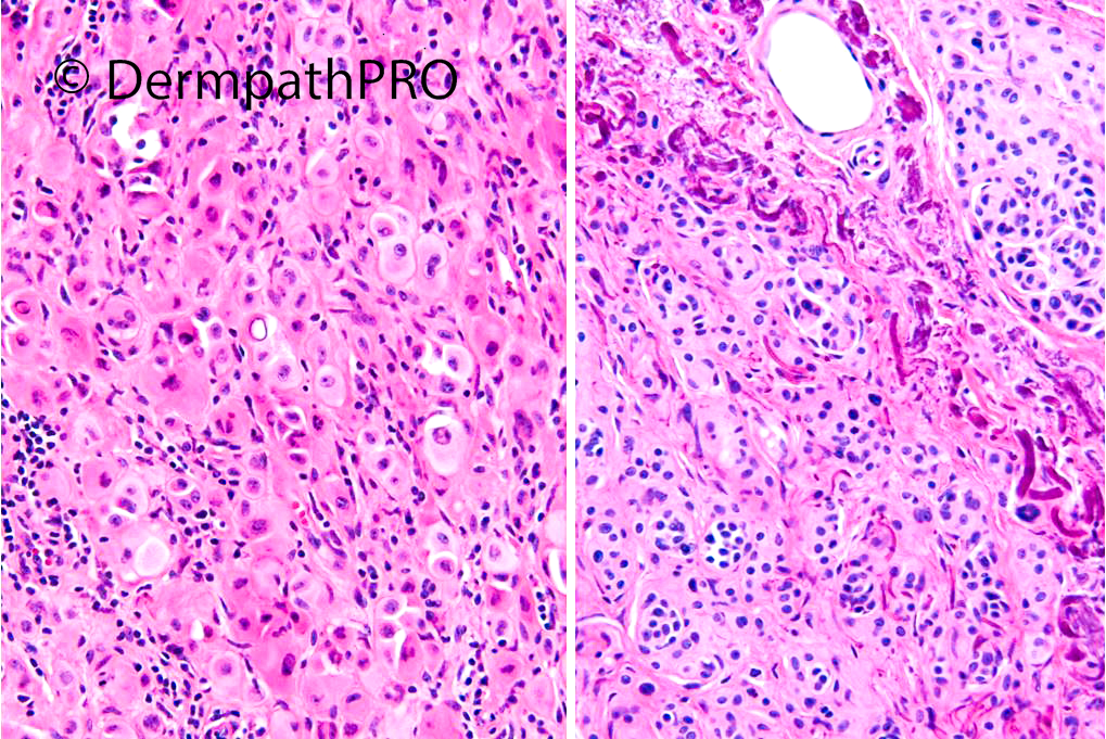
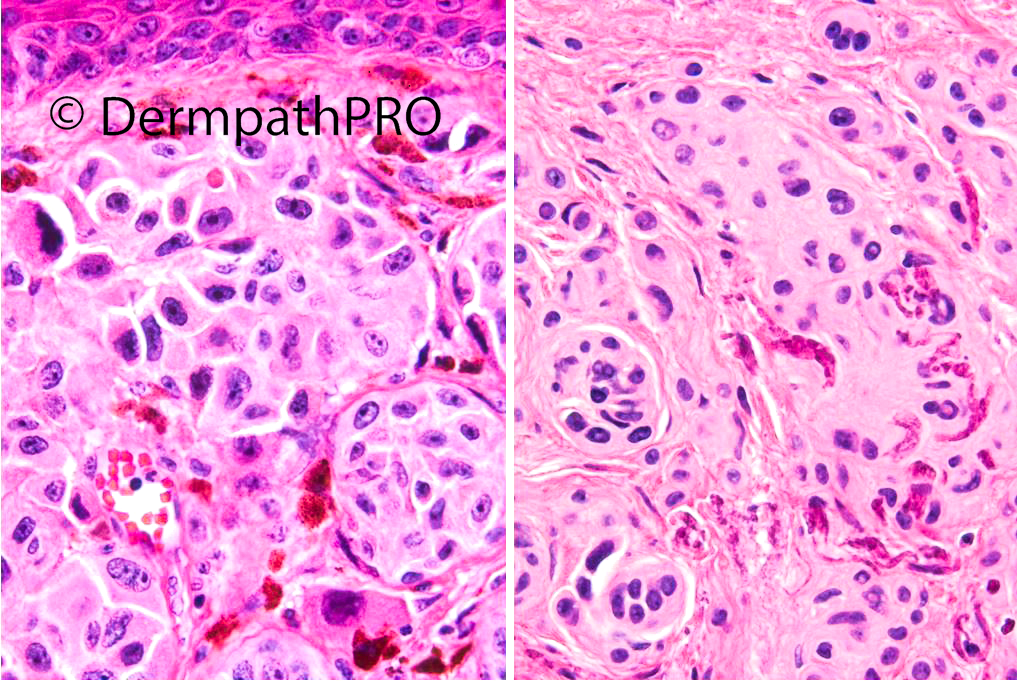
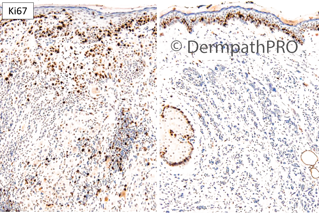
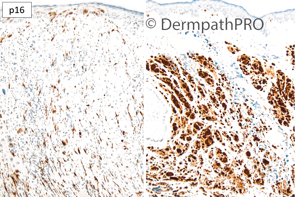
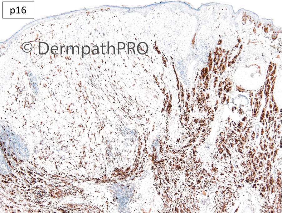
F70. Lifelong papular naevus on check. On half has become darker in last 3/12.


  Report Record

User Feedback


Left--> Melanoma, balloon cell type, sprung by a Lentigo Maligna. Right--> nevus. 

Melanoma has an expansive nodular growth and pushes the elastic fibers, while nevus has an infiltrative one, between collagen and elastic fibers. 

Share this comment


Link to comment
share_externally

Eman El-Nabarawy

Posted

Beauty!!

The DD here is balloon cell change in a nevus (with overlying junctional component) vs balloon cell melanoma. Age of the pt, nuclear pleomorphism, lack of (maturation, pigmentation, intervening stroma) and prominent nucleoli favor the 2nd. Also atypical mitosis, if present.

Share this comment


Link to comment
share_externally

Meenakshi Batrani

Posted (edited)

There appears to be expansile nodule with cytological atypia. Loss of p16 in this zone compared to the rest. I would favour nodular melanoma arising in a nevus. 

Edited by Meenakshi Batrani

Share this comment


Link to comment
share_externally

Looks like a different clone, melanoma (p16 loss) arising in nevus (p16 preserved )

Share this comment


Link to comment
share_externally

Krishnakumar subramanian

Posted

AGREE MELANOMA IN A NEVUS

Share this comment


Link to comment
share_externally

agree with melanoma in a nevus.  also curious to know what is the BAP-1 status? it does have that BAPoma look of those epithelioid melanocytes with binucleation and lymphocytes response.

Share this comment


Link to comment
share_externally

Dr. Richard Carr

Posted

Agree with melanoma in a naevus. p16 was helpful for assessing the depth. Nice example of a background Miescher type naevus with exaggerated elastic fibres not seen in the melanoma part. p16 null and a small lateral SSMM were present (not shown) so I did not do BAP1 but agree worth considering in all biphenotypic naevi (although the "sptizoid" component here is a little too pleomorphic with quite irregular chromatin). 

Share this comment


Link to comment
share_externally


Join the conversation

You can post now and register later. If you have an account, sign in now to post with your account.

Guest
Add a comment...

×   Pasted as rich text.   Paste as plain text instead

  Only 75 emoji are allowed.

×   Your link has been automatically embedded.   Display as a link instead

×   Your previous content has been restored.   Clear editor

×   You cannot paste images directly. Upload or insert images from URL.

Loading...

×
×
  • Create New...