Jump to content
In this section we have spot diagnoses posted on a daily basis since June 2010, now over 4000! You can review the archived cases and read the suggested diagnoses by users and the final comment by the contributors.
Case are uploaded each week day by 10 am UK time with the correct diagnosis will generally be posted at 8 pm UK time. Why not view the most recent spot diagnosis and proffer a diagnosis?

Case Number : Case 2896 - 12 August 2021 Posted By: Saleem Taibjee

Please read the clinical history and view the images by clicking on them before you proffer your diagnosis.
Submitted Date :
   (0 reviews)

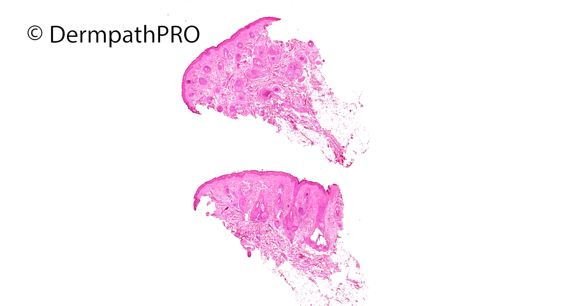
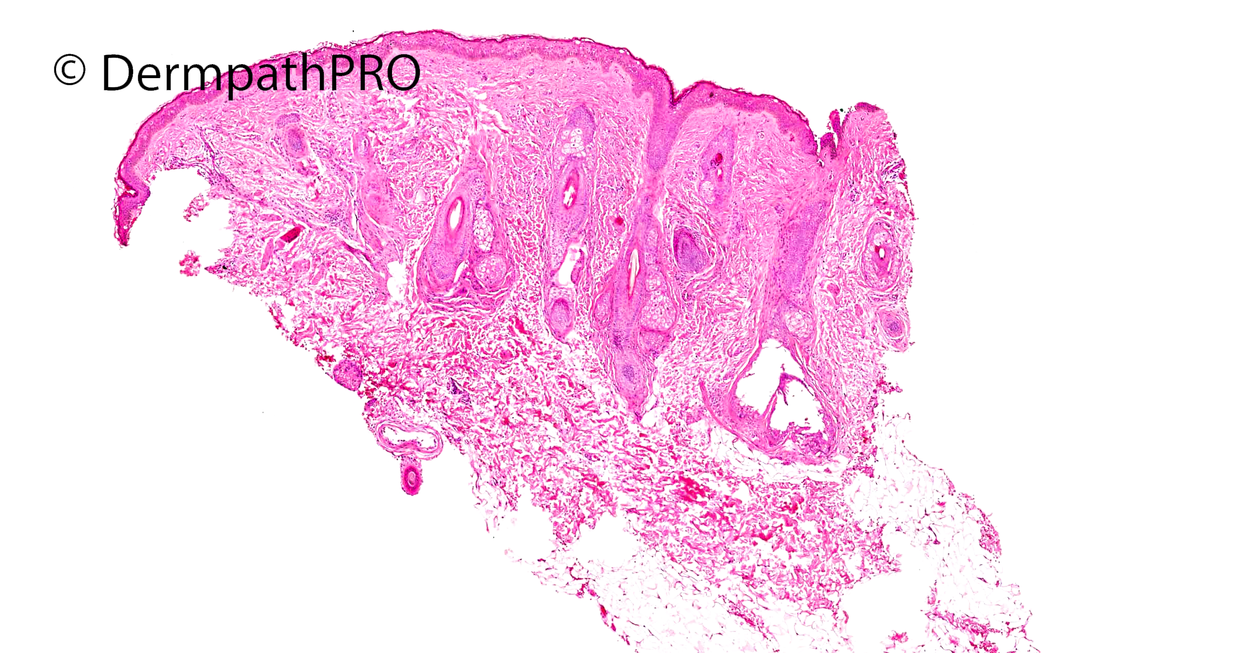
12F Persistent pink erythema bilateral cheeks, painful and warm intermittently, sometimes eyelid erythema ?dermatomyositis ?connective tissue disease ??other

  Report Record

User Feedback


Richard Logan

Posted

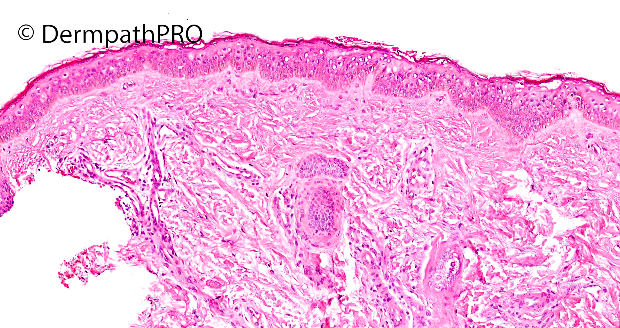
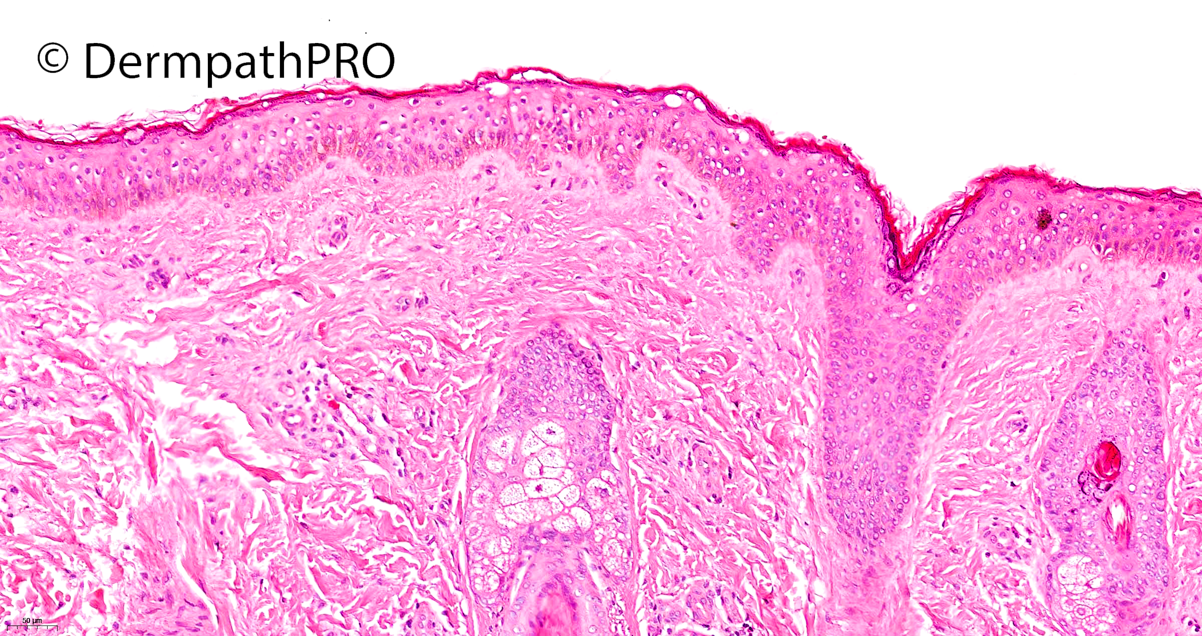
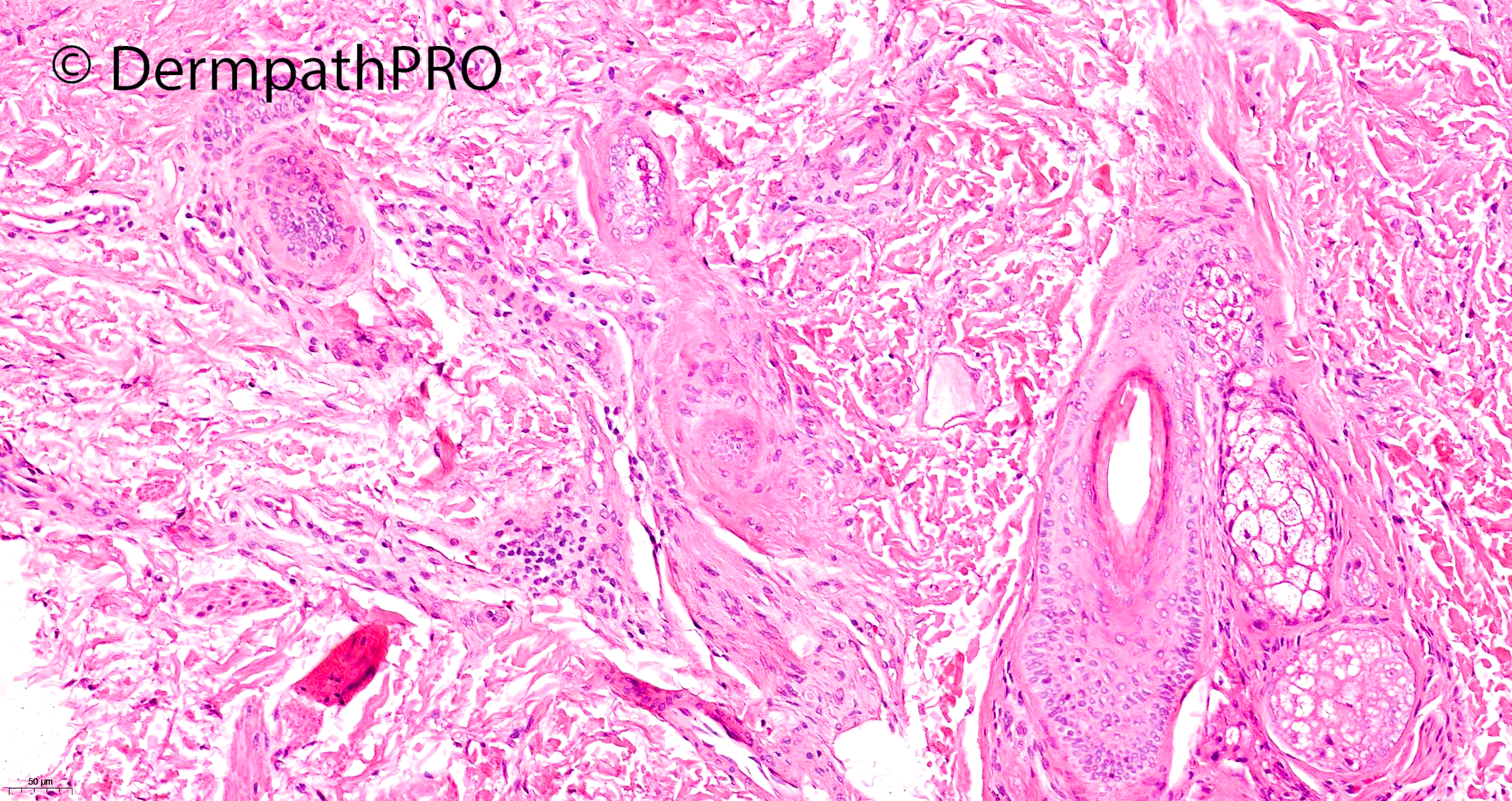
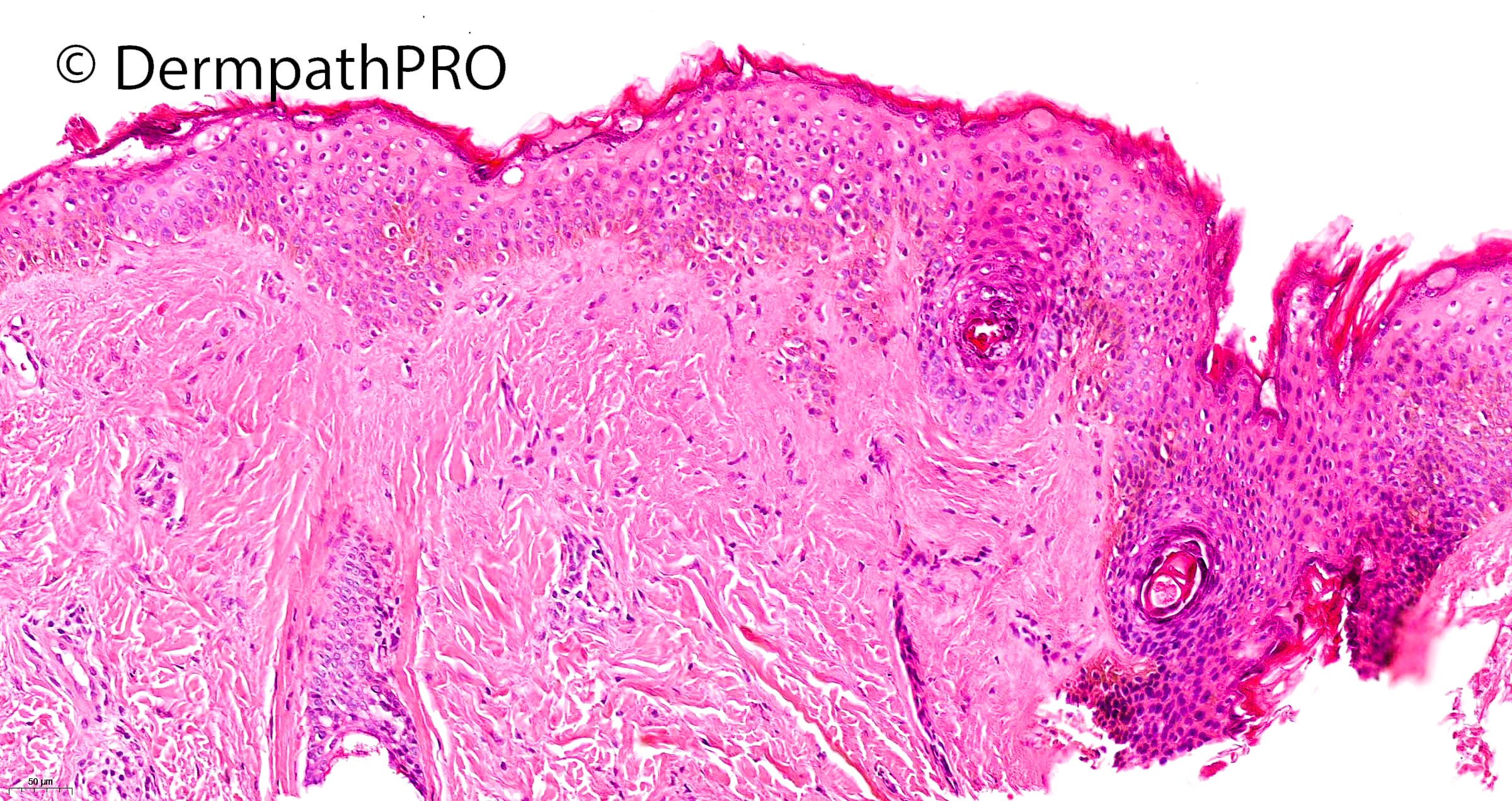
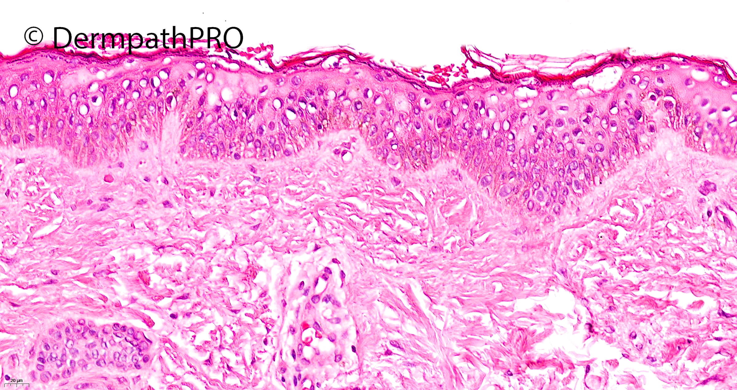
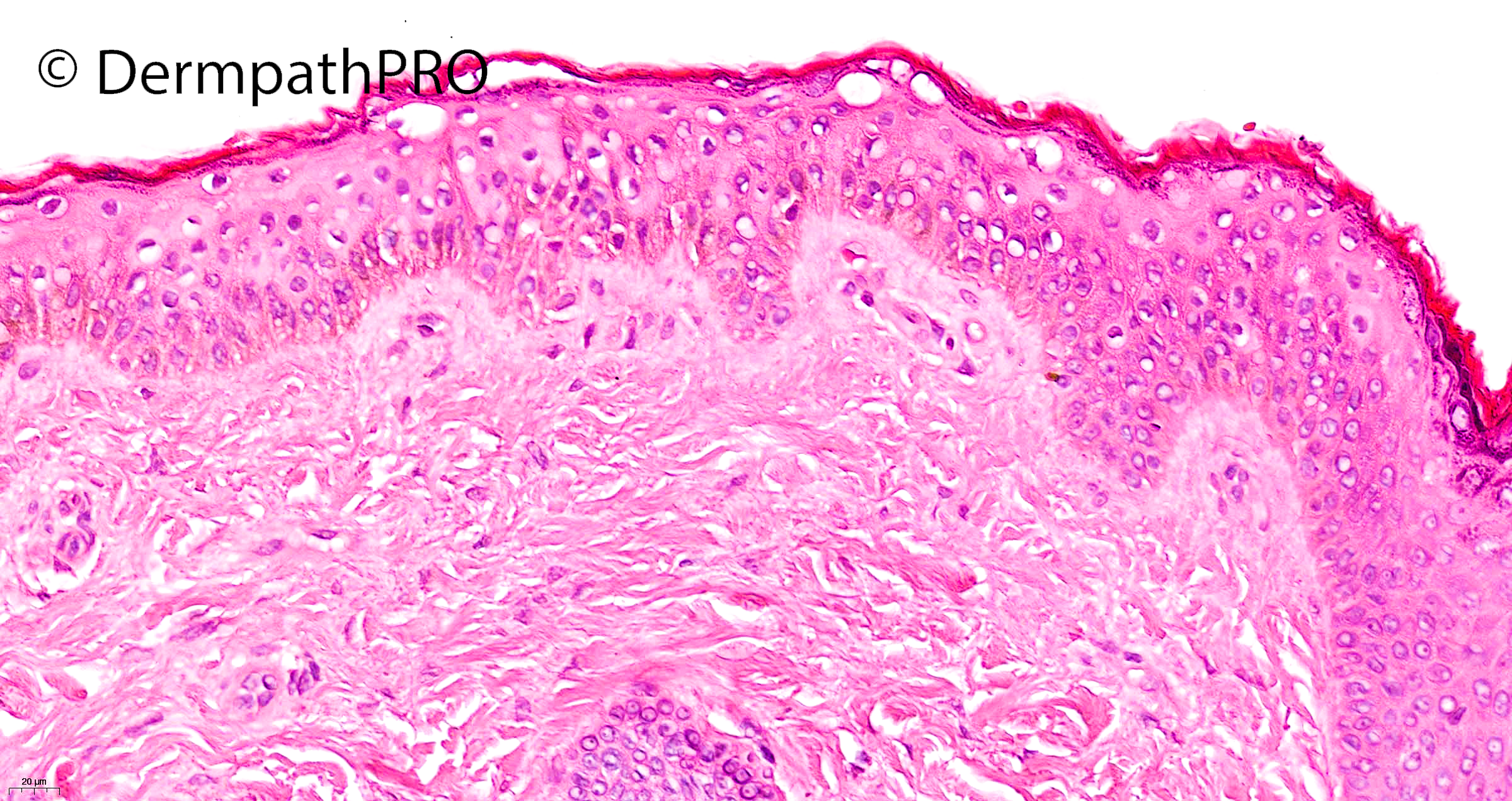
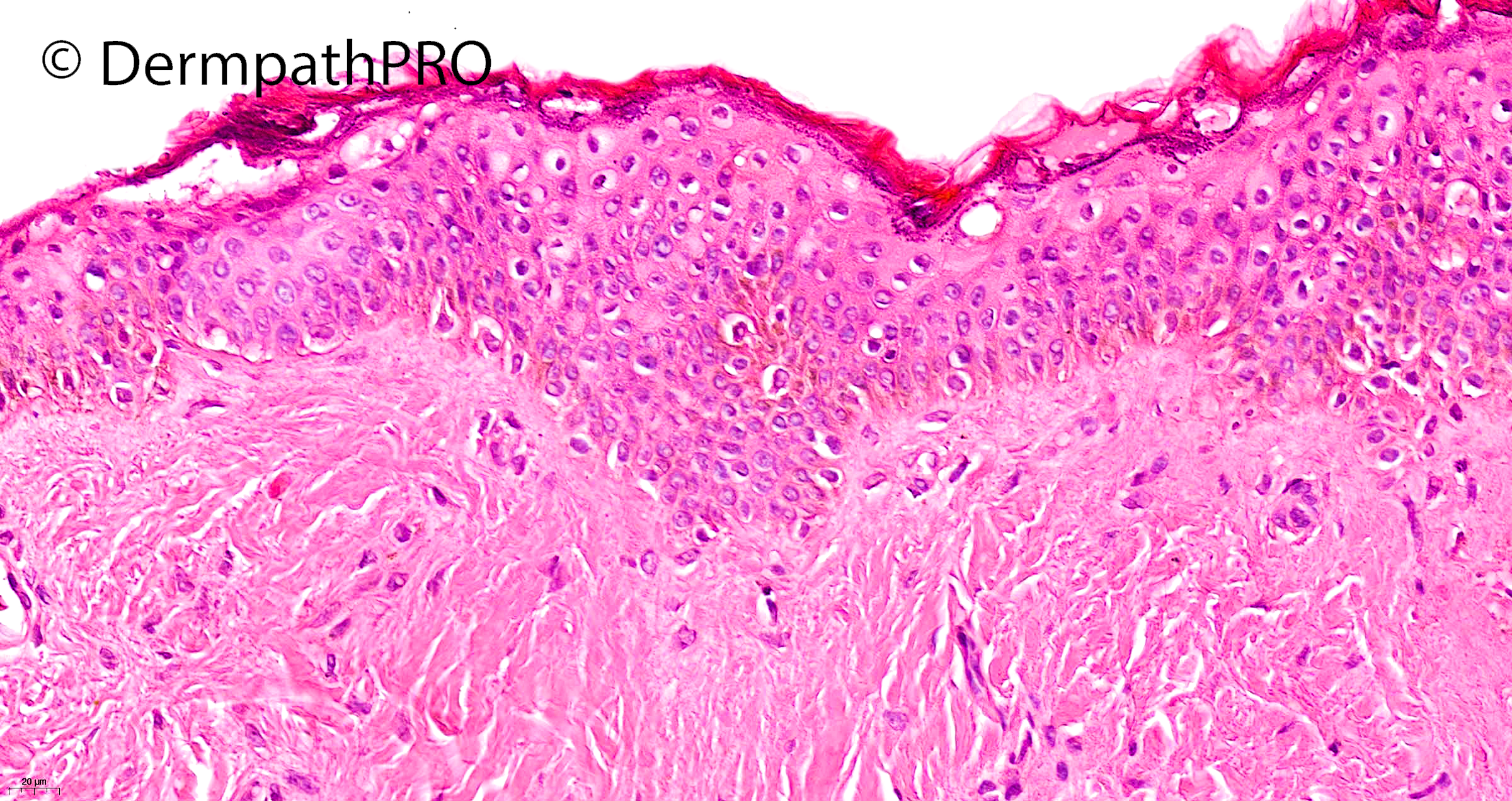
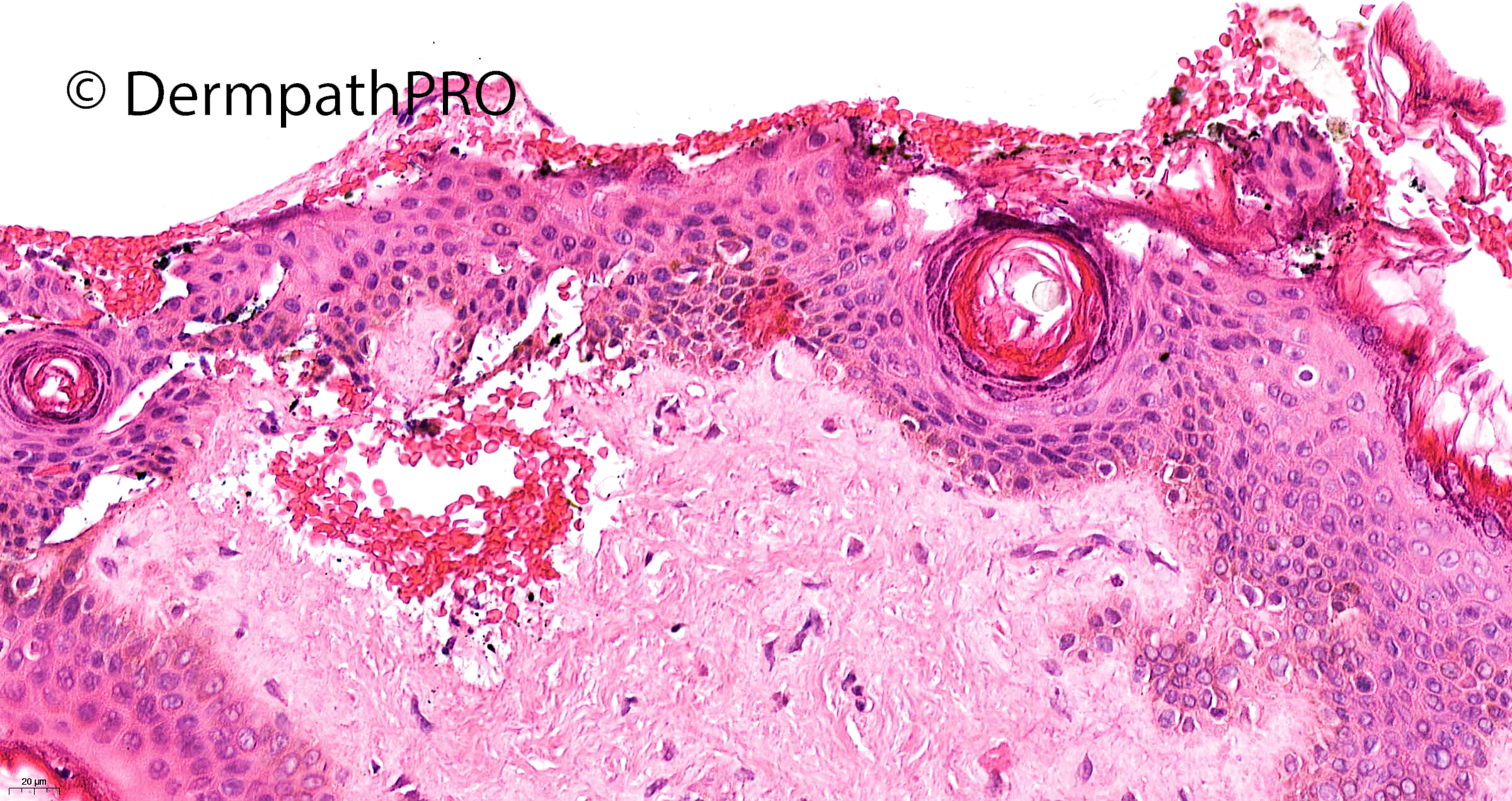
This is a top-heavy histology with upper epidermal pallor, and peri-nuclear vacuolation at all levels of the epidermis.  The stratum corneum is disrupted, absent in places and there is haemorrhage both on the surface and sub-epidermally.

I would tend to agree with Eman El-Nabarawy that this is some form of nutritional deficiency.  I was also considering dermatitis artefacta, common in girls of this age.  However, it sounds as though the symptoms are persistent rather than episodic which makes a nutritional deficiency more likely.  I look forward to hearing the clinical details.

Share this comment


Link to comment
share_externally

Krishnakumar subramanian

Posted

epidermal pallor mild and no inflammation in dermis. No mucin in dermis

need CPC to confirm pellagra

Share this comment


Link to comment
share_externally

Saleem Taibjee

Posted

Not too many comments this time, perhaps many are on summer holidays.....

This is a recent case. I am almost certain it is dermatitis artefacta. As Richard states, it is the clinicopathological correlation which is also crucial, and recognising that dermatitis artefacta is a real possibility in this demographic. Essentially, she is a well girl, and investigations have been normal.

With regards to the pathology, there is a suggestion of 'outside in' aetiology, with alteration of the upper levels of the epidermis, and relative sparing of the lower epidermis and follicular epithelium. There is more than just pallor, but actually a mummified appearance to the epidermis which is a useful clue to DA. I would like to acknowledge Wolfgang Weyers who brought this histological sign to my attention in one of his many excellent talks. There is additional upper epidermis vacuolisation and some red cell extravasation.

There aren't many publications on histopathology of DA, but here is one useful article: Joe EK et al. Cutis 1999;63:209-14

We should not underestimate the important role of the histopathologist/dermatopathologist in cases of DA, in suggesting or reinforcing the possible diagnosis to the clinician. I tend to report such cases as indicating to 'consider external factors/aetiology', to avoid a direct label or confrontation. This nonetheless really helps the clinician, especially in those paediatric/adolescent cases in which the parents may find it difficult to accept the diagnosis.

BW

Saleem

Share this comment


Link to comment
share_externally


Join the conversation

You can post now and register later. If you have an account, sign in now to post with your account.

Guest
Add a comment...

×   Pasted as rich text.   Paste as plain text instead

  Only 75 emoji are allowed.

×   Your link has been automatically embedded.   Display as a link instead

×   Your previous content has been restored.   Clear editor

×   You cannot paste images directly. Upload or insert images from URL.

Loading...

×
×
  • Create New...