Jump to content
In this section we have spot diagnoses posted on a daily basis since June 2010, now over 4000! You can review the archived cases and read the suggested diagnoses by users and the final comment by the contributors.
Case are uploaded each week day by 10 am UK time with the correct diagnosis will generally be posted at 8 pm UK time. Why not view the most recent spot diagnosis and proffer a diagnosis?

Case Number : Case 2900 - 18 August 2021 Posted By: Saleem Taibjee

Please read the clinical history and view the images by clicking on them before you proffer your diagnosis.
Submitted Date :
   (0 reviews)

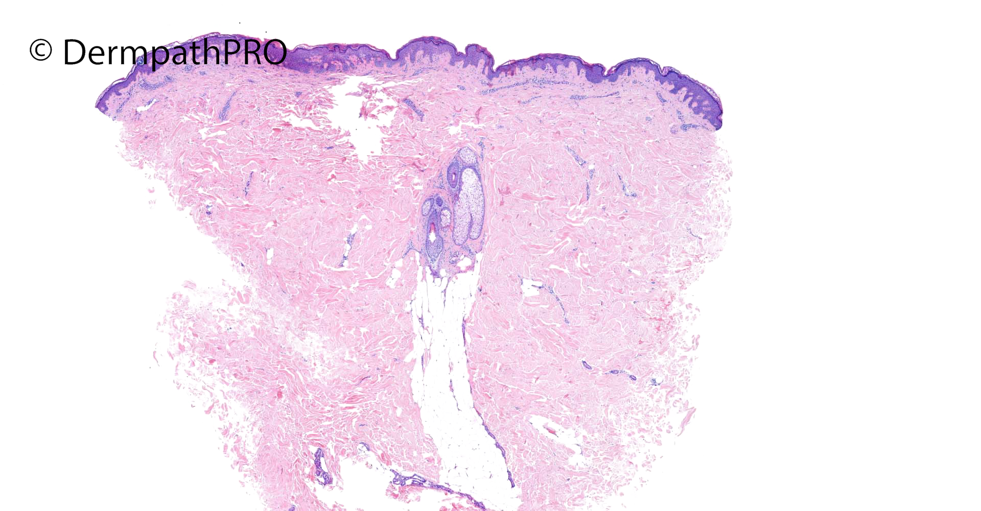
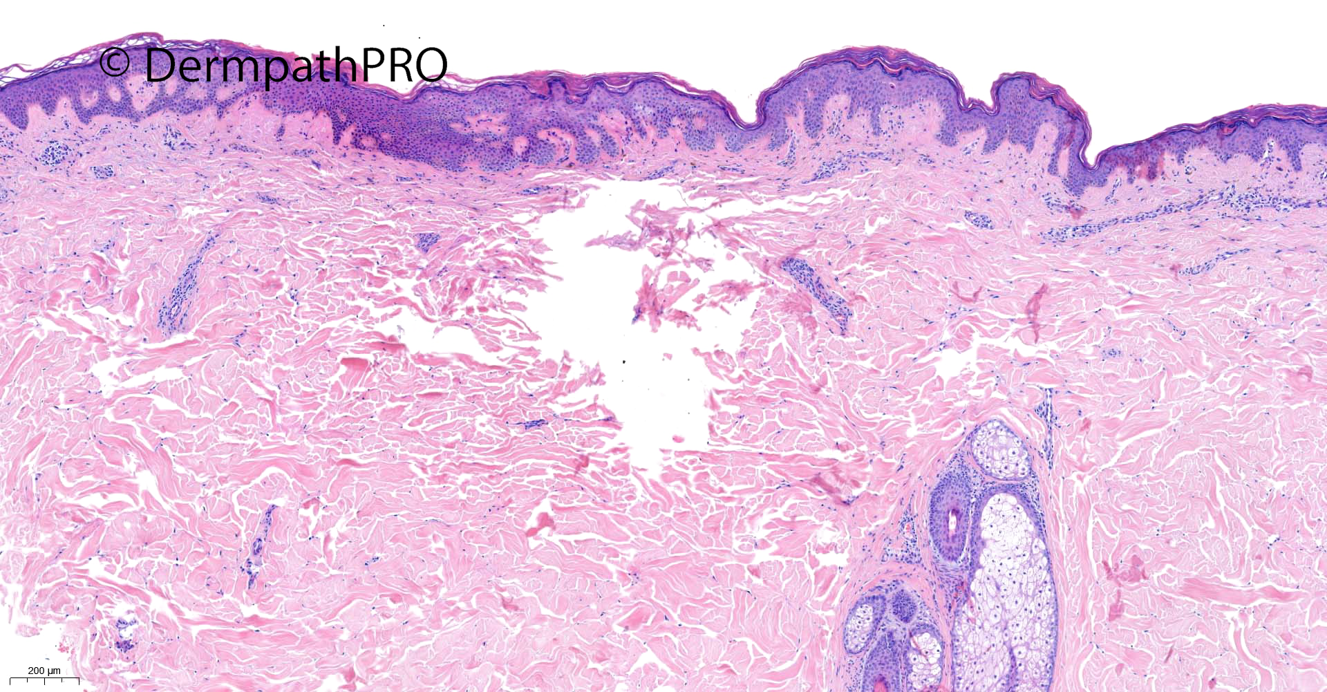
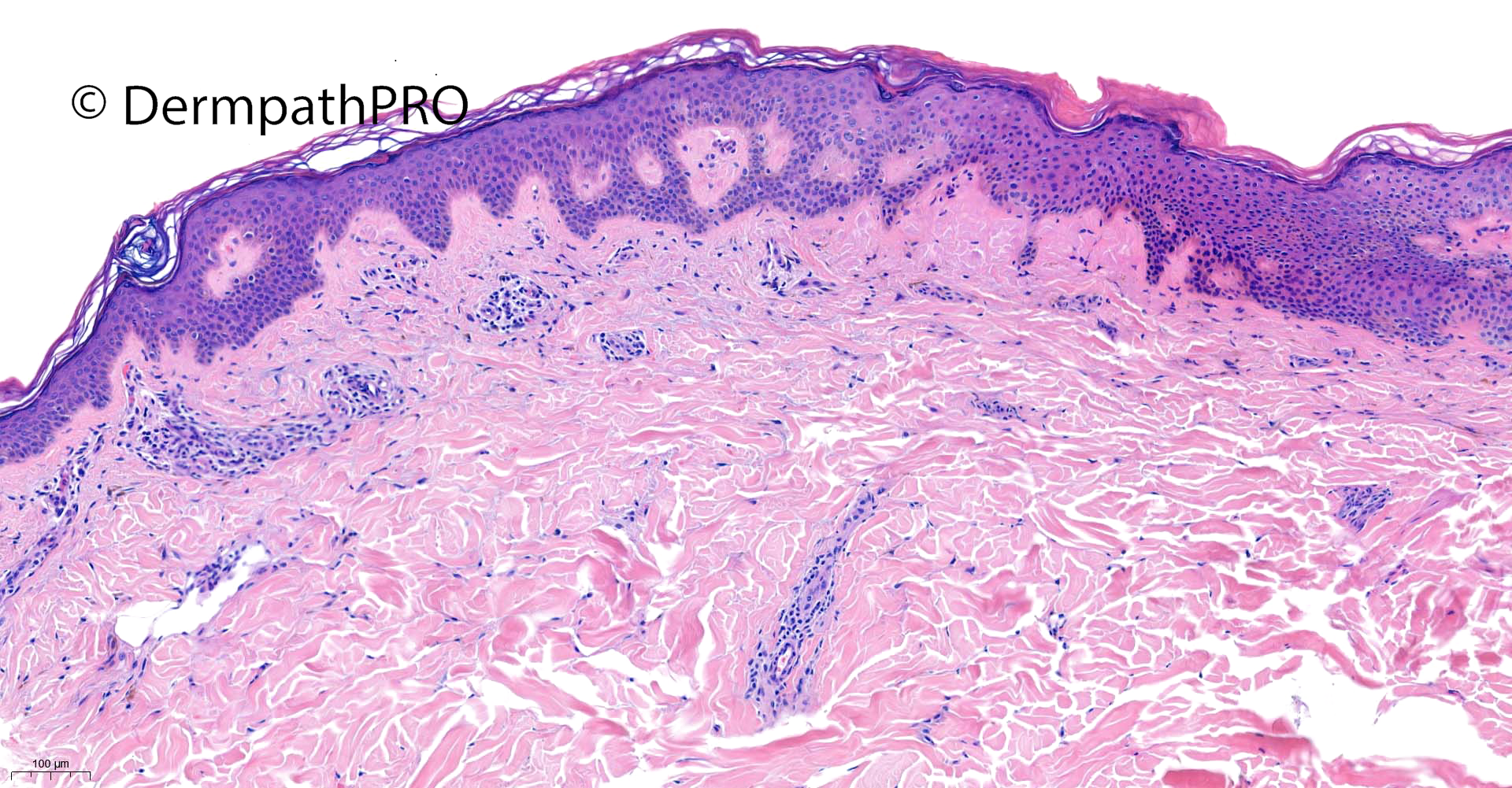
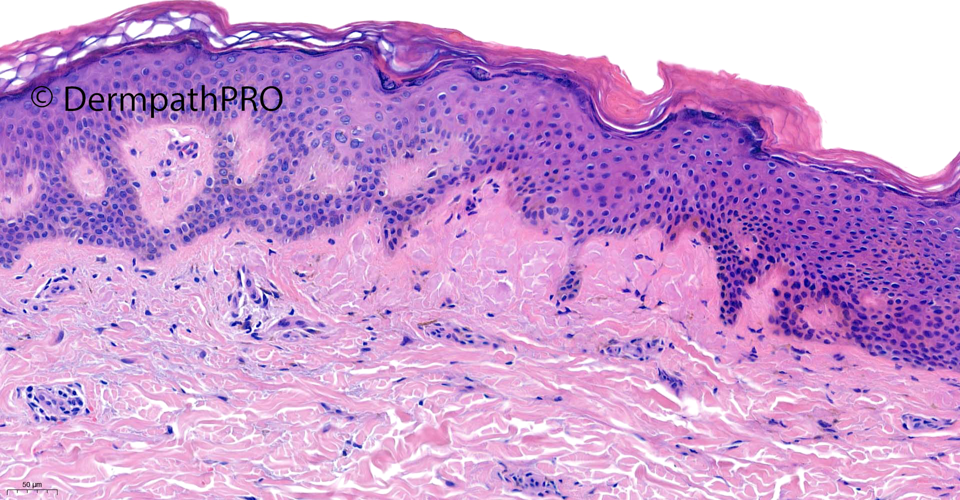
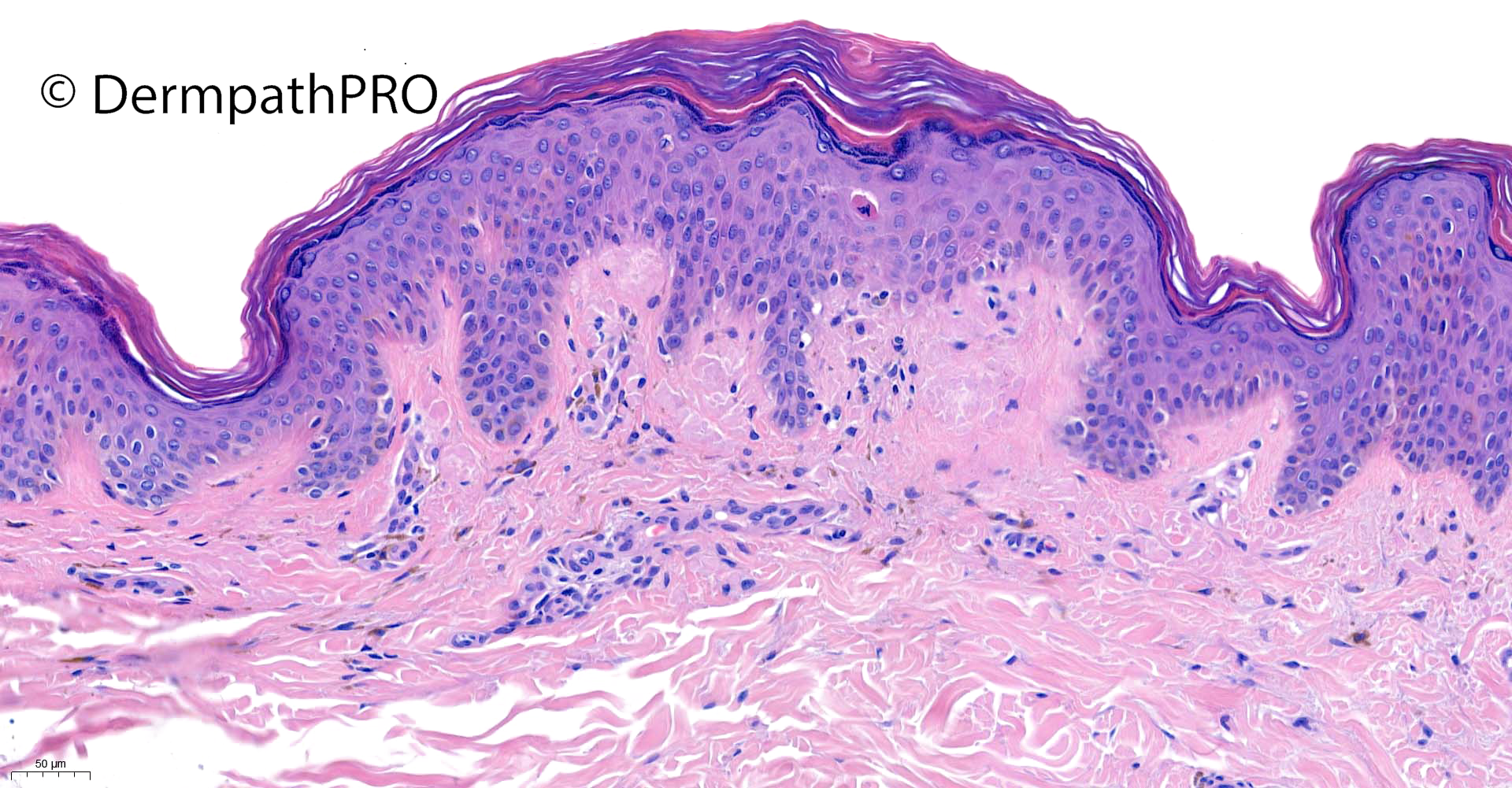
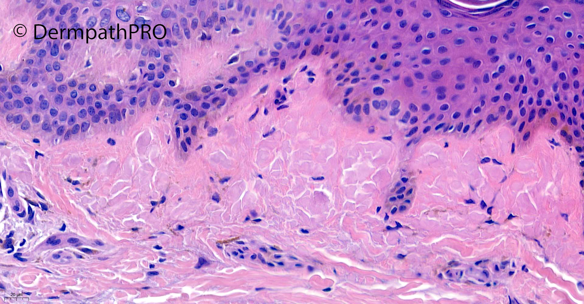
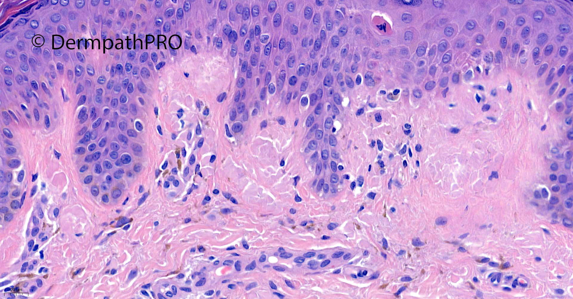
29F, incisional biopsy mid-back: Progressive pigmentation on mid-back


  Report Record

User Feedback


Krishnakumar subramanian

Posted

necrotic keratinocytes and pigment incontinence in dermis

could it be a drug rash

Share this comment


Link to comment
share_externally

Richard Logan

Posted

The low-power views suggest confluent and reticulated papillomatosis, with blunt-topped papillomatosis and a degree of epidermal invagination by orthokeratotic keratin.  However, at higher power there is pigmentary incontinence and a lot of amorphous bodies in the papillary dermis.  Therefore I think I would want to do amyloid stains, with amyloid being my preferred diagnosis.

I noticed the necrotic keratinocyte but there was only one that i could see, so the features above seem more pertinent.

Share this comment


Link to comment
share_externally

Krishnakumar subramanian

Posted

agree sir, could be lichen amyloid

Share this comment


Link to comment
share_externally

Amorphous hyaline material in the papillary dermis, favor amyloid.

Share this comment


Link to comment
share_externally

Fav amyloid, mid back usually macular amyloid 

Share this comment


Link to comment
share_externally

Saleem Taibjee

Posted

Hi. Yes well done, this is a recent case of macular (cutaneous) amyloid. As Volha mentions, this is the subtype of cutaneous amyloid typically seen on the back, and shows a characteristic rippled macular  pigmentation pattern clinically. It is another example of 'invisible' dermatosis (or easily missed pathology).

It is also worth noting that keratin stains are usually more helpful than traditional amyloid stains such as Congo red in such cases. I include the relevant stains below. Interestingly, the choice of keratin stain does seem to matter, some cases seem to stain better for one pankeratin (e.g. MNF116, AE1/AE3, 34BE12) than another. But I am yet to make a firm conclusion as to which is the best, and might suggest requesting a couple of IHC stains in such cases. For example, AE1/AE3 was rather weak in this case compared to MNF116 and 34BE12.

BW
Saleem

34579_20.0x AE1AE3 labelled.jpg

34579_20.0x 34BE12 labelled.jpg

34579_20.0x MNF116b labelled.jpg

34579_20.0x Congo red labelled.jpg


34579_20.0x AE1AE3 labelled.jpg

Share this comment


Link to comment
share_externally


Join the conversation

You can post now and register later. If you have an account, sign in now to post with your account.

Guest
Add a comment...

×   Pasted as rich text.   Paste as plain text instead

  Only 75 emoji are allowed.

×   Your link has been automatically embedded.   Display as a link instead

×   Your previous content has been restored.   Clear editor

×   You cannot paste images directly. Upload or insert images from URL.

Loading...

×
×
  • Create New...