Diagnostic Pearls : Case 2901 - 19 August 2021
F18. Cheek. IDN?
Dr. Richard Carr
Posted 18/08/21
Posted 18/08/21
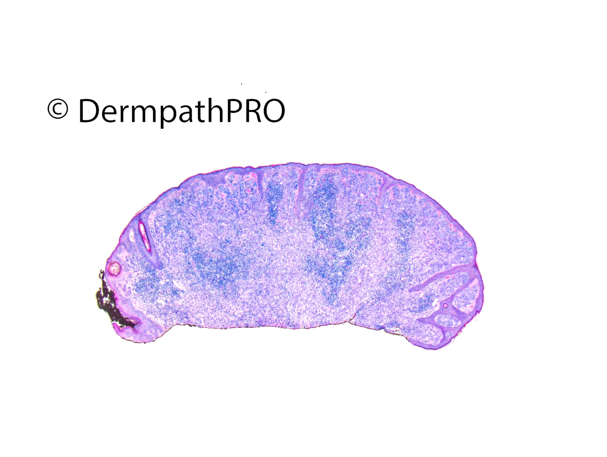
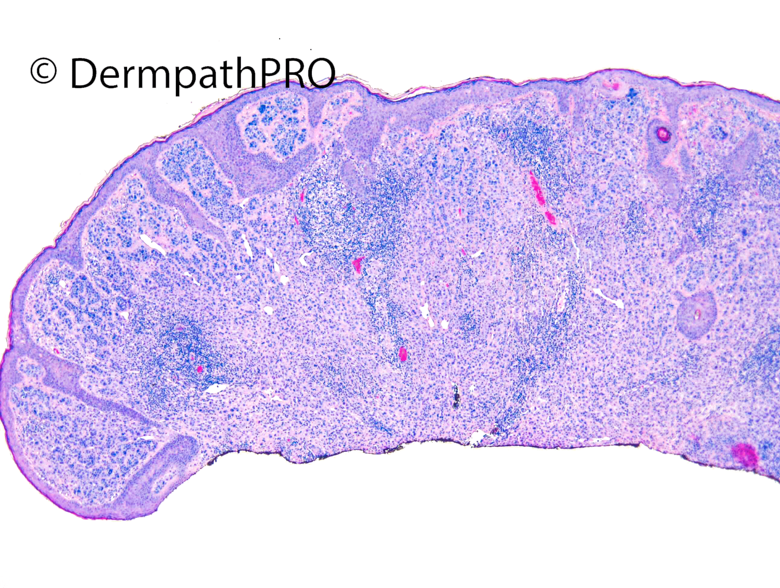

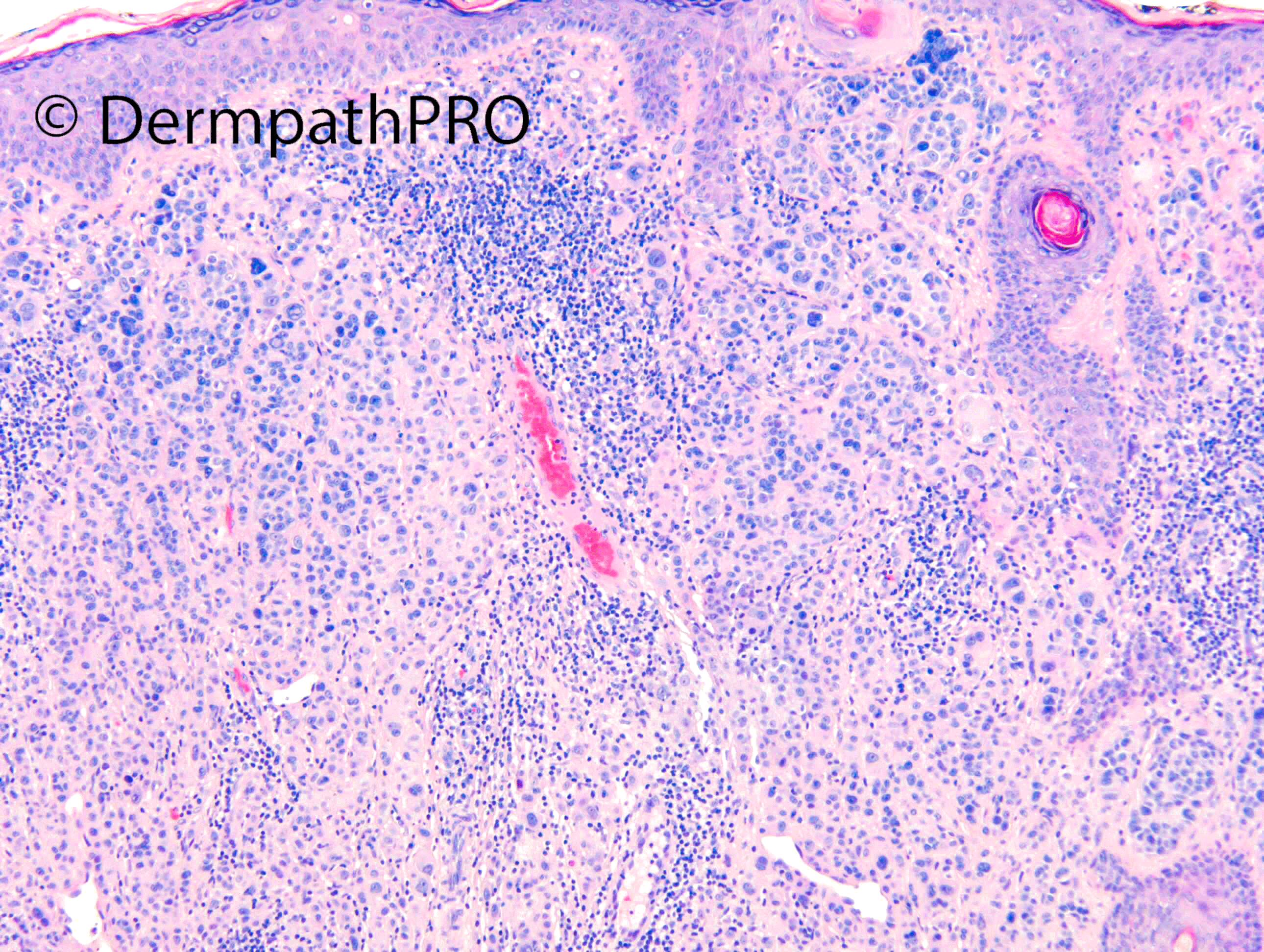
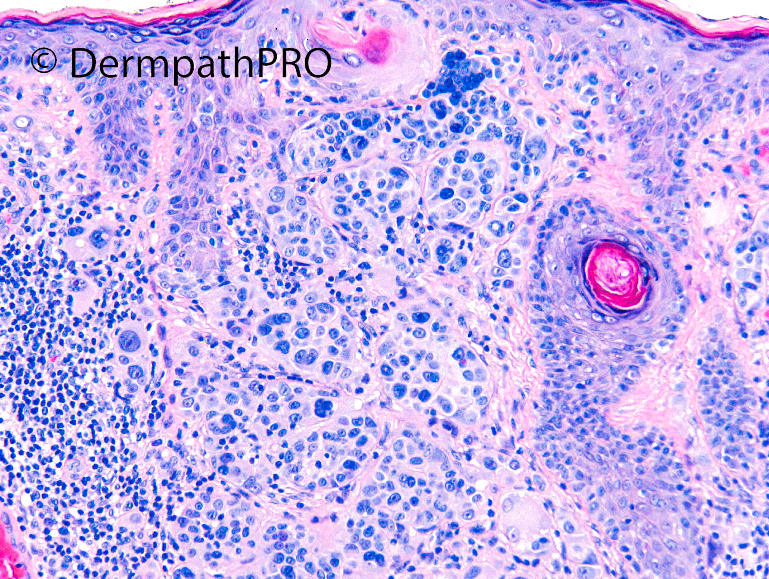
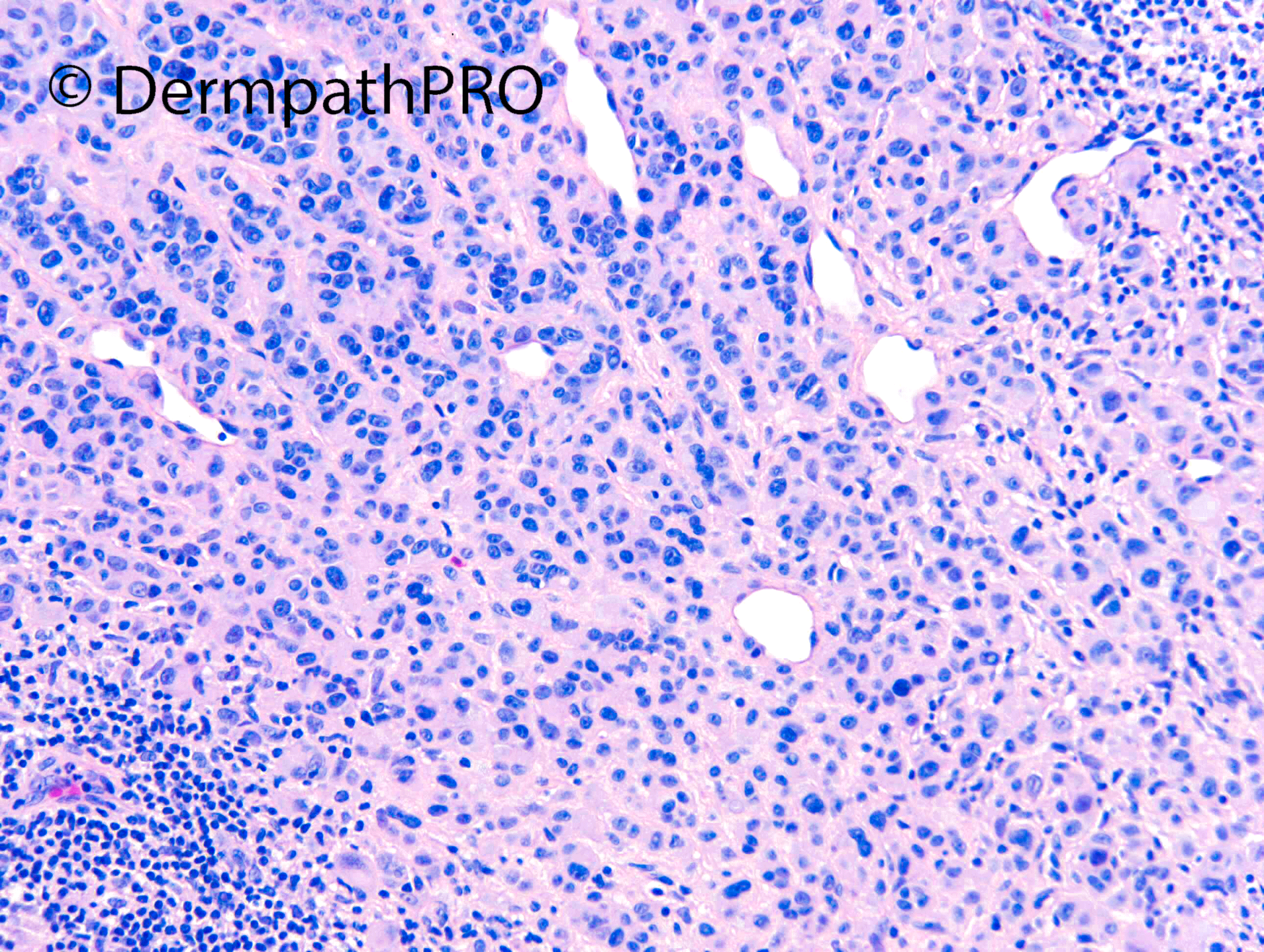
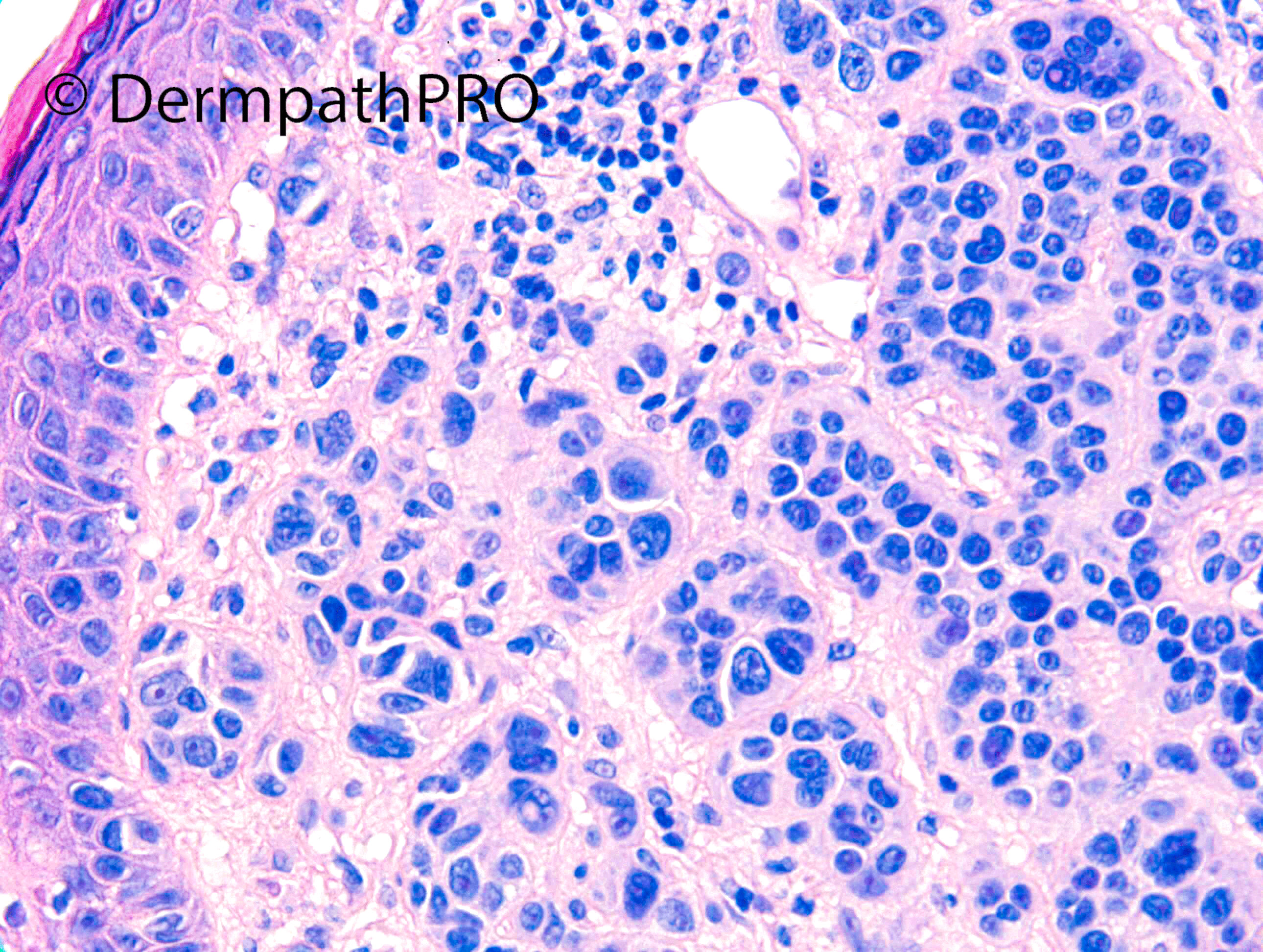
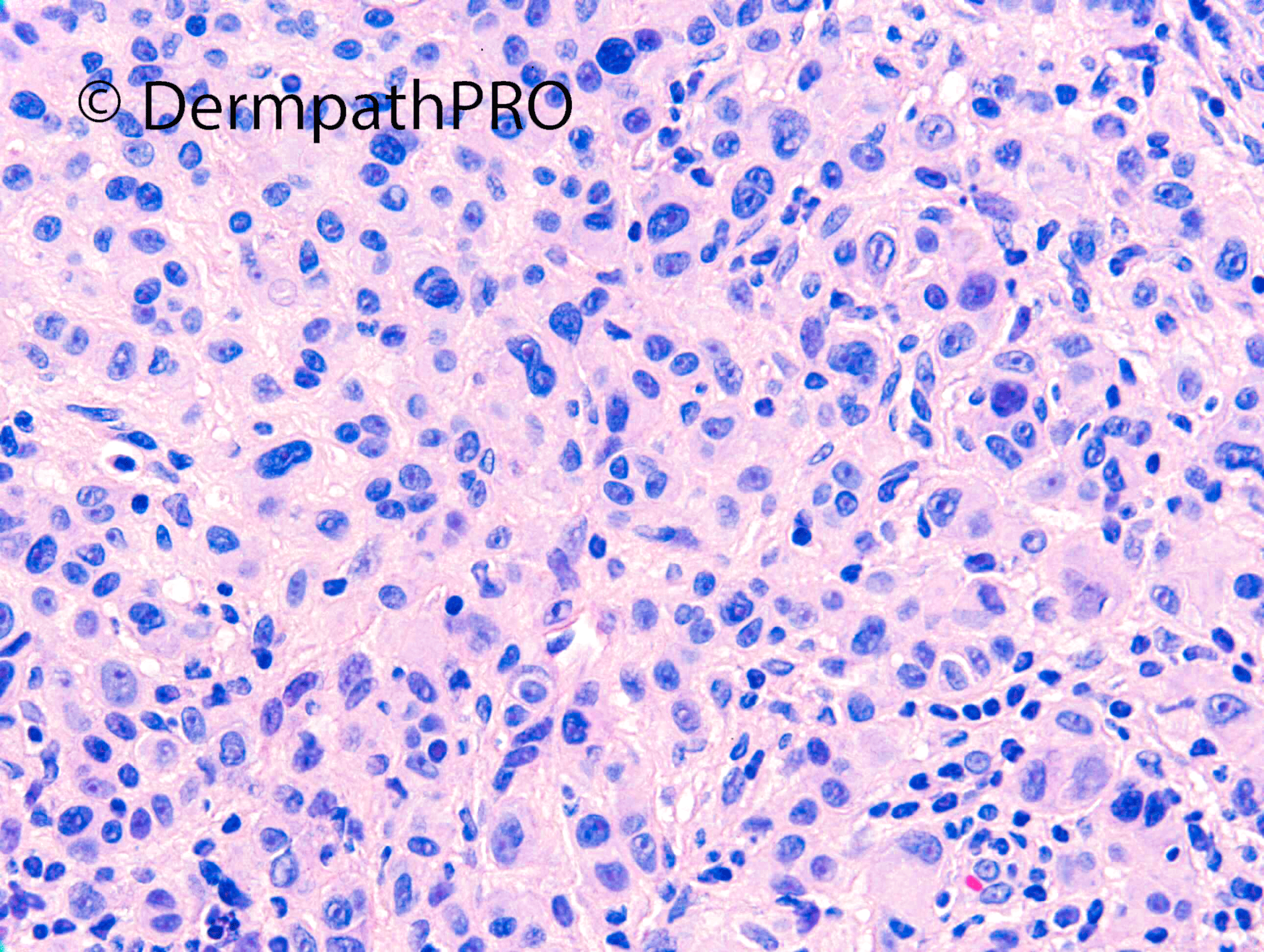
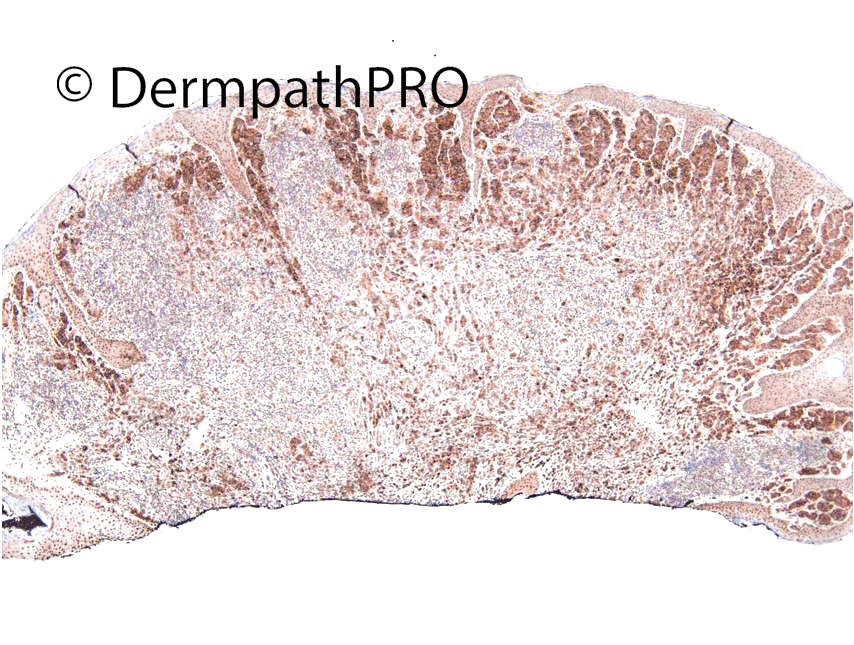
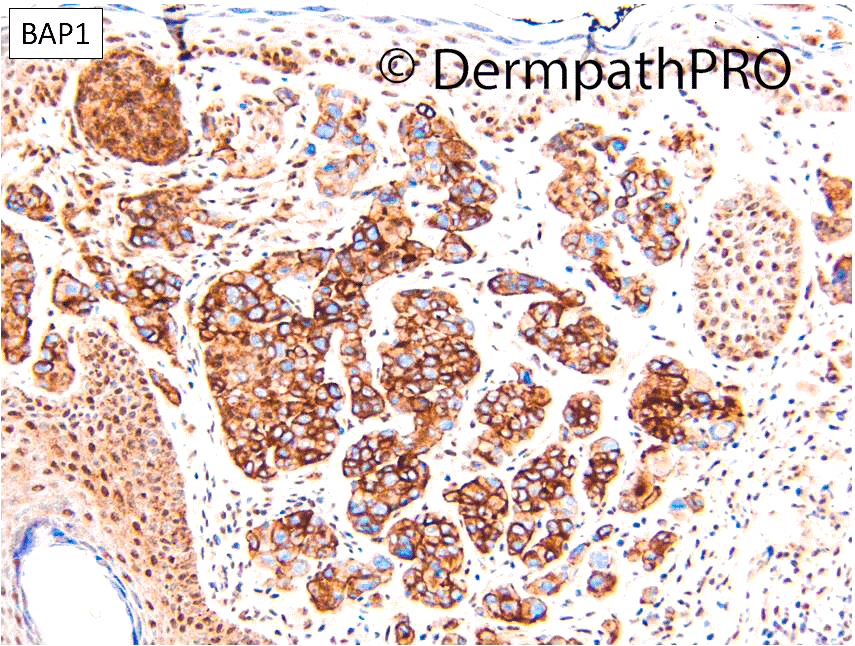
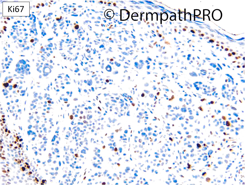
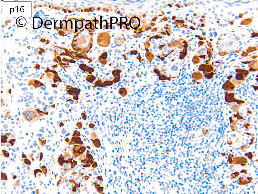
F18. Cheek. IDN?
Join the conversation
You can post now and register later. If you have an account, sign in now to post with your account.