Jump to content
In this section we have spot diagnoses posted on a daily basis since June 2010, now over 4000! You can review the archived cases and read the suggested diagnoses by users and the final comment by the contributors.
Case are uploaded each week day by 10 am UK time with the correct diagnosis will generally be posted at 8 pm UK time. Why not view the most recent spot diagnosis and proffer a diagnosis?

Case Number : Case 2904 - 24 August 2021 Posted By: Saleem Taibjee

Please read the clinical history and view the images by clicking on them before you proffer your diagnosis.
Submitted Date :
   (0 reviews)

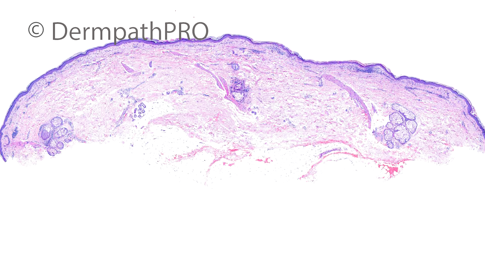
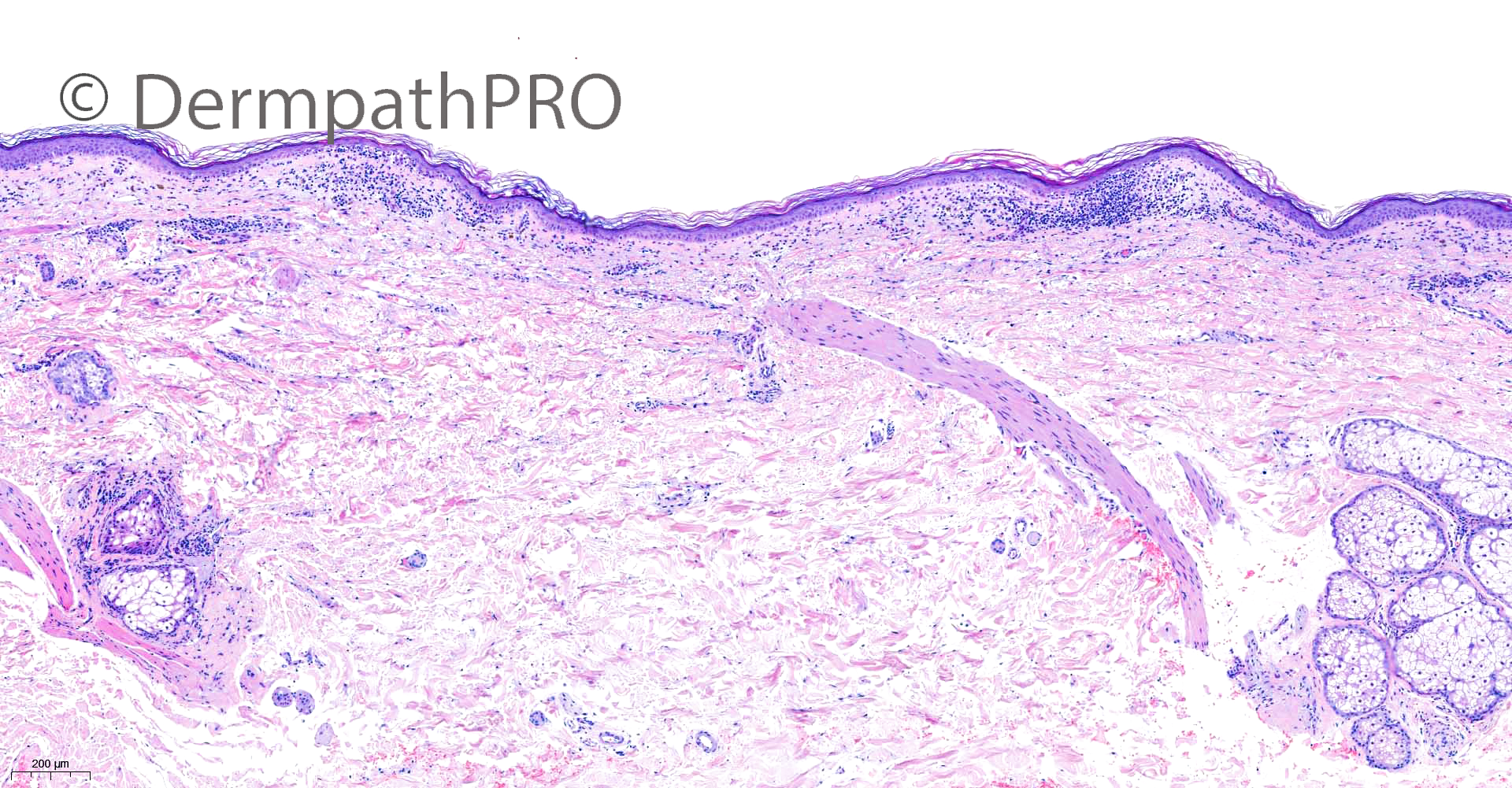
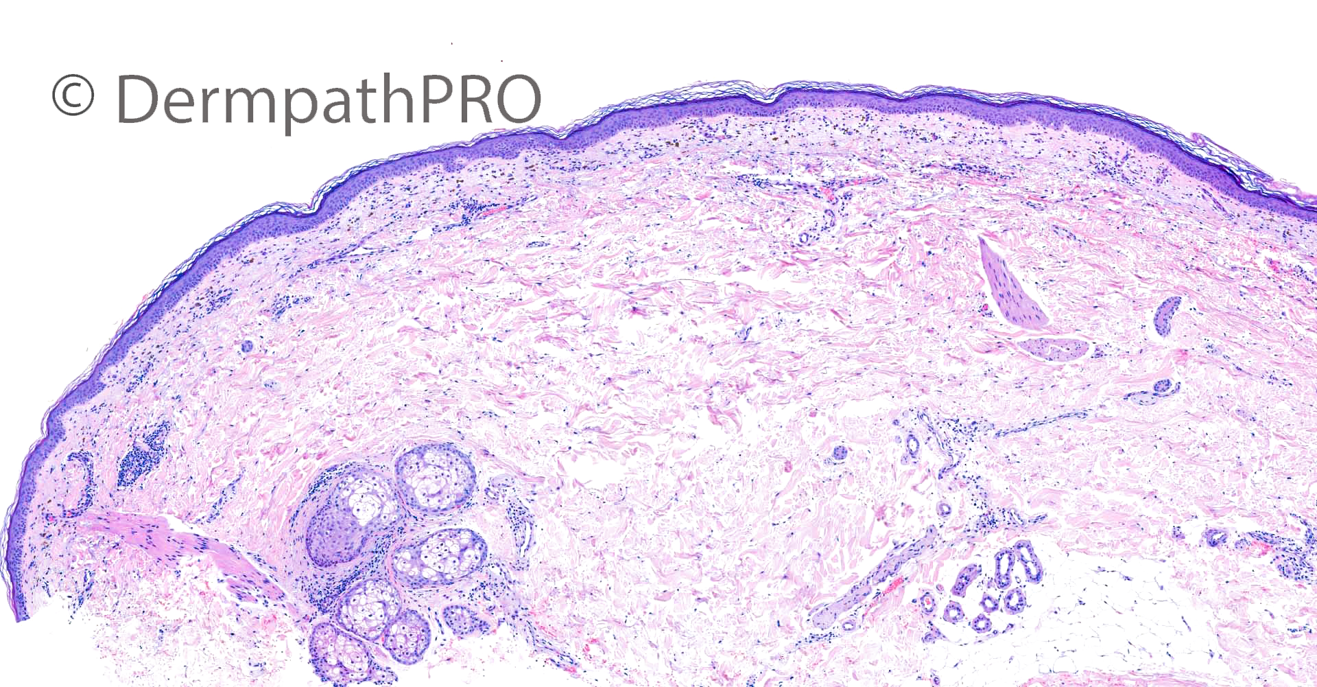
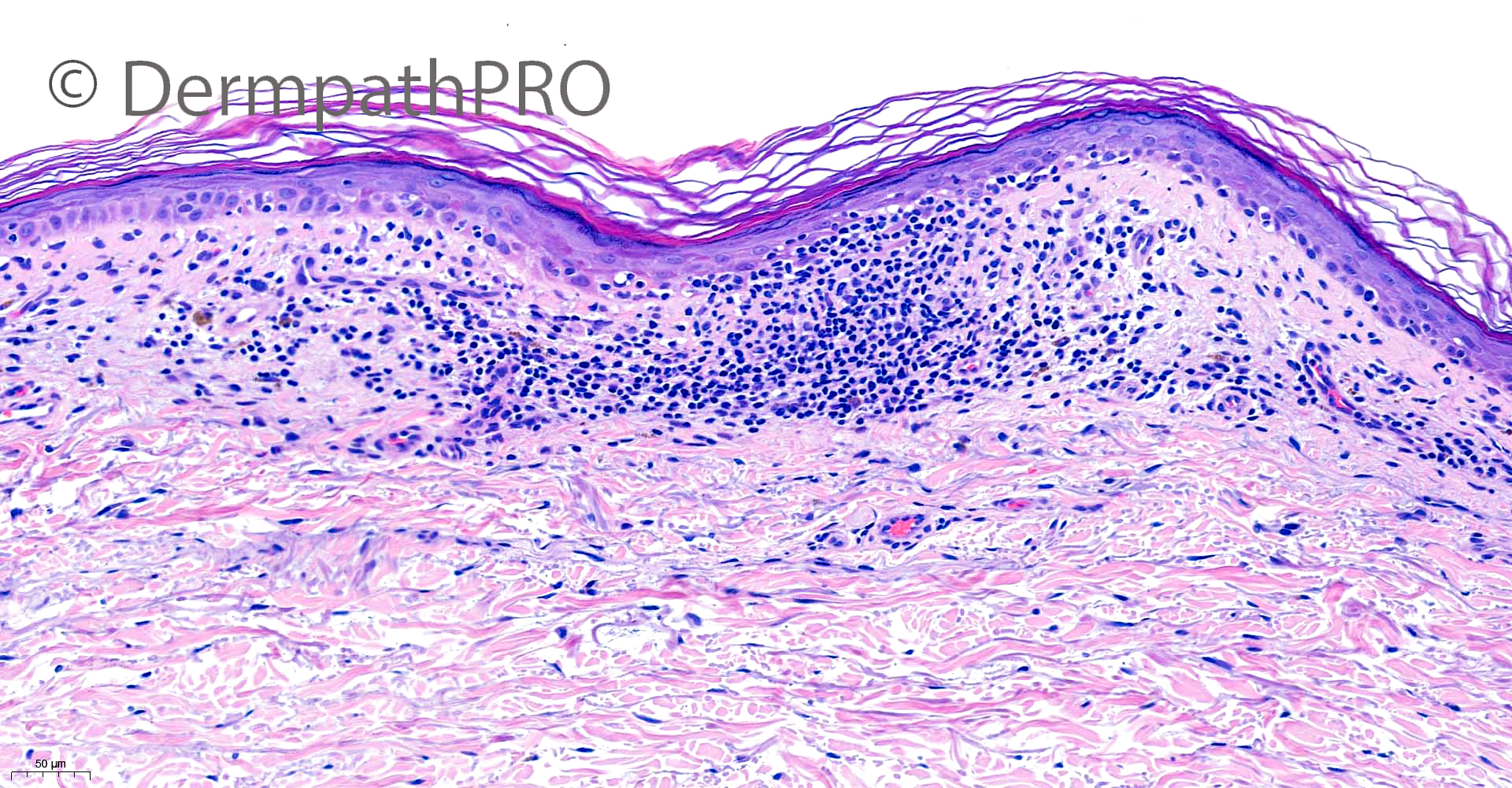
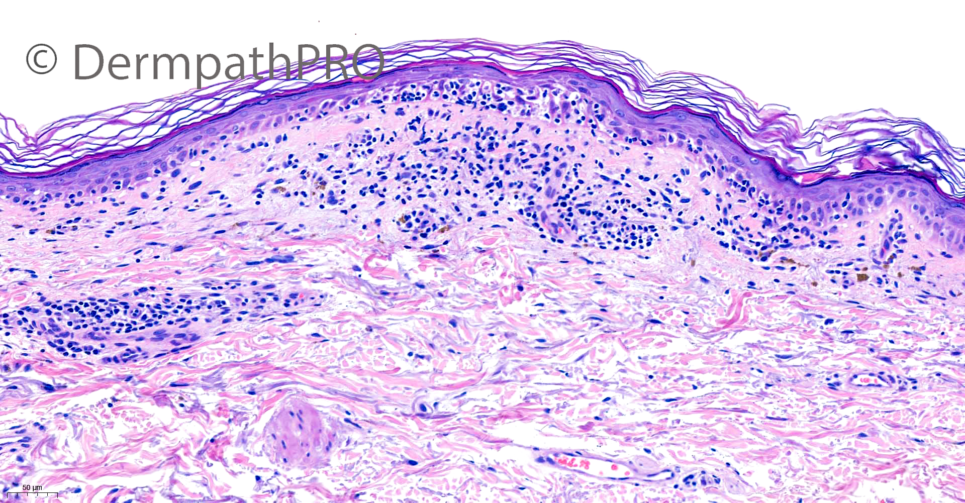
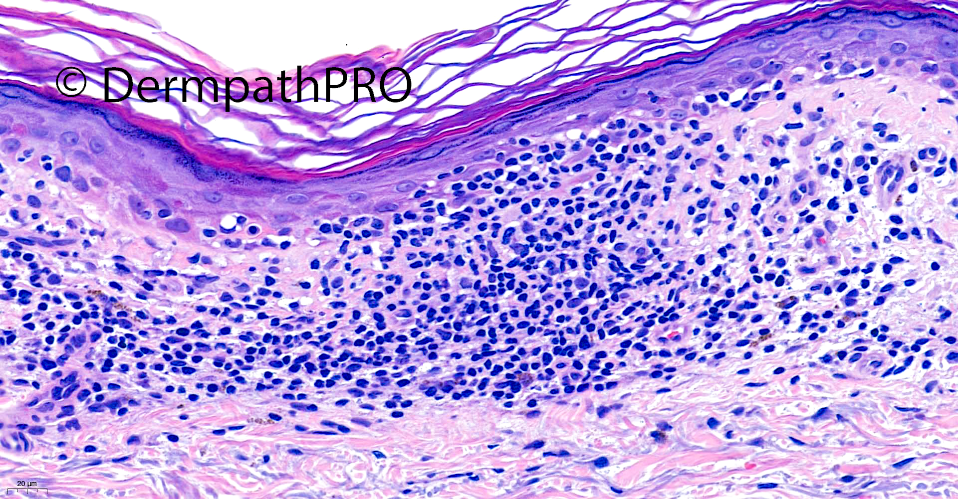
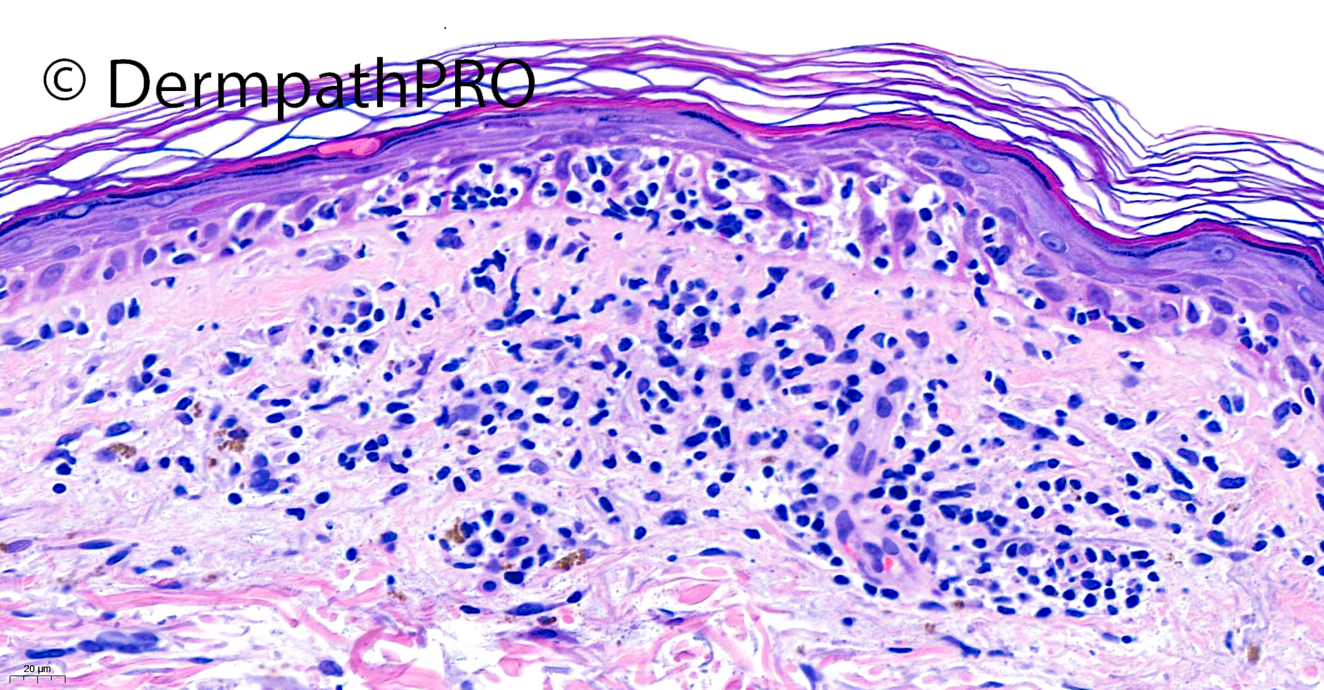
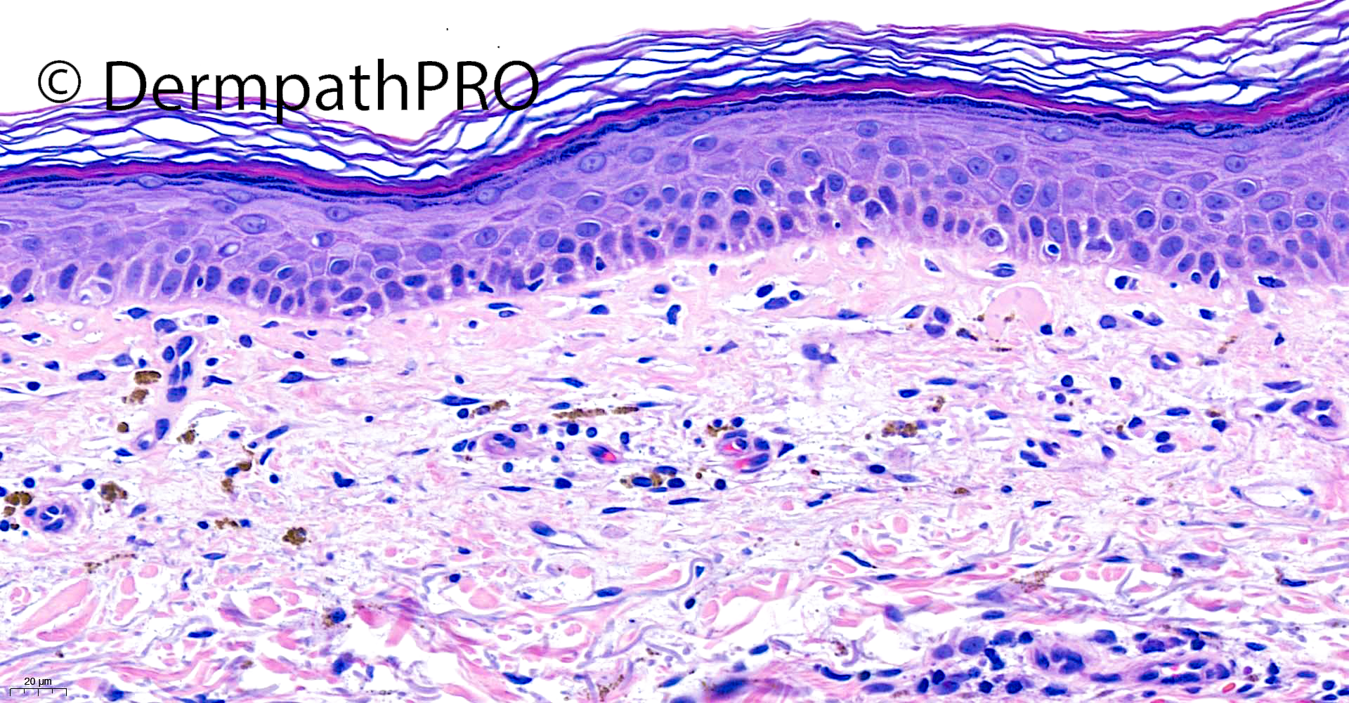
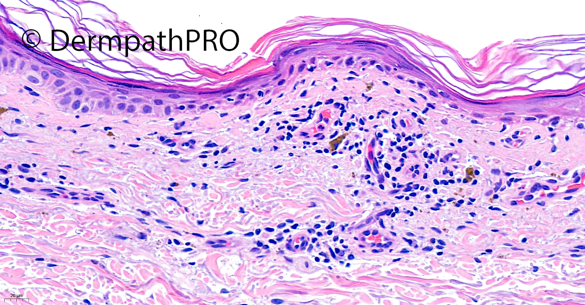
59F Incisional biopsy left breast ?seborrhoeic keratosis, exclude melanoma


  Report Record

User Feedback


Anil Patki

Posted

Looks like unilesional MF. Needs IHC for confirmation. 

Share this comment


Link to comment
share_externally

Krishnakumar subramanian

Posted

some sort of epidermal tropism is seen. IHC and some more clinical information such as is it single patch or any patches elsewhere.

Share this comment


Link to comment
share_externally

Meenakshi Batrani

Posted

There is epidermotropism but in view of clinical I would consider lichen planus like keratosis which has an atrophic variant and may also shows epidermotropism or pseudo- MF like histological features. 

Benign lichenoid keratosis with histologic features of mycosis fungoides: Clinicopathologic description of a clinically significant histologic pattern. J Cutan Pathol. 29:291-294 2002.

Share this comment


Link to comment
share_externally

Lichenoid keratosis with melanin incontinence, either atrophic or regressing.

Share this comment


Link to comment
share_externally

Agree with some kind of BLK, or bette yet LPL-AK, but would see levels, to rule out a superficial type of BCC...

Share this comment


Link to comment
share_externally

Saleem Taibjee

Posted

Yes, well done, I also favoured lichenoid keratosis with unusually prominent epidermotropism / pseudolymphomatous features. I did not request immunostains. Arguably I could have requested additional levels, but did not do so given that this is a large and generous biopsy and could see a suggestion of pre-existing solar lentigo in areas, but I would have done so if a smaller biopsy. Of course, this case is absolutely dependent on clinical correlation. In this instance, the clinical information provided seems to fit well for this, and does not at all suggest mycosis fungoides (although unilesional MF can occur uncommonly). Thanks to Meenakshi for pointing out this useful article from Journal of Cutaneous Pathology. Of course, there are other situations in which we might see prominent epidermotropism such as lichen sclerosus (I have come across one case of epidermotropic lichen sclerosus which was signed out by another pathologist as mycosis fungoides). Even in those cases which are likely to be lymphoma, we should not simply sign out as mycosis fungoides but emphasise the need for clinicopathological correlation and MDT discussion given the various other lymphomas which may show epidermotropism including gamma-delta lymphoma, primary cutaneous aggressive epidermotropic CD8+ lymphoma, etc.

I did draw attention to the epidermotropism in my histology report, shown below. BW, Saleem

"The epidermis shows areas with elongated rete ridges and basal epidermal pigmentation. This contrasts with atrophic areas with associated lichenoid damage and colloid bodies. There is a moderately dense band-like and interface lymphohistiocytic infiltrate with superficial fibrosis and pigment incontinence. There are foci of lymphocyte basal epidermotropism/exocytosis and even a hint of mild lymphoid atypia.

The features are in keeping with solar lentigo with lichenoid reaction/lichenoid keratosis. No melanocytic proliferation seen. The solitary nature of the lesion and clinical context with background changes suggestive of solar lentigo would indicate a 'pseudolymphomatoid' lichenoid keratosis pattern, rather than cutaneous T-cell lymphoma, but clinical correlation is required."

Share this comment


Link to comment
share_externally

Krishnakumar subramanian

Posted

thanks a lot for this case sir

Share this comment


Link to comment
share_externally


Join the conversation

You can post now and register later. If you have an account, sign in now to post with your account.

Guest
Add a comment...

×   Pasted as rich text.   Paste as plain text instead

  Only 75 emoji are allowed.

×   Your link has been automatically embedded.   Display as a link instead

×   Your previous content has been restored.   Clear editor

×   You cannot paste images directly. Upload or insert images from URL.

Loading...

×
×
  • Create New...