Jump to content
In this section we have spot diagnoses posted on a daily basis since June 2010, now over 4000! You can review the archived cases and read the suggested diagnoses by users and the final comment by the contributors.
Case are uploaded each week day by 10 am UK time with the correct diagnosis will generally be posted at 8 pm UK time. Why not view the most recent spot diagnosis and proffer a diagnosis?

Case Number : Case 2976 - 2 December 2021 Posted By: Saleem Taibjee

Please read the clinical history and view the images by clicking on them before you proffer your diagnosis.
Submitted Date :
   (0 reviews)

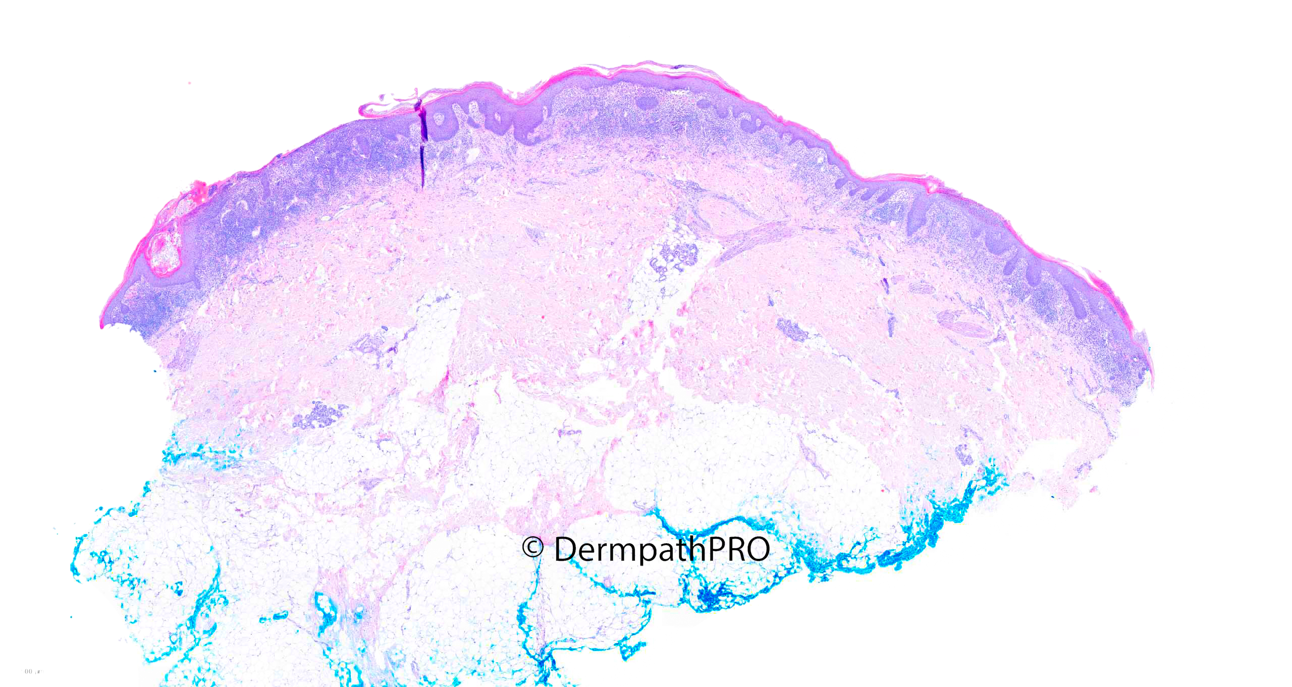
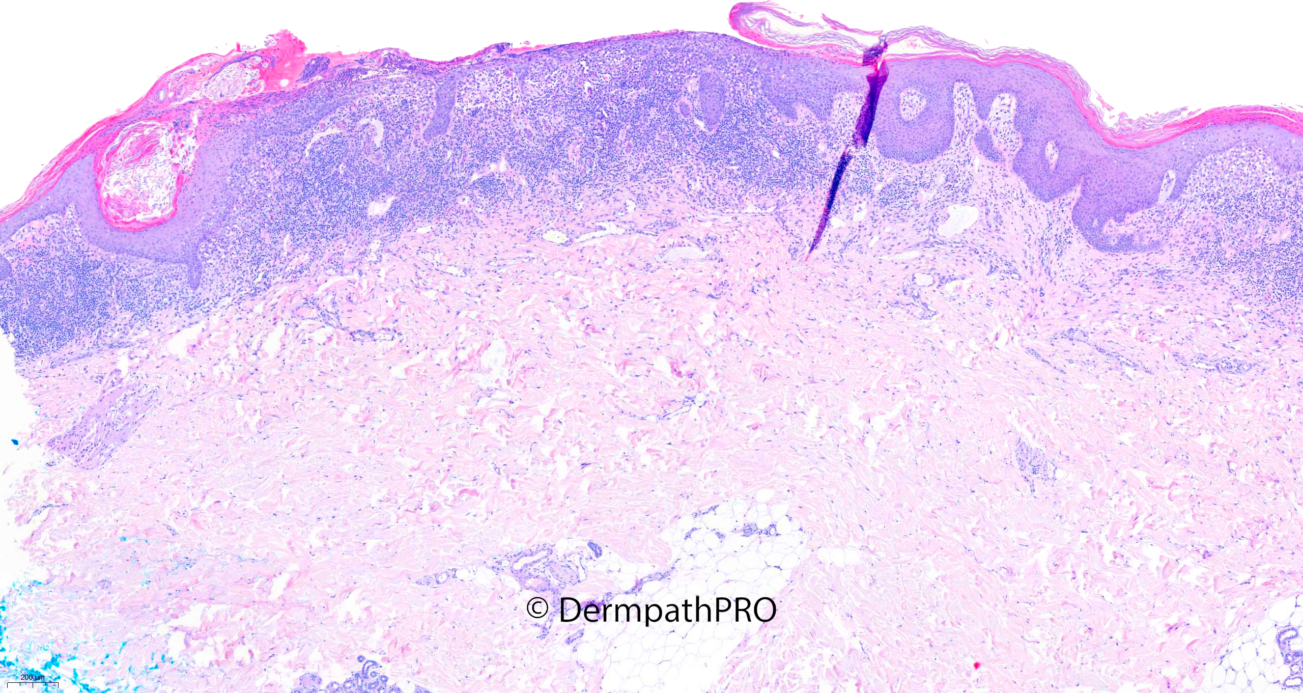
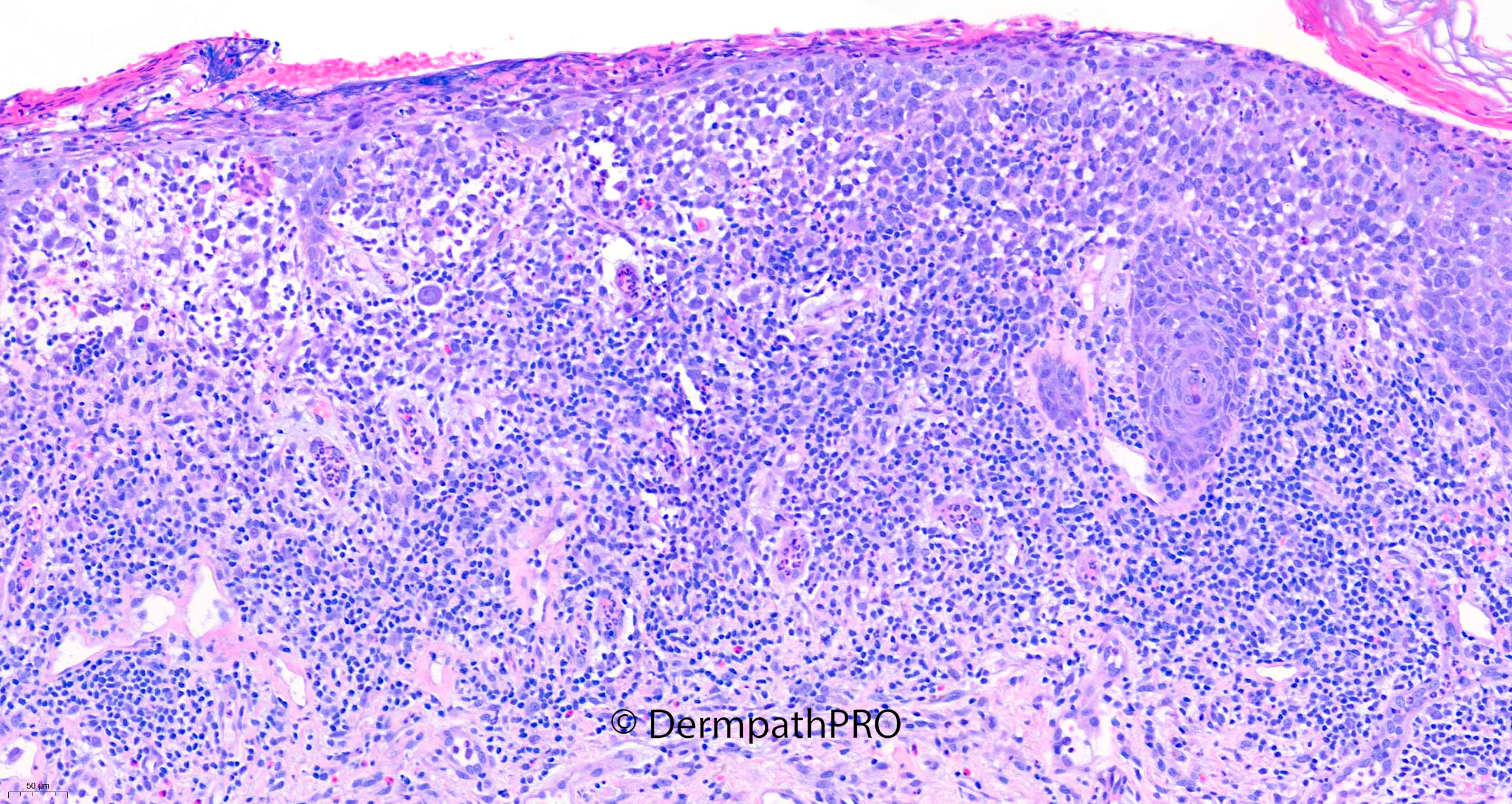
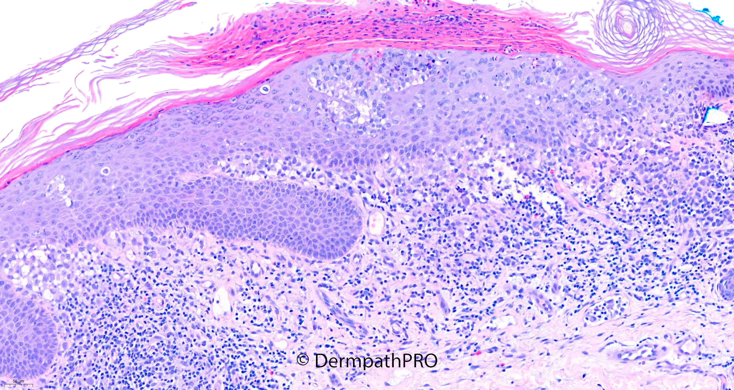
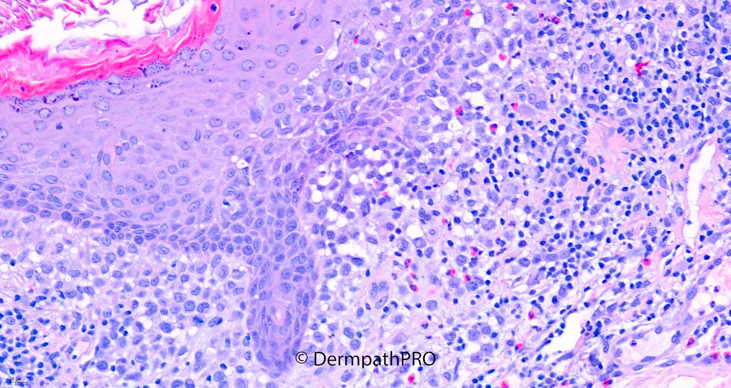
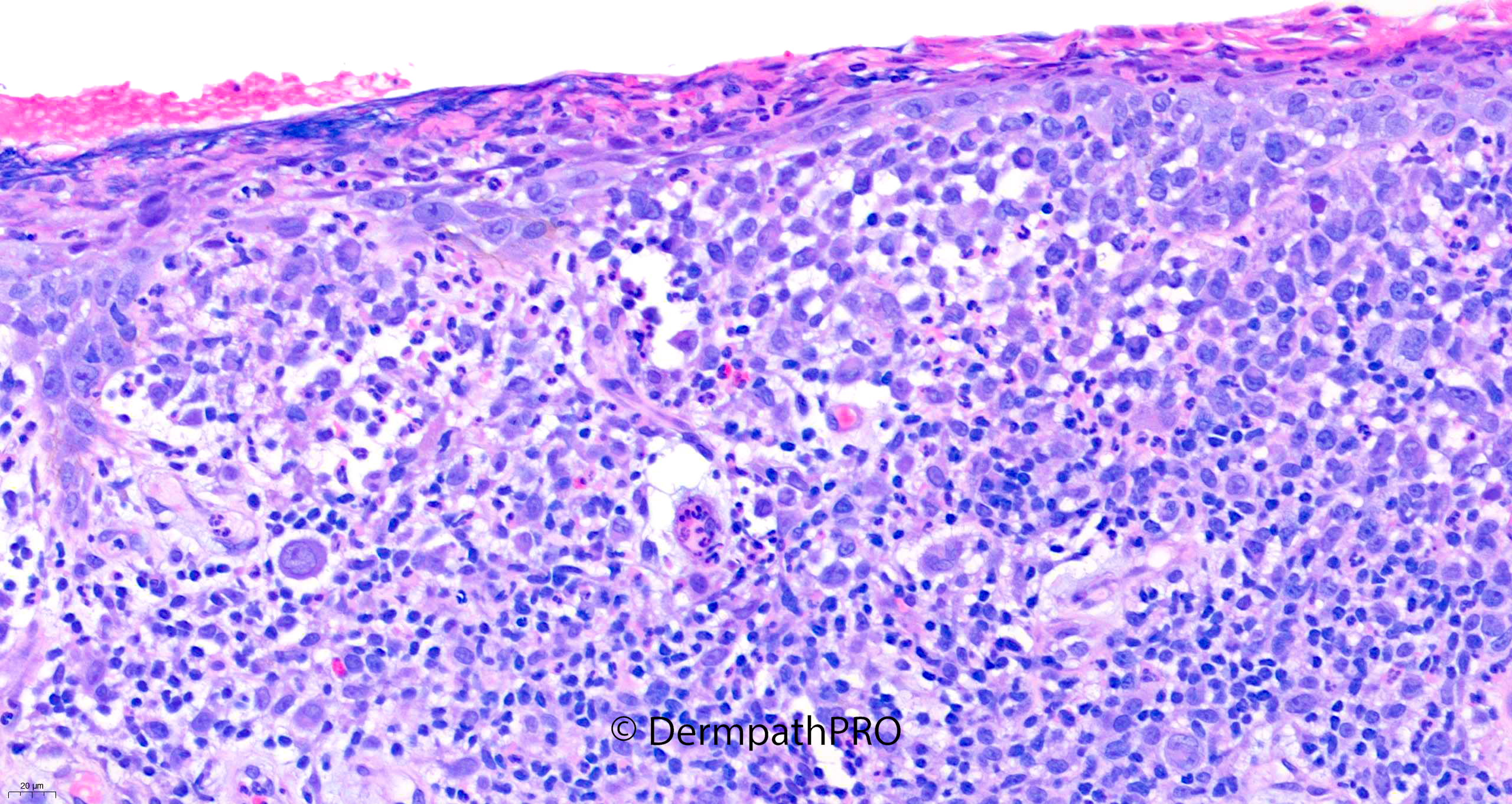
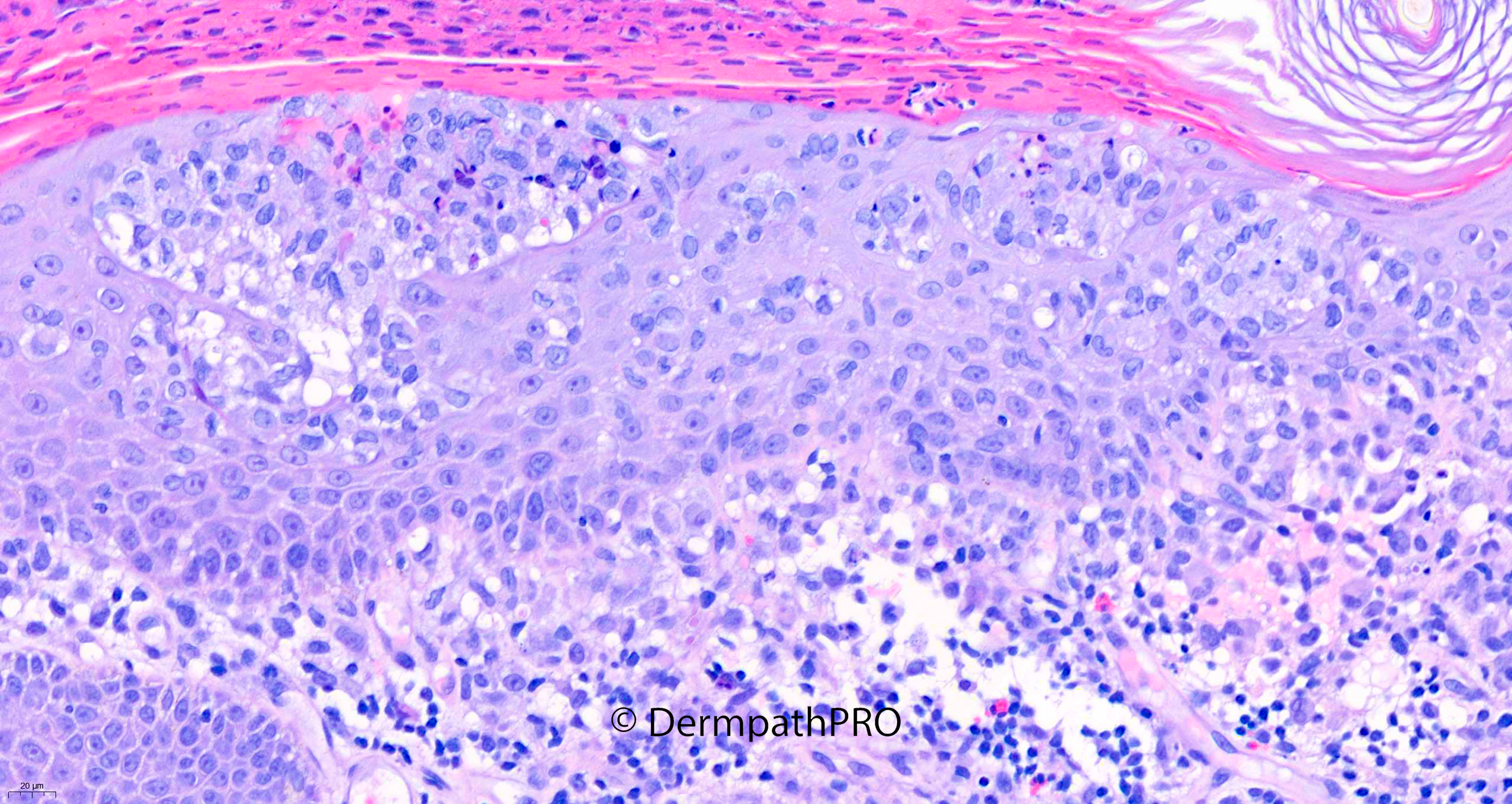
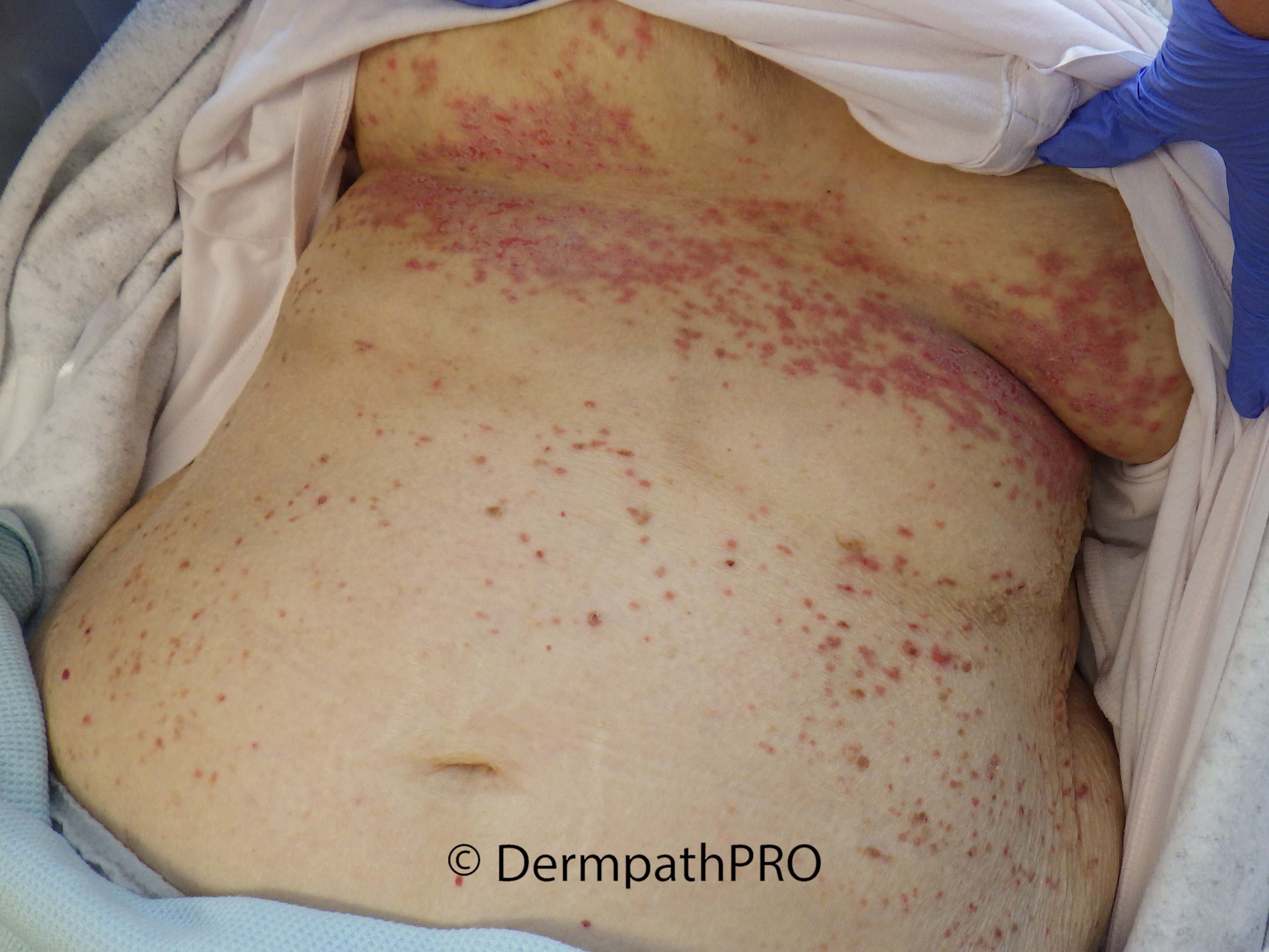
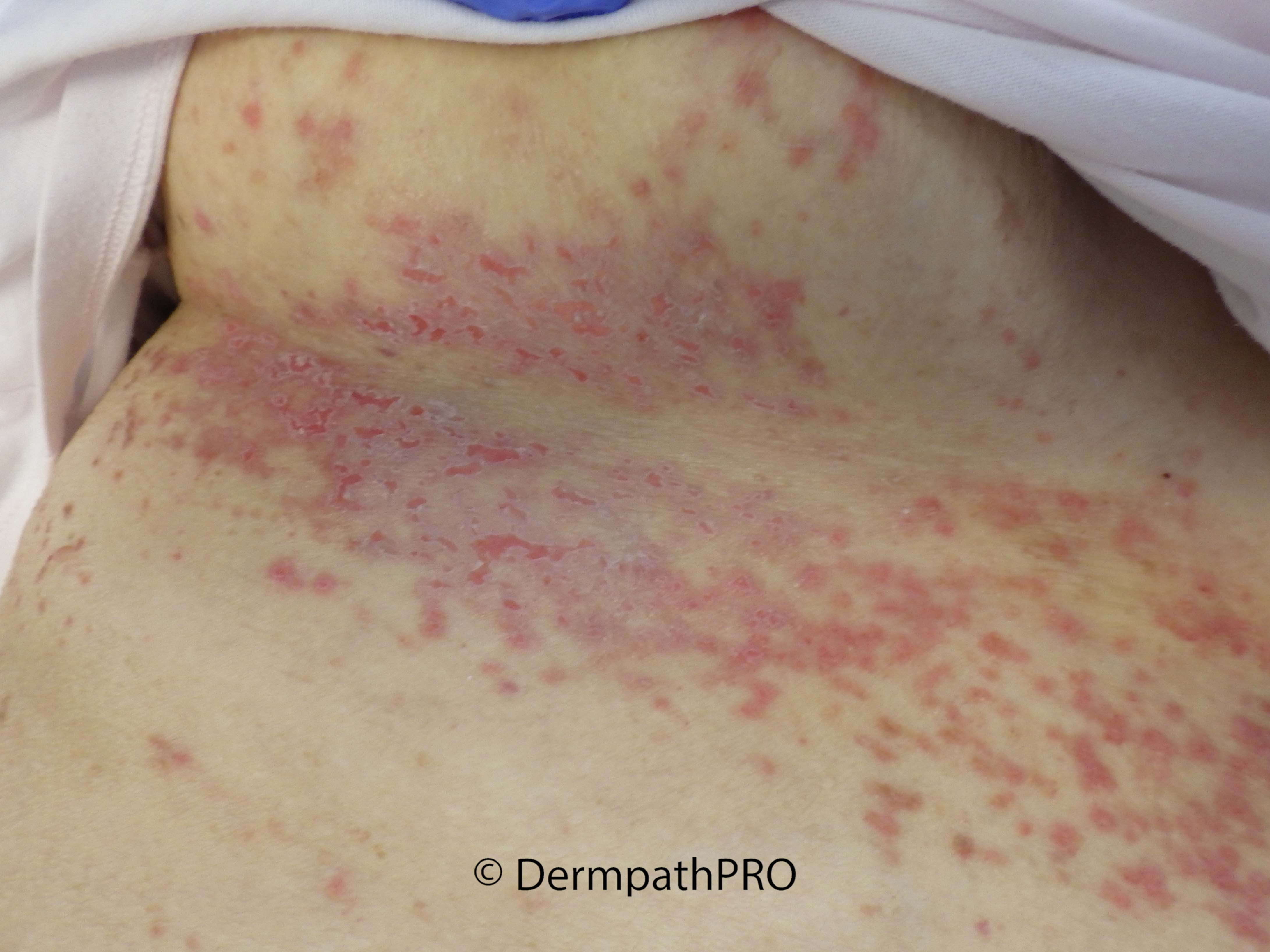
90F Incisional biopsy left submammary skin: suspected lichenoid inflammation, PMH: recent transformation of chronic myelomonocytic leukaemia

  Report Record

User Feedback


Probably involvement by the patient's known CMML. Need IHC to confirm.

Share this comment


Link to comment
share_externally

Richard Logan

Posted

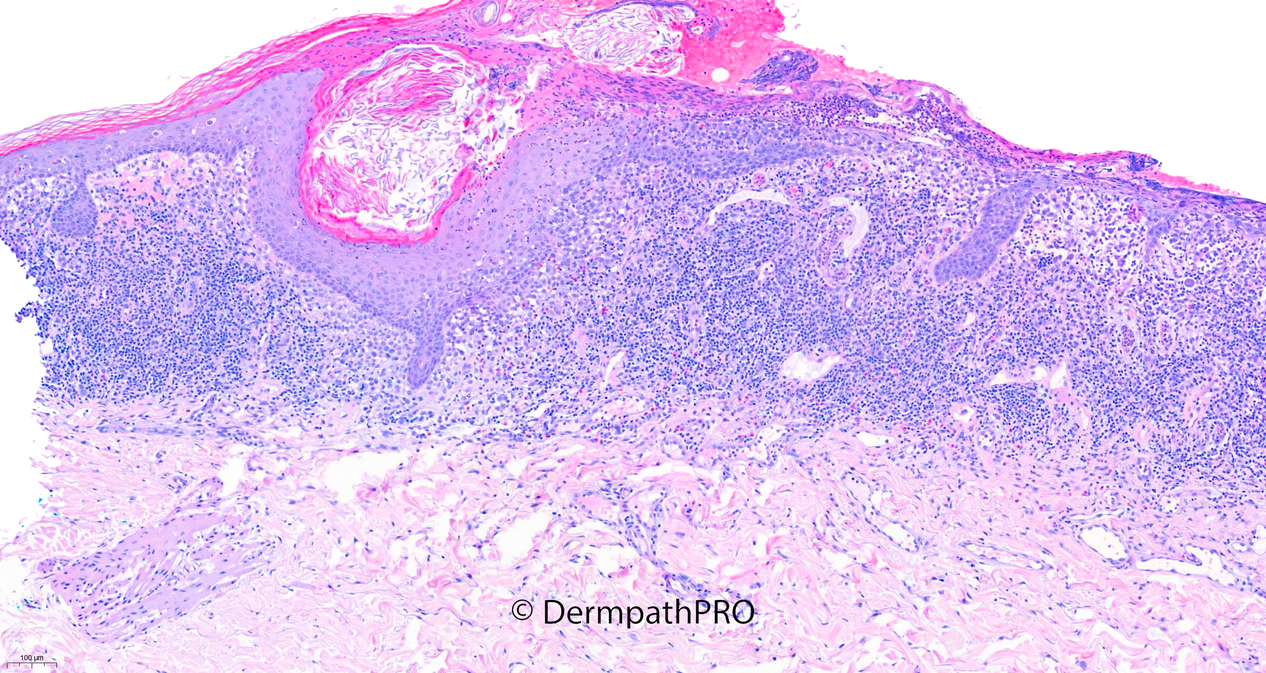
There's rather a lot of abnormality in the stratum corneum for leukaemia cutis.  Combined with the clinical pictures, I would be wondering about extensive Candidiasis and would like to see a PAS stain.  Also, eosinophils are conspicuous in the infiltrate, so a drug reaction would need to be considered.

Share this comment


Link to comment
share_externally

Meenakshi Batrani

Posted (edited)

14 hours ago, sfwenson said:

Probably involvement by the patient's known CMML. Need IHC to confirm.


Edited by Meenakshi Batrani

Share this comment


Link to comment
share_externally

Meenakshi Batrani

Posted (edited)

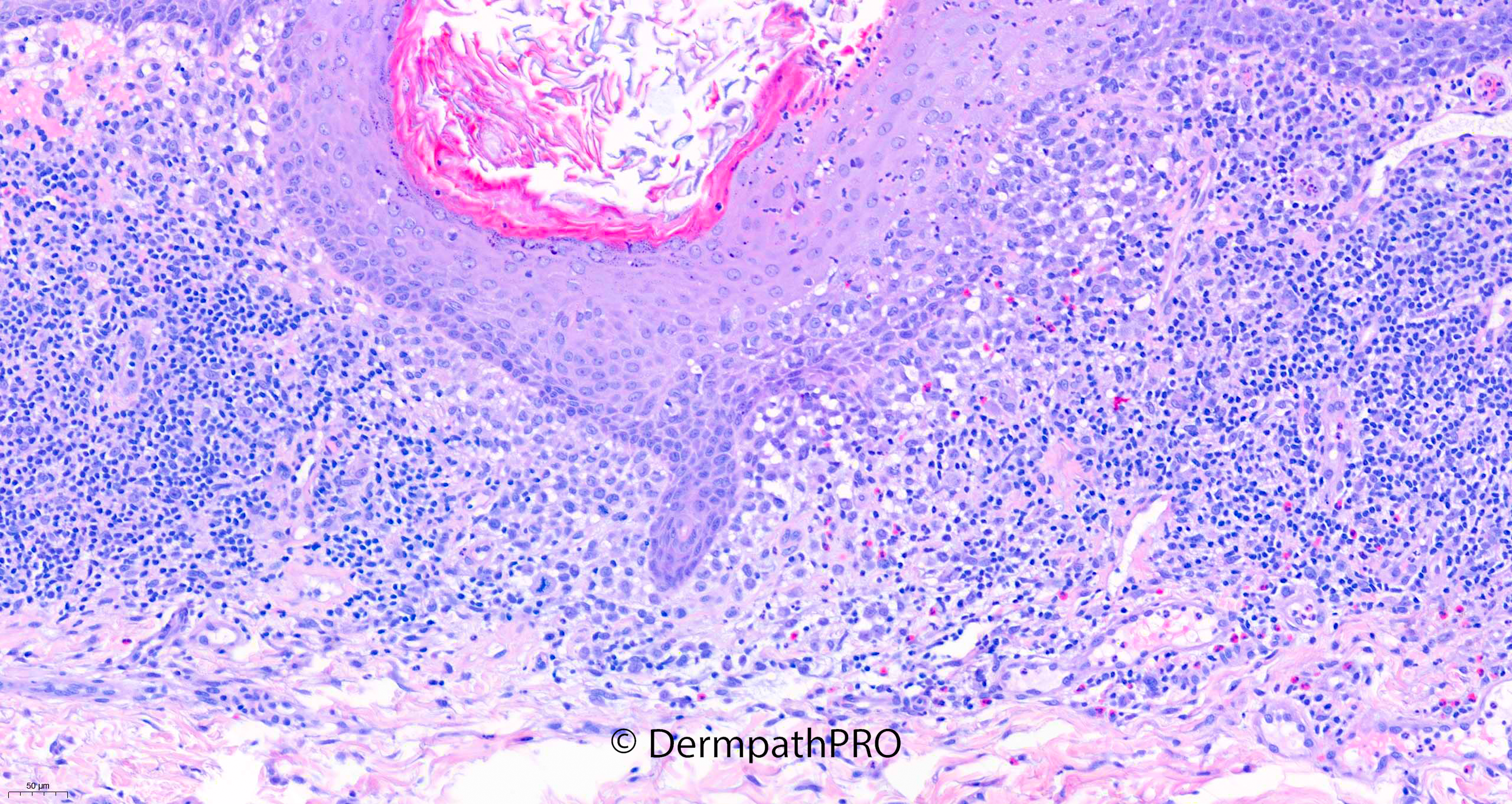
To me cells appear like Langerhans cell. Could it be Langerhans or indeterminate cell histiocytosis.  There also appear to be some atypical cells which could be atypical Langerhans cells itself or myeloid cells. There are reports of Langerhans cells histiocytosis and Langerhans cell sarcoma, as well as Indeterminate cells histiocytosis associated with myeloid leukemia, in one report both types Langerhans and myeloid cells coexisted in cutaneous lesions. 


1. Yohe SL, Chenault CB, Torlakovic EE, Asplund SL, McKenna RW. Langerhans cell histiocytosis in acute leukemias of ambiguous or myeloid lineage in adult patients: support for a possible clonal relationship. Mod Pathol. 2014 May;27(5):651-6.

2. Pina-Oviedo S, Torres-Cabala CA, Miranda RN, Tetzlaff MT, Singh S, Rapini RP, Prieto VG, Aung PP. Concomitant Cutaneous Langerhans Cell Histiocytosis and Leukemia Cutis. Am J Dermatopathol. 2017 May;39(5):388-392.

Edited by Meenakshi Batrani

Share this comment


Link to comment
share_externally

Krishnakumar subramanian

Posted

agree, it is interesting case

Histiocytic infiltrates in Myeloid leukemia

other thing is Myeloid Neoplasms with Elevated Plasmacytoid Dendritic Cell Differentiation with skin lesions

IHC is panel is needed


Share this comment


Link to comment
share_externally

On 02/12/2021 at 08:39, Meenakshi Batrani said:

To me cells appear like Langerhans cell. Could it be Langerhans or indeterminate cell histiocytosis.  There also appear to be some atypical cells which could be atypical Langerhans cells itself or myeloid cells. There are reports of Langerhans cells histiocytosis and Langerhans cell sarcoma, as well as Indeterminate cells histiocytosis associated with myeloid leukemia, in one report both types Langerhans and myeloid cells coexisted in cutaneous lesions. 


1. Yohe SL, Chenault CB, Torlakovic EE, Asplund SL, McKenna RW. Langerhans cell histiocytosis in acute leukemias of ambiguous or myeloid lineage in adult patients: support for a possible clonal relationship. Mod Pathol. 2014 May;27(5):651-6.

2. Pina-Oviedo S, Torres-Cabala CA, Miranda RN, Tetzlaff MT, Singh S, Rapini RP, Prieto VG, Aung PP. Concomitant Cutaneous Langerhans Cell Histiocytosis and Leukemia Cutis. Am J Dermatopathol. 2017 May;39(5):388-392.

I was about to write that in my opinion, in the absence of a clinical history, it was LCH. Then I saw Meenakshi's comment, which I am in tune with. 

It needs IHC, of course, as CD1a Langherine CD4 CD56 CD123 CD34 CD11c CD14 CD68. 

Share this comment


Link to comment
share_externally

The clinical picture suggests candidiasis. I think that this is leukemia cutis with superadded candidiasis. Would suggest myeloperoxidase stain in addition to those suggested by Vincenzo.

Share this comment


Link to comment
share_externally

I think i recall this case presented elsewhere and LCH was the diagnosis? 

Share this comment


Link to comment
share_externally

Saleem Taibjee

Posted

This is a patient from our hospital only seen in the past few weeks. The histology has not been presented elsewhere. I will allow more people to respond before showing the immunostains and revealing the answer.

Share this comment


Link to comment
share_externally

Furthermore epidermotropism is typical of Langherans cells...so my first spot, without immune, is LCH. Another diff could be an exceedingly strong reaction as allergic contact dermatitis, but never seen so many lang-cells. Waiting for the answer.  

Share this comment


Link to comment
share_externally

Saleem Taibjee

Posted

The immunos are shown below.

This is a very recent case which I reported at our local hospital of Langerhans cell histiocytosis which is quite rare overall, but especially in adults. It is the first case I have seen since working in Dorset over the past 8 years or so.

As Vincenzo points out, the epidermotropism is a very good clue. Here the infiltrate is really too dense for this to be reactive (Langerhans cell hyperplasia), and furthermore the cells lack the dendritic morphology which is usually more typical of reactive Langerhans cells. But I have certainly seen inflammatory dermatoses and neoplasms with very prominent Langerhans cell hyperplasia, and that should certainly be considered. With this in mind in can be helpful to request Cyclin-D1, or as in this case IHC for BRAFV600E which is clearly positive in this case providing evidence of a clonal expansion. In some situations this can also highlight a potential therapeutic target with BRAF inhibitors, but unfortunately this patient's other haematological issues were already a significant factor, and the skin was a minor issue. 

I can see that Richard had spotted the accompanying eosinophils, and it is worth remembering that these can be present in LCH.

As Meenakshi and Vincenzo point out, there is not infrequent co-existence of LCH and myeloproliferative disease when LCH presents in adults (and have already provided references!) Perhaps this reflects a common progenitor cell of origin. In this case a subpopulation of cells did express myeloperoxidase. Given the transformation state, this could certainly indicate some spill over of neoplastic myeloid cells within the skin, but even so, the Langherans cell population is the dominant and also correlates well with the seborrhoeic dermatitis/intertrigo-like cutaneous presentation.

I did also request CD123, but the staining was minimal in this case. No fungi seen on PAS.

BW, Saleem

09518_5.0x CD1a labelled.jpg

09518_20.0x Langerin b labelled.jpg09518_20.0x MPO labelled.jpg55366_20.0x BRAFV600E labelled.jpg


Share this comment


Link to comment
share_externally

I usually learn something making a misdiagnosis, however this time learned a lot having come very close to the correct diagnosis, and all this thanks to to you, Saleem and thanks to DermpatPro. 

Share this comment


Link to comment
share_externally

Saleem Taibjee

Posted

Thanks for this lovely feedback. It reassures me that it is worthwhile posting these cases on DermpathPro.

Share this comment


Link to comment
share_externally


Join the conversation

You can post now and register later. If you have an account, sign in now to post with your account.

Guest
Add a comment...

×   Pasted as rich text.   Paste as plain text instead

  Only 75 emoji are allowed.

×   Your link has been automatically embedded.   Display as a link instead

×   Your previous content has been restored.   Clear editor

×   You cannot paste images directly. Upload or insert images from URL.

Loading...

×
×
  • Create New...