Jump to content
In this section we have spot diagnoses posted on a daily basis since June 2010, now over 4000! You can review the archived cases and read the suggested diagnoses by users and the final comment by the contributors.
Case are uploaded each week day by 10 am UK time with the correct diagnosis will generally be posted at 8 pm UK time. Why not view the most recent spot diagnosis and proffer a diagnosis?

Case Number : Case 2981 - 9 December 2021 Posted By: Saleem Taibjee

Please read the clinical history and view the images by clicking on them before you proffer your diagnosis.
Submitted Date :
   (0 reviews)

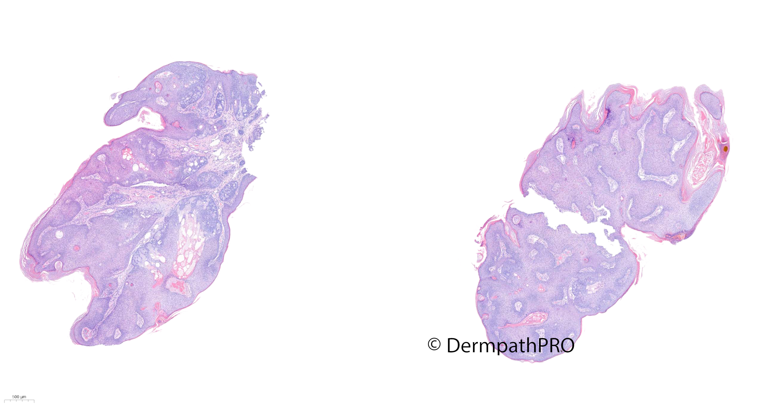
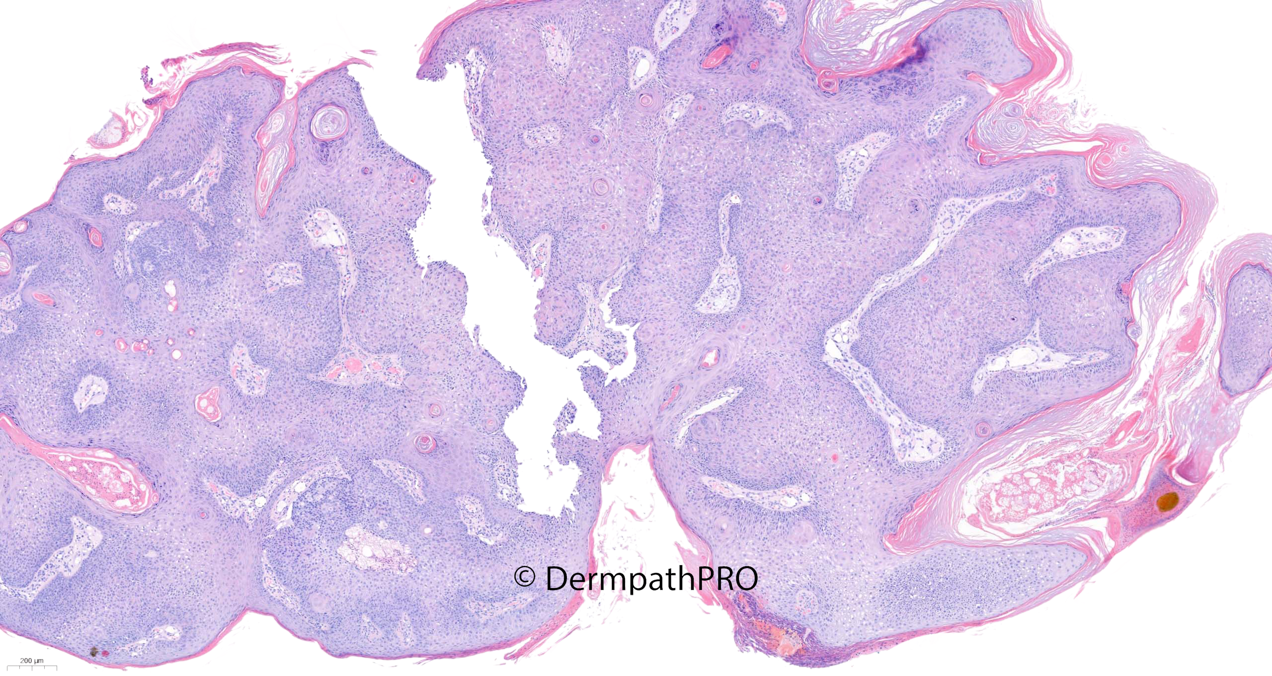
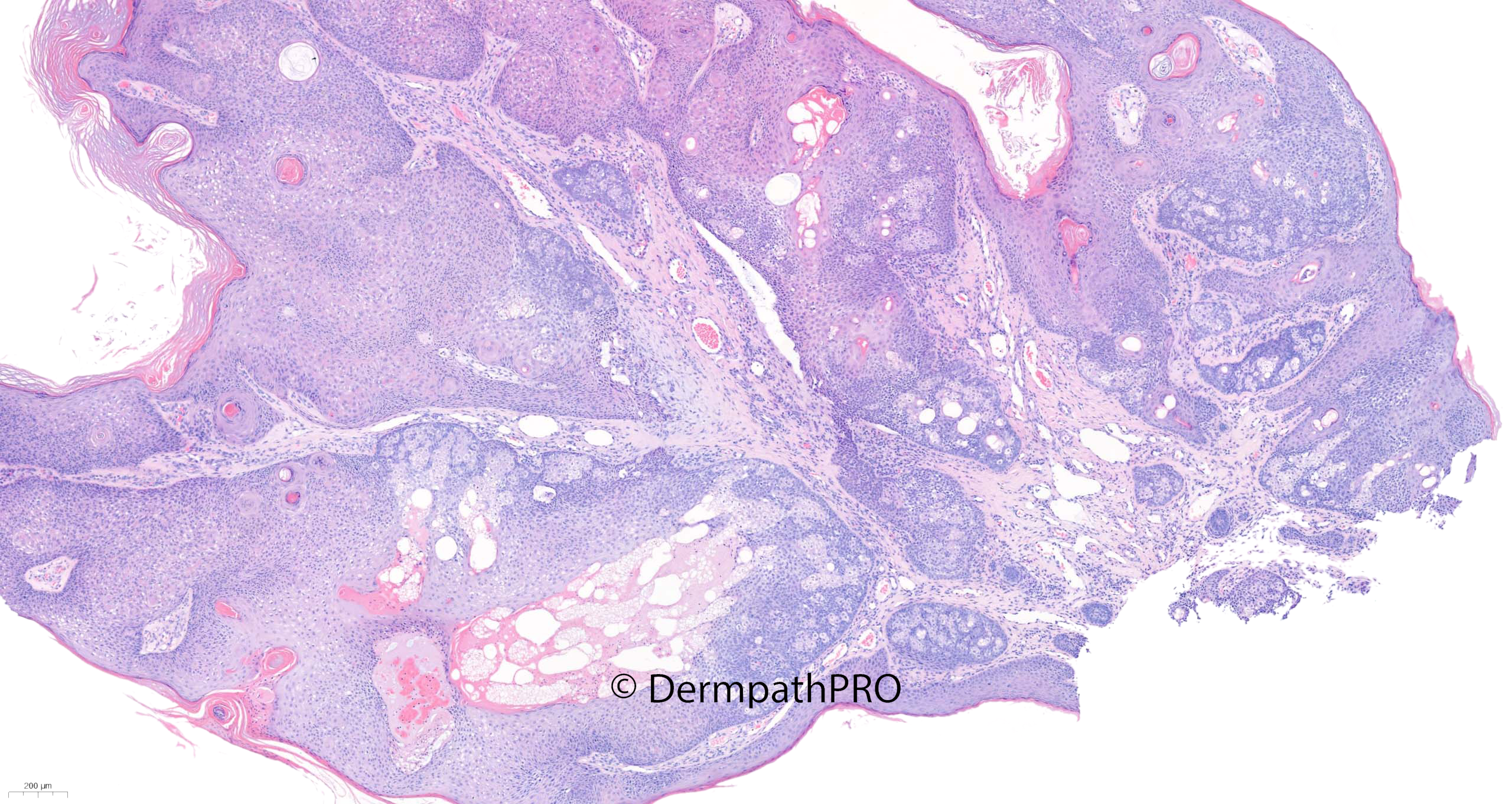
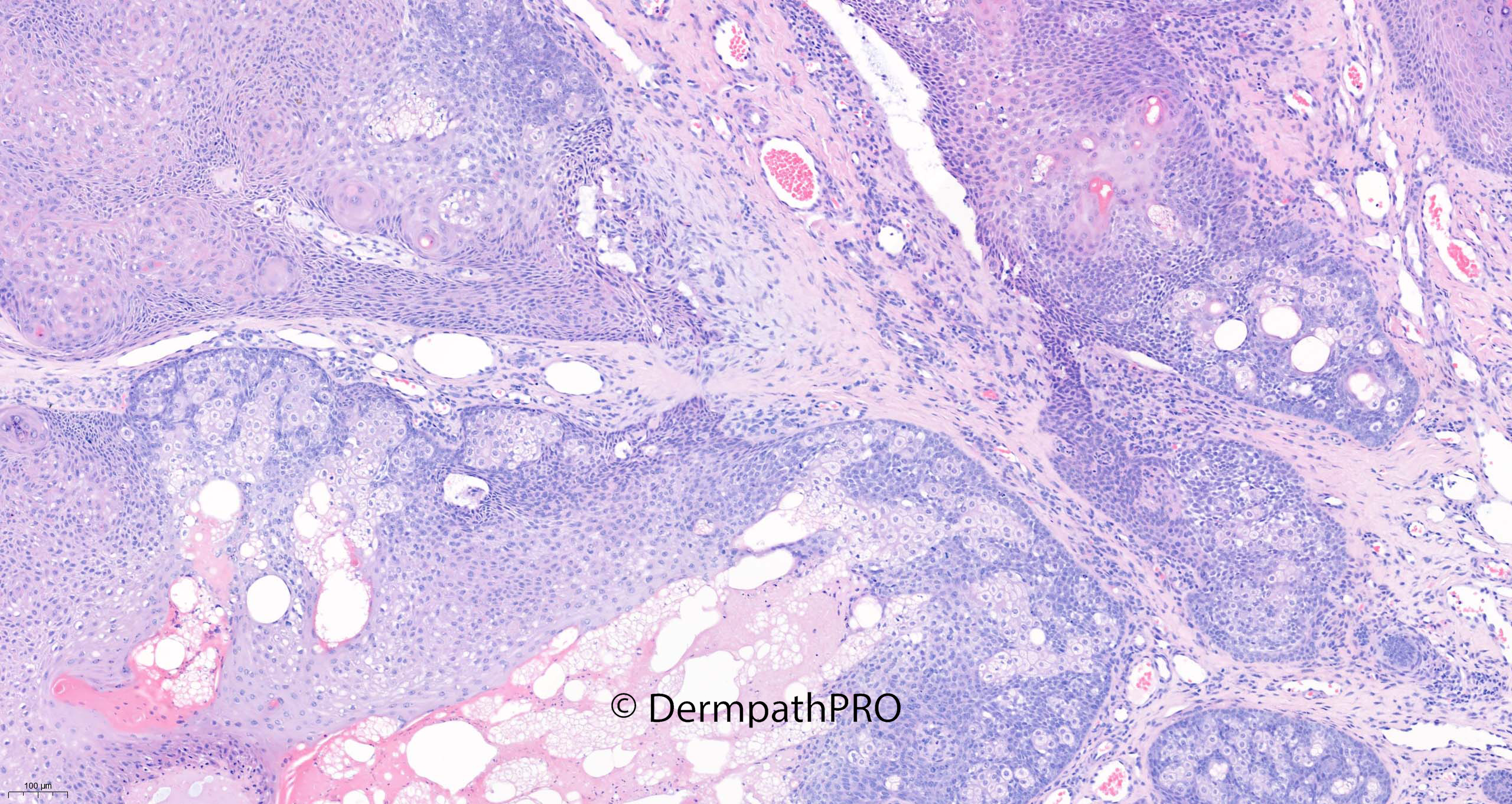
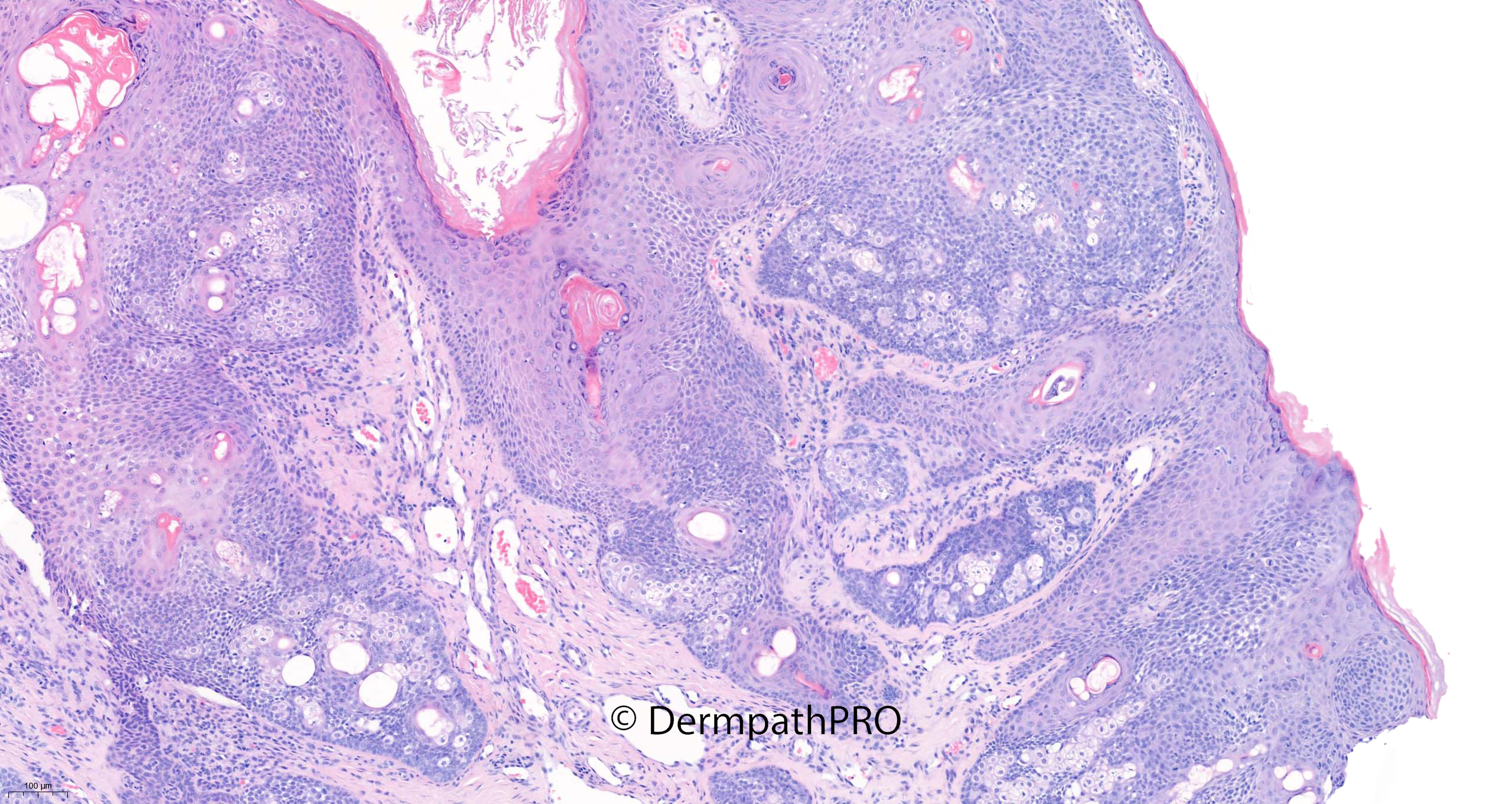
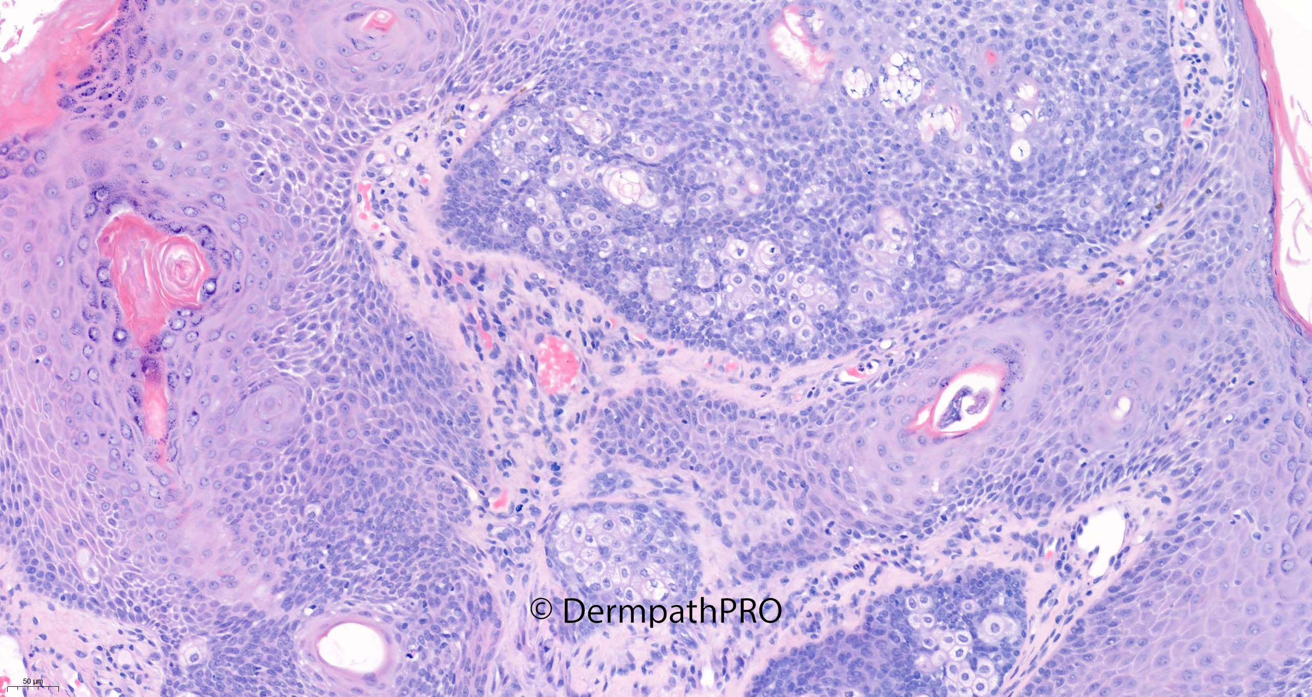
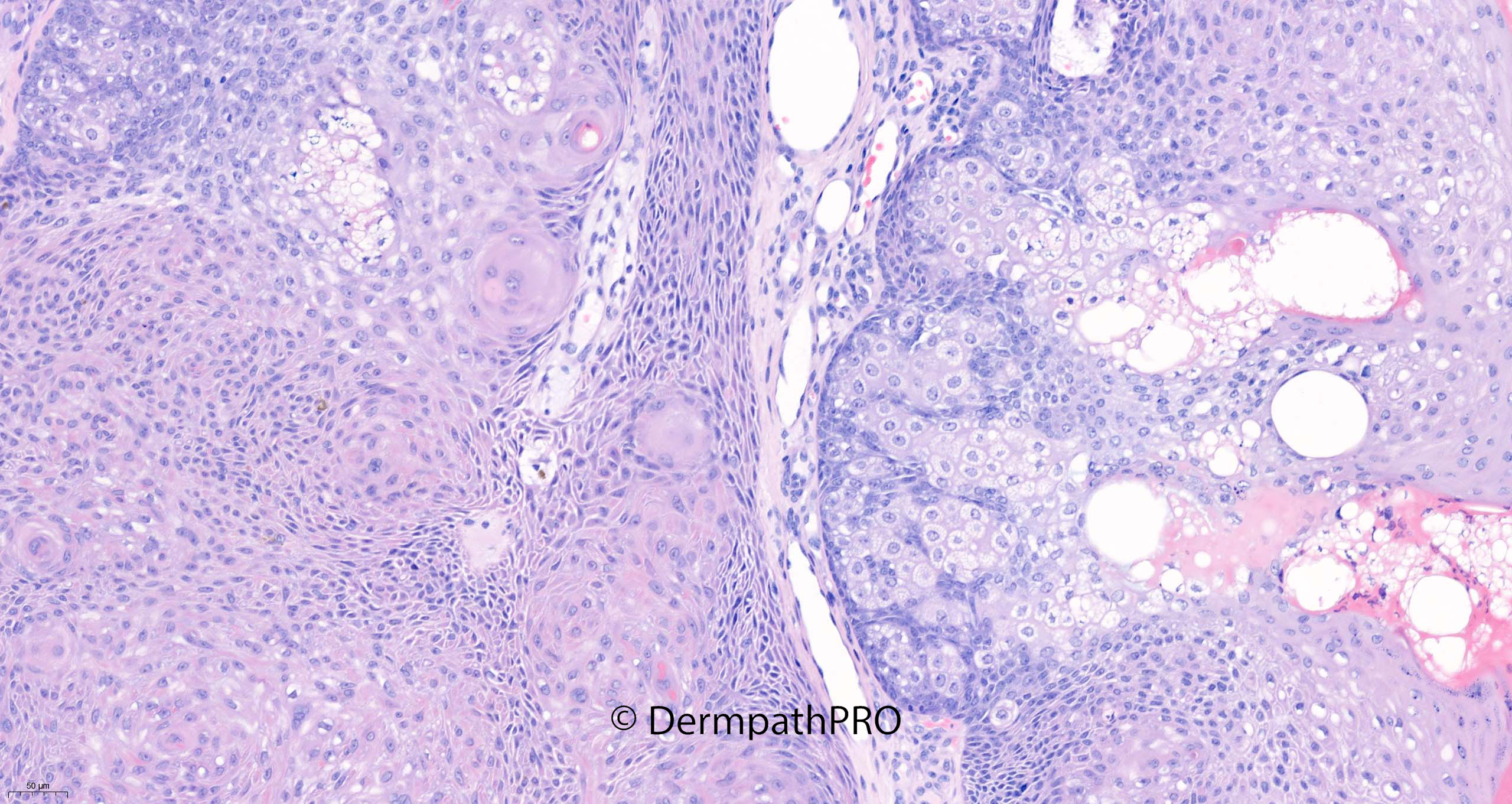
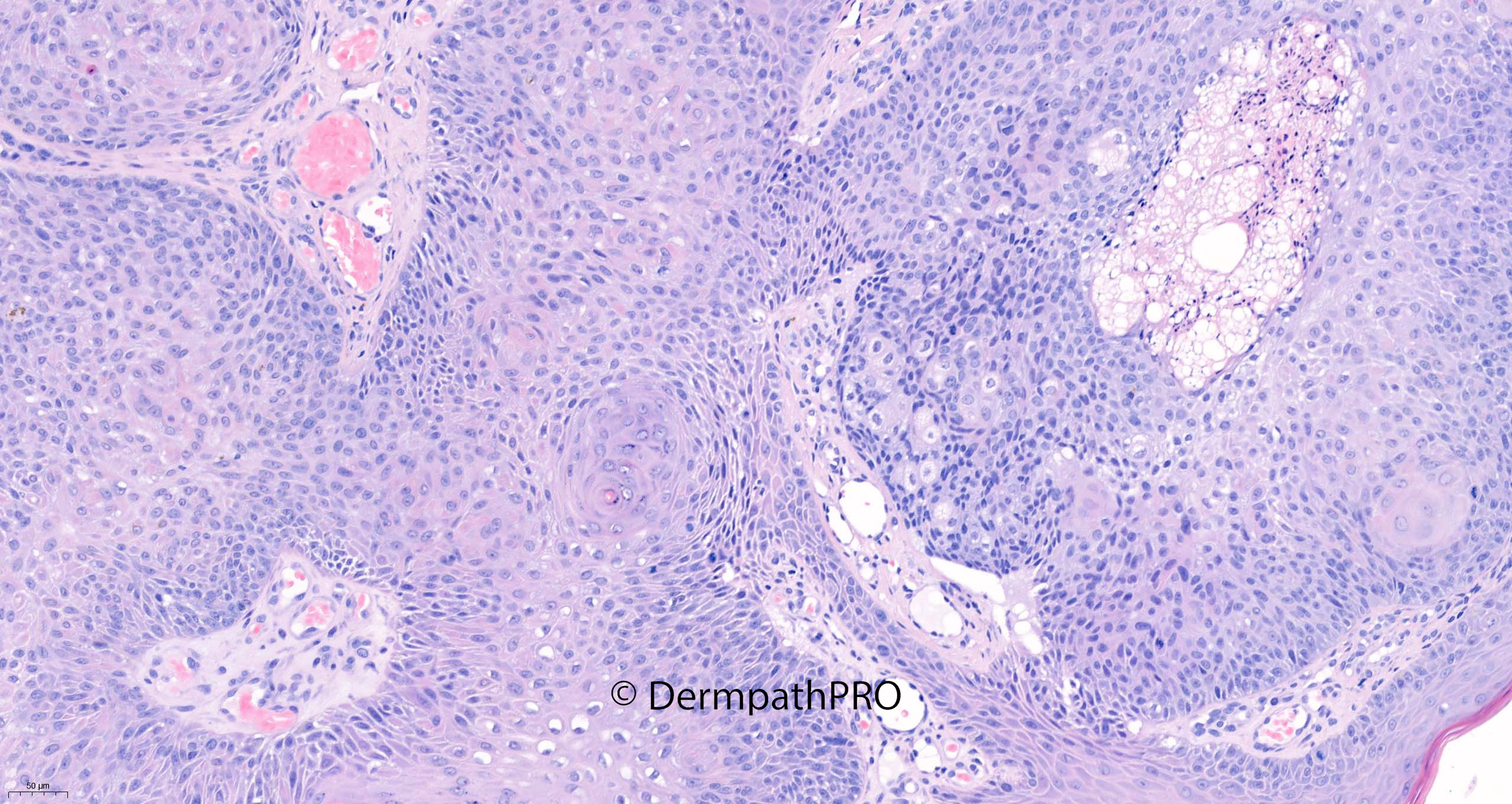
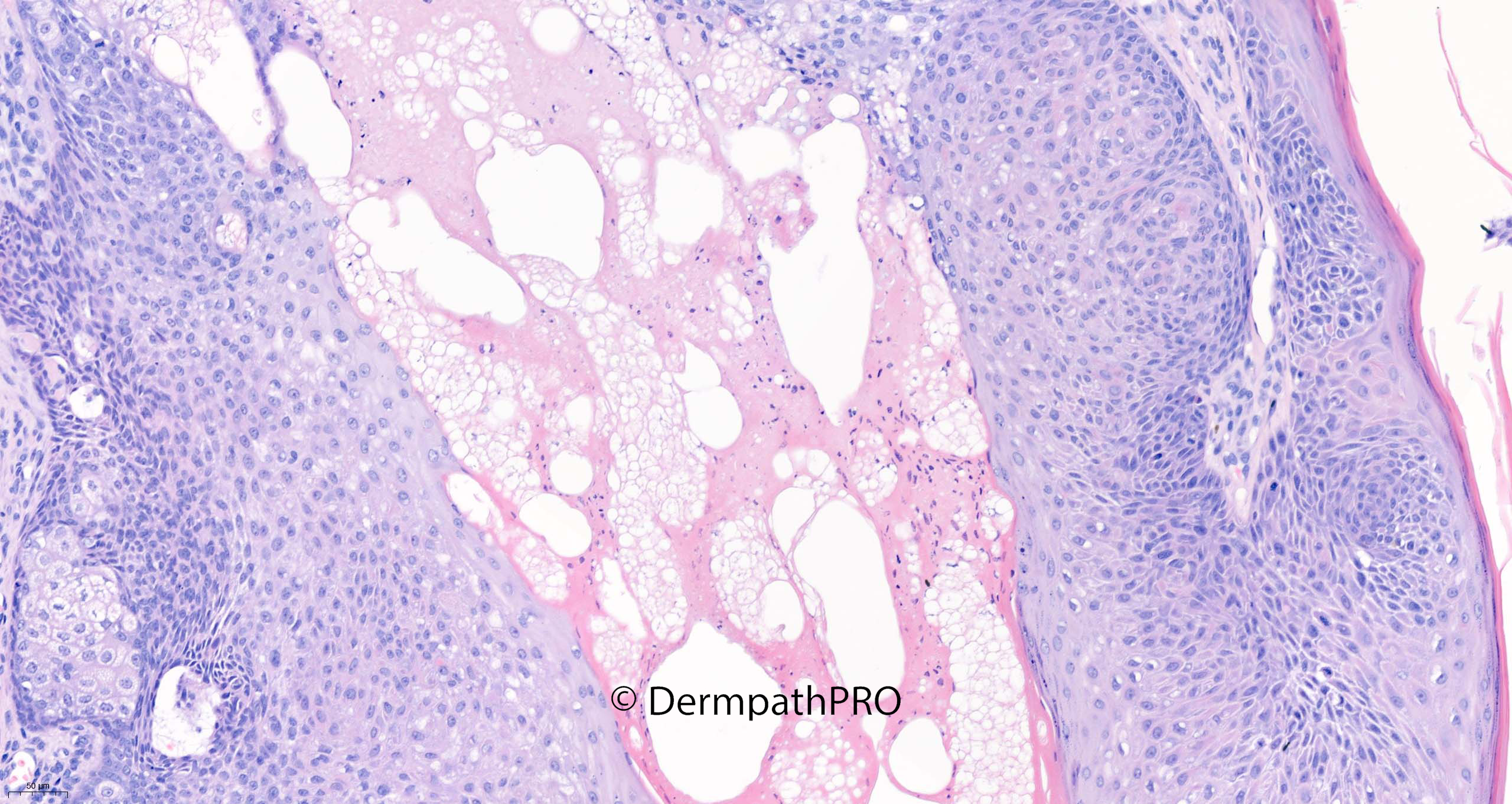
82M Curettage pigmented lesion right nostril


  Report Record

User Feedback


Inverted follicular keratosis with sebaceous differentiation in some areas.

Share this comment


Link to comment
share_externally

Krishnakumar subramanian

Posted

agree to Inverted follicular keratosis of helwig with some amount of sebaceous differentiation

Share this comment


Link to comment
share_externally

Meenakshi Batrani

Posted

Agree with seborrhoeic keratosis / IFK with sebaceous differentiation. According to a recent November 2021 article in AJDP, seborrhoeic keratosis is the preferred term unifying the entities variably labeled as superficial epithelioma with sebaceous differentiation and reticulated acanthoma with sebaceous differentiation. The same article also advocates exclusion of  Muir–Torre syndrome in such cases. 

Share this comment


Link to comment
share_externally

Saleem Taibjee

Posted

Yes, well done. I also thought this is a great example of seborrheic keratosis/inverted follicular keratosis with sebaceous differentiation. The areas of typical seborrheic keratosis, including nice squamous eddies as shown in the images above, helps to distinguish between a primary sebaceous neoplasm (sebaceoma) in which the cells in the basal component tend to look more primitive and larger.

I’ve seen a few examples of this over the years, but this is the most striking example I’ve come across just recently. And as Meenakshi points out, it coincided rather fortuitously with the recent publication in American Journal of Dermatopathology, so I could not resist including it on Dermpath Pro.

In this particular case, the sebaceous differentiation was so prominent that I did request IHC for mismatch repair proteins. This showed no evidence of loss on MSH2, MSH6, MLH1 and PMS2.

BW, Saleem

Share this comment


Link to comment
share_externally


Join the conversation

You can post now and register later. If you have an account, sign in now to post with your account.

Guest
Add a comment...

×   Pasted as rich text.   Paste as plain text instead

  Only 75 emoji are allowed.

×   Your link has been automatically embedded.   Display as a link instead

×   Your previous content has been restored.   Clear editor

×   You cannot paste images directly. Upload or insert images from URL.

Loading...

×
×
  • Create New...