Jump to content
In this section we have spot diagnoses posted on a daily basis since June 2010, now over 4000! You can review the archived cases and read the suggested diagnoses by users and the final comment by the contributors.
Case are uploaded each week day by 10 am UK time with the correct diagnosis will generally be posted at 8 pm UK time. Why not view the most recent spot diagnosis and proffer a diagnosis?

Case Number : Case 2982 - 10 December 2021 Posted By: Dr. Richard Carr

Please read the clinical history and view the images by clicking on them before you proffer your diagnosis.
Submitted Date :
   (0 reviews)

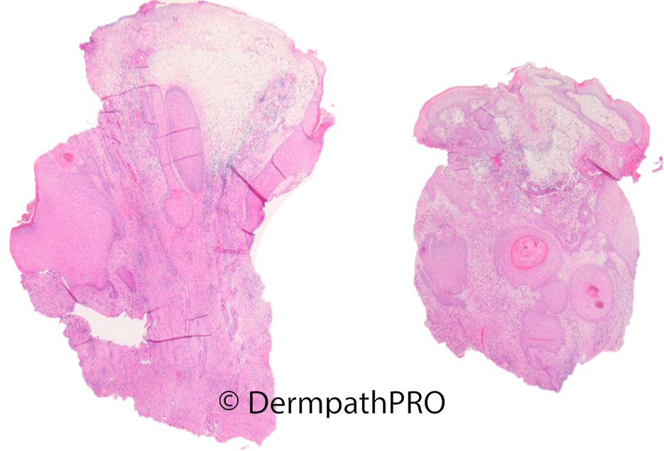
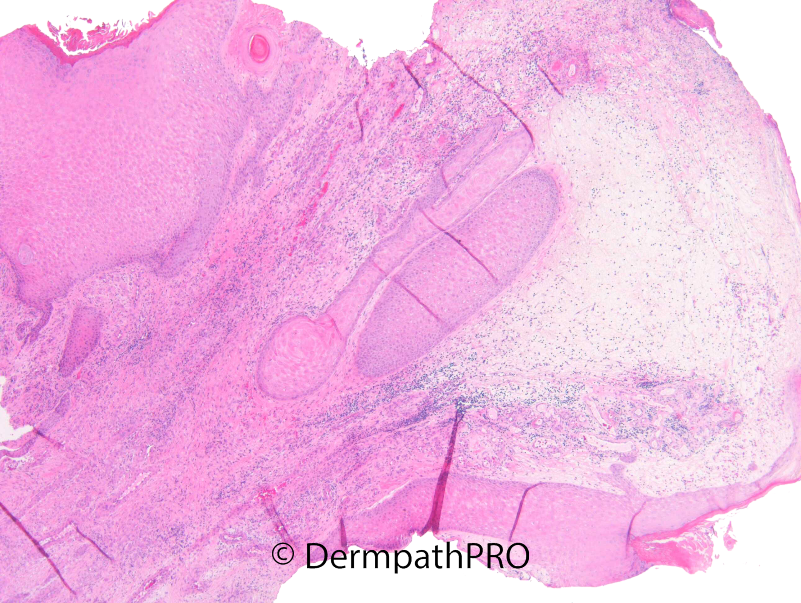
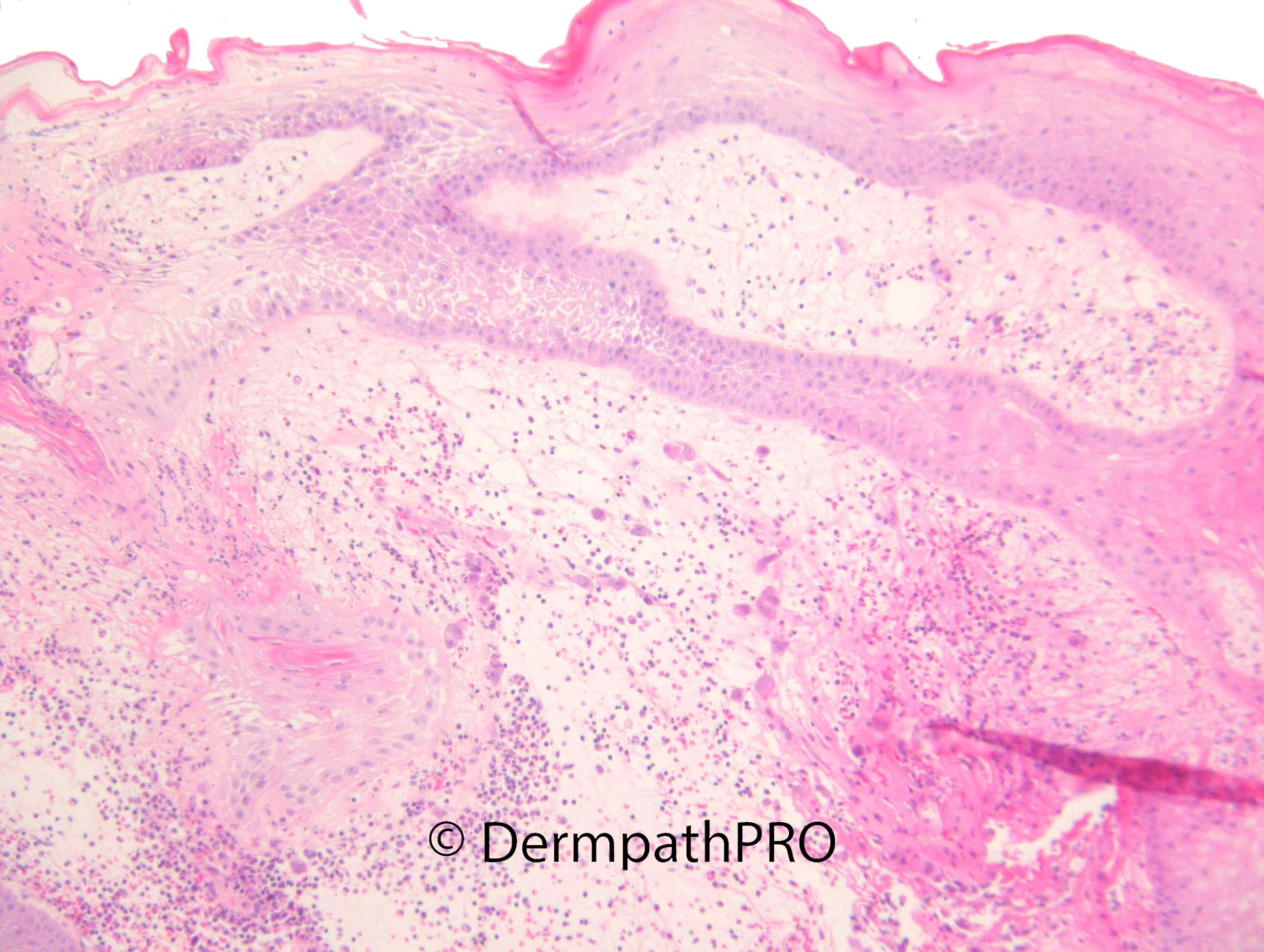
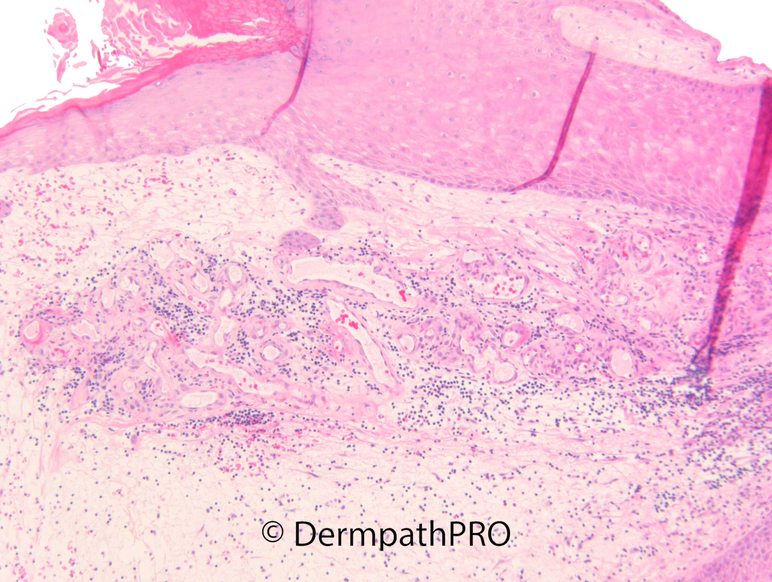
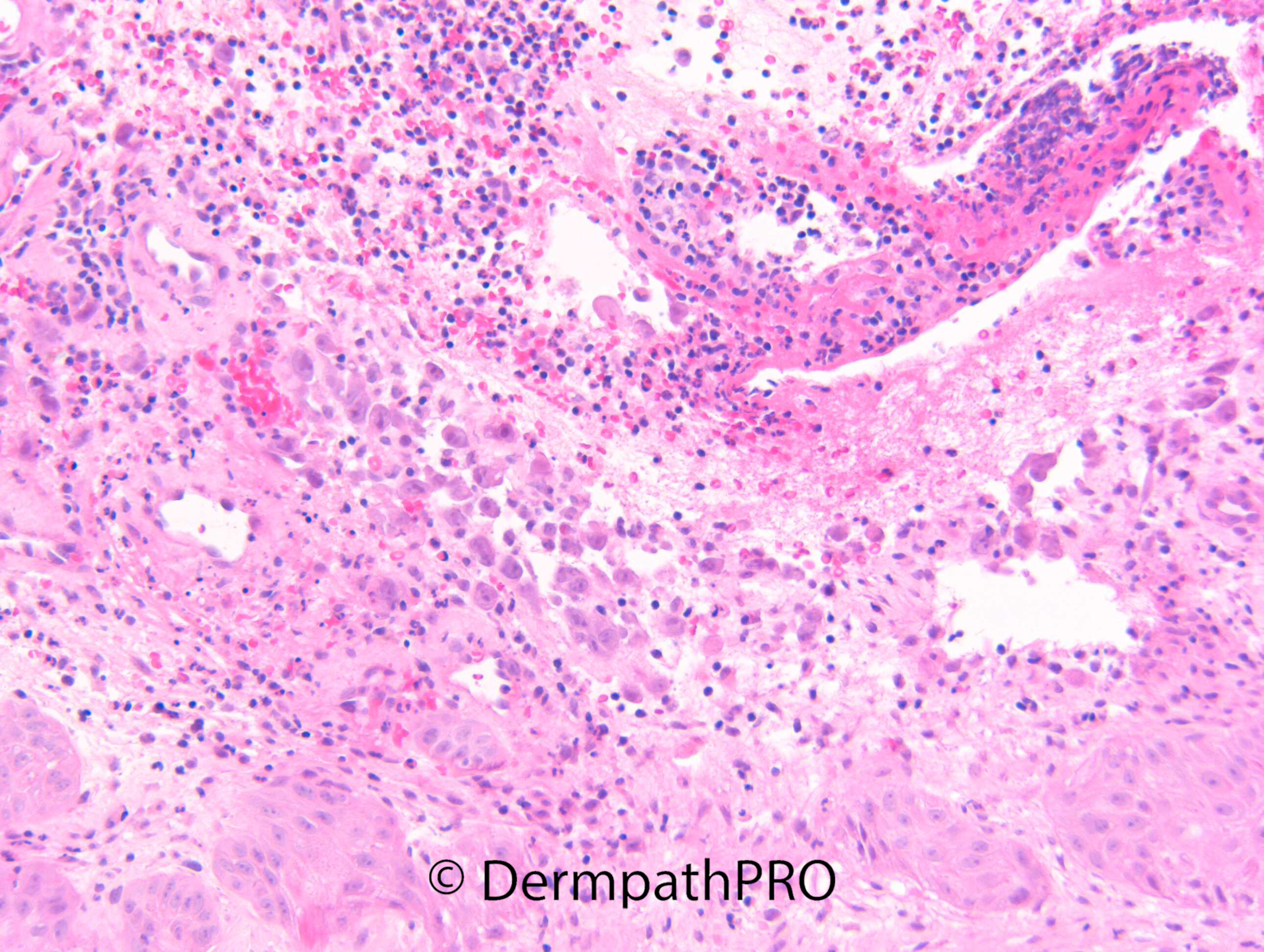
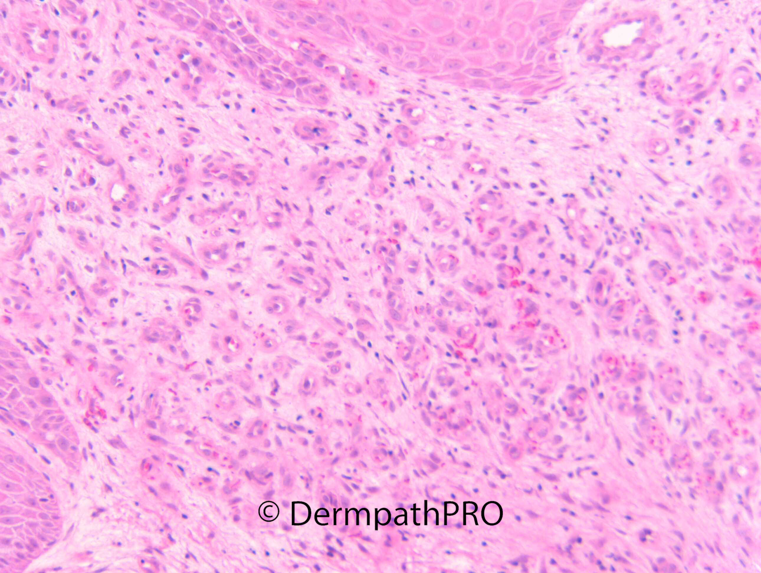
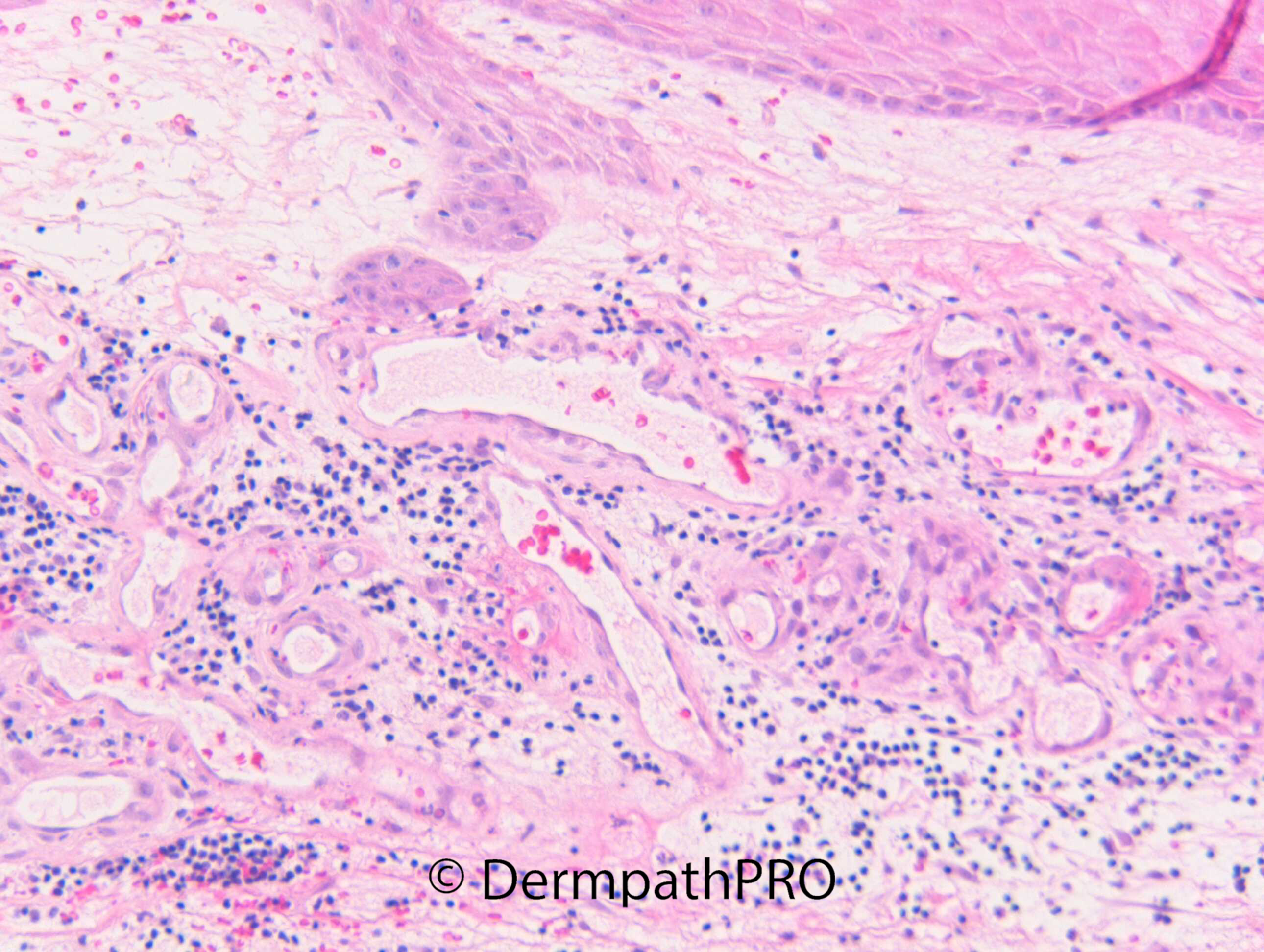
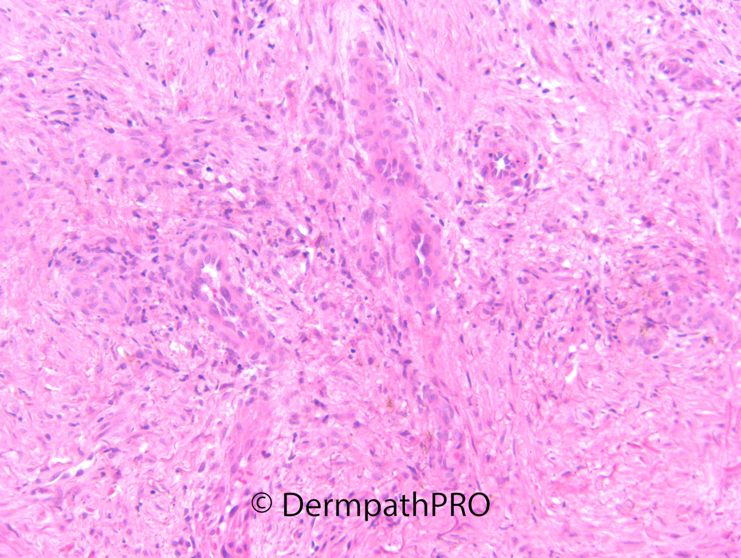
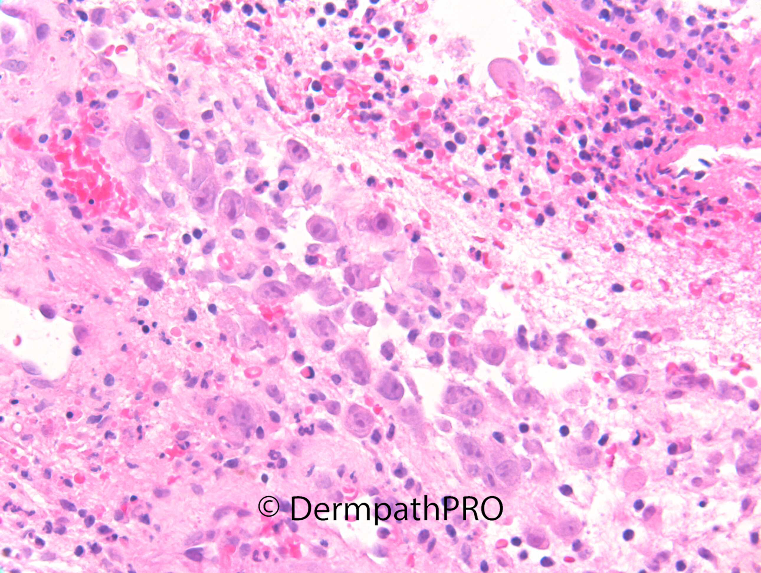
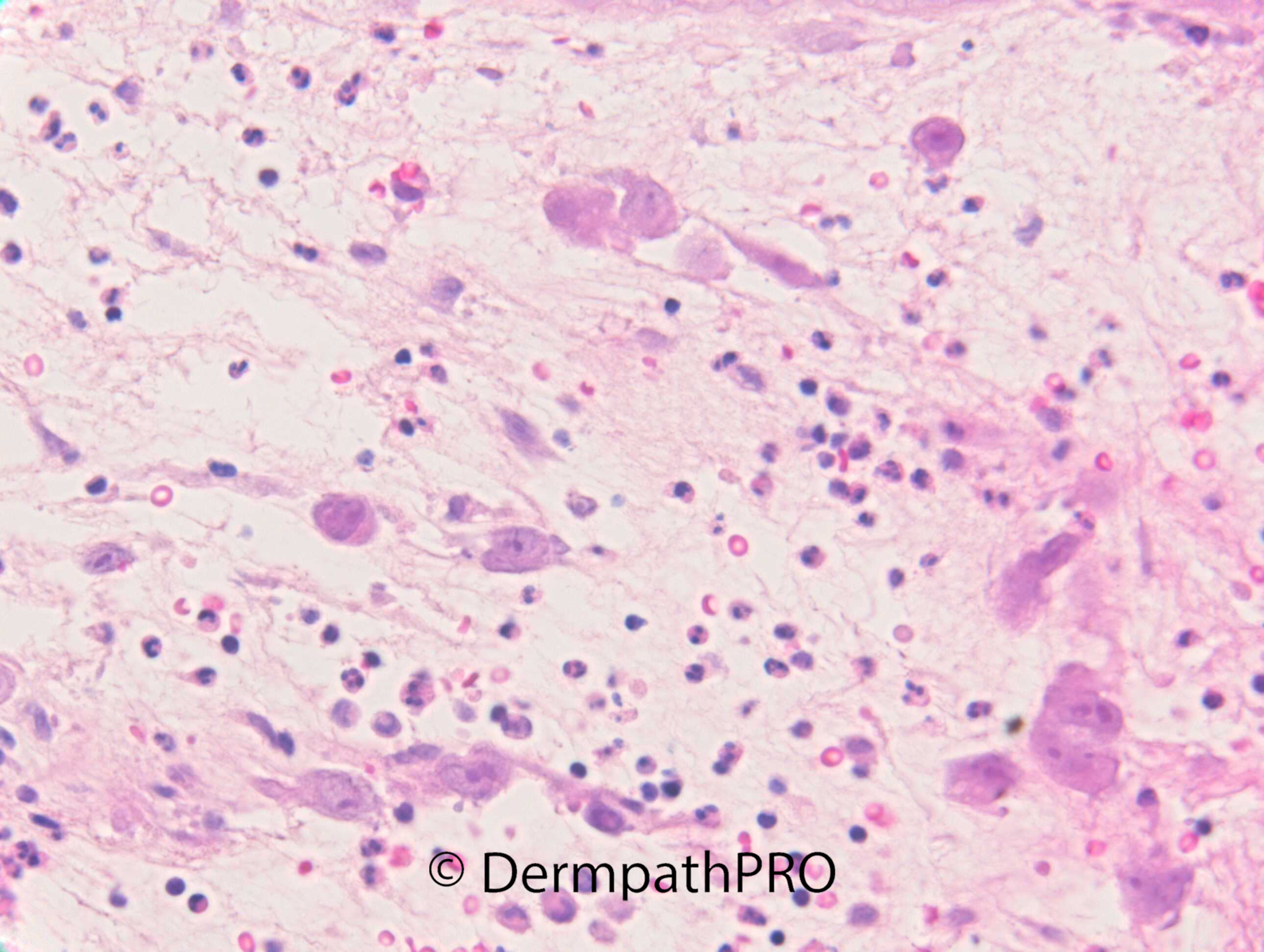
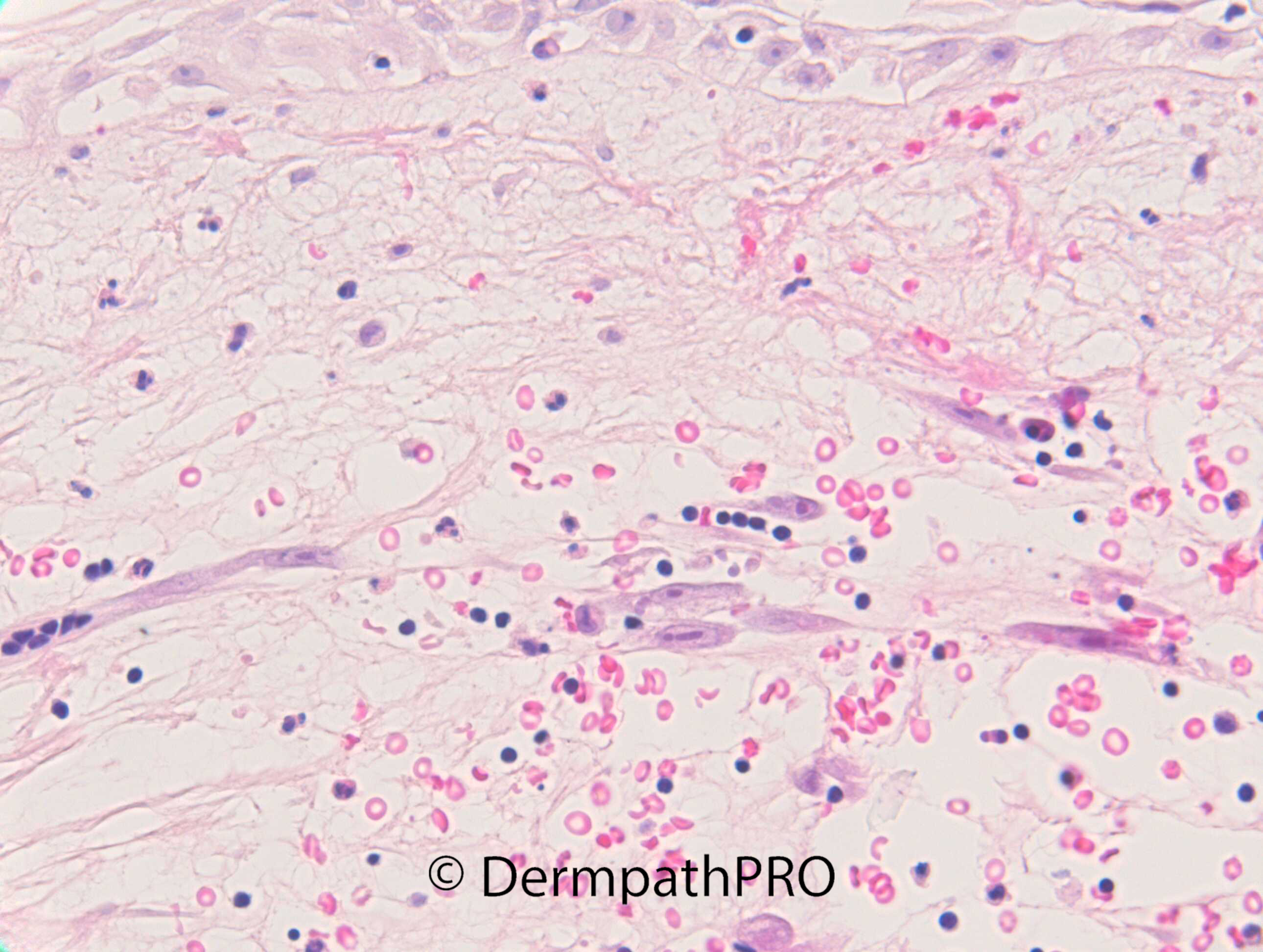
"F85. Left Calf: 6 – 7 weeks skin lesion on L calf. Non-healing, bleeds, fast growing
DD: ?SCC."


  • Like 1
  Report Record

User Feedback


Dr. Richard Carr

Posted

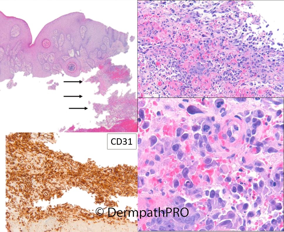
Apologies to Meenakshi and Anil. I'm going to ask the dermpathpro team to remove the intended 6pm montage and your diagnoses! 

Share this comment


Link to comment
share_externally

Krishnakumar subramanian

Posted

sir, i agree with them it is angiosarcoma

Share this comment


Link to comment
share_externally

I am favoring something reactive like ischemic fasciitis as well. It just doesn't look neoplastic or malignant to me.

Share this comment


Link to comment
share_externally

Meenakshi Batrani

Posted

Agree. I was quick to overcall it angiosarcoma. May be some reactive pseudosarcomatous lesion with reactive vascular proliferation, myfibroblasts and ganglion like cells which can be seen in ischemic or proliferative fascitis 

Share this comment


Link to comment
share_externally

Well never mind then! Looks like angiosarcoma now.

Share this comment


Link to comment
share_externally

Dr. Richard Carr

Posted

Thanks and actually your responses were quite helpful - showing the difficulty of the case. The dermpath pro team inadvertantly posted the montage with CD31 (from a later specimen) with the punch biopsy images. I reported the punch biopsy as showing atypical cells of uncertain histogenesis after doing S100, epithelial markers and even lymphoid markers but no vascular markers. We discussed the case at MDM and in fact the legs were lymphoedematous but none of the experienced team of clinicians or myself considered the possibility of an angiosarcoma. Excision of the ulcer, due to clinical suspicion of malignancy, was undertaken and the specimen was sent to a nearby hospital without mention of the punch biopsy showing atypical cells of uncertain histogenesis with a clinical query of BCC or SCC. Unfortunately subtle vasogenic angiosarcoma was missed including a small focus with similar atypical cells as seen on the punch biopsy. The biopsy did also show marked, verrucous epithelial hyperplasia & reactive vascular hyperplasia of venous stasis and changes compatible with lymphoedema. The area never healed which was not surprising to the clinician as this was an elderly frail woman with chronic lymphoedema. It was not until a curettage was undertaken several months later that the high grade area of angiosarcoma became apparent although even then the pathologist only included a vascular marker in the second round of immunostains. Some cases are just very difficult and we have to learn from them.

Share this comment


Link to comment
share_externally

Meenakshi Batrani

Posted

21 hours ago, Dr. Richard Carr said:

Thanks and actually your responses were quite helpful - showing the difficulty of the case. The dermpath pro team inadvertantly posted the montage with CD31 (from a later specimen) with the punch biopsy images. I reported the punch biopsy as showing atypical cells of uncertain histogenesis after doing S100, epithelial markers and even lymphoid markers but no vascular markers. We discussed the case at MDM and in fact the legs were lymphoedematous but none of the experienced team of clinicians or myself considered the possibility of an angiosarcoma. Excision of the ulcer, due to clinical suspicion of malignancy, was undertaken and the specimen was sent to a nearby hospital without mention of the punch biopsy showing atypical cells of uncertain histogenesis with a clinical query of BCC or SCC. Unfortunately subtle vasogenic angiosarcoma was missed including a small focus with similar atypical cells as seen on the punch biopsy. The biopsy did also show marked, verrucous epithelial hyperplasia & reactive vascular hyperplasia of venous stasis and changes compatible with lymphoedema. The area never healed which was not surprising to the clinician as this was an elderly frail woman with chronic lymphoedema. It was not until a curettage was undertaken several months later that the high grade area of angiosarcoma became apparent although even then the pathologist only included a vascular marker in the second round of immunostains. Some cases are just very difficult and we have to learn from them.

One of those great learning cases. Seeing montage it was easy to call it angiosarcoma. But when I checked the case again, montage was removed offcourse, and without it when I looked at the rest of the images, I thought may be I overcalled it in haste. 

Share this comment


Link to comment
share_externally


Join the conversation

You can post now and register later. If you have an account, sign in now to post with your account.

Guest
Add a comment...

×   Pasted as rich text.   Paste as plain text instead

  Only 75 emoji are allowed.

×   Your link has been automatically embedded.   Display as a link instead

×   Your previous content has been restored.   Clear editor

×   You cannot paste images directly. Upload or insert images from URL.

Loading...

×
×
  • Create New...