Diagnostic Pearls : Case 2985 - 15 December 2021
80M Excision scalp
Saleem Taibjee
Posted 14/12/21
Posted 14/12/21
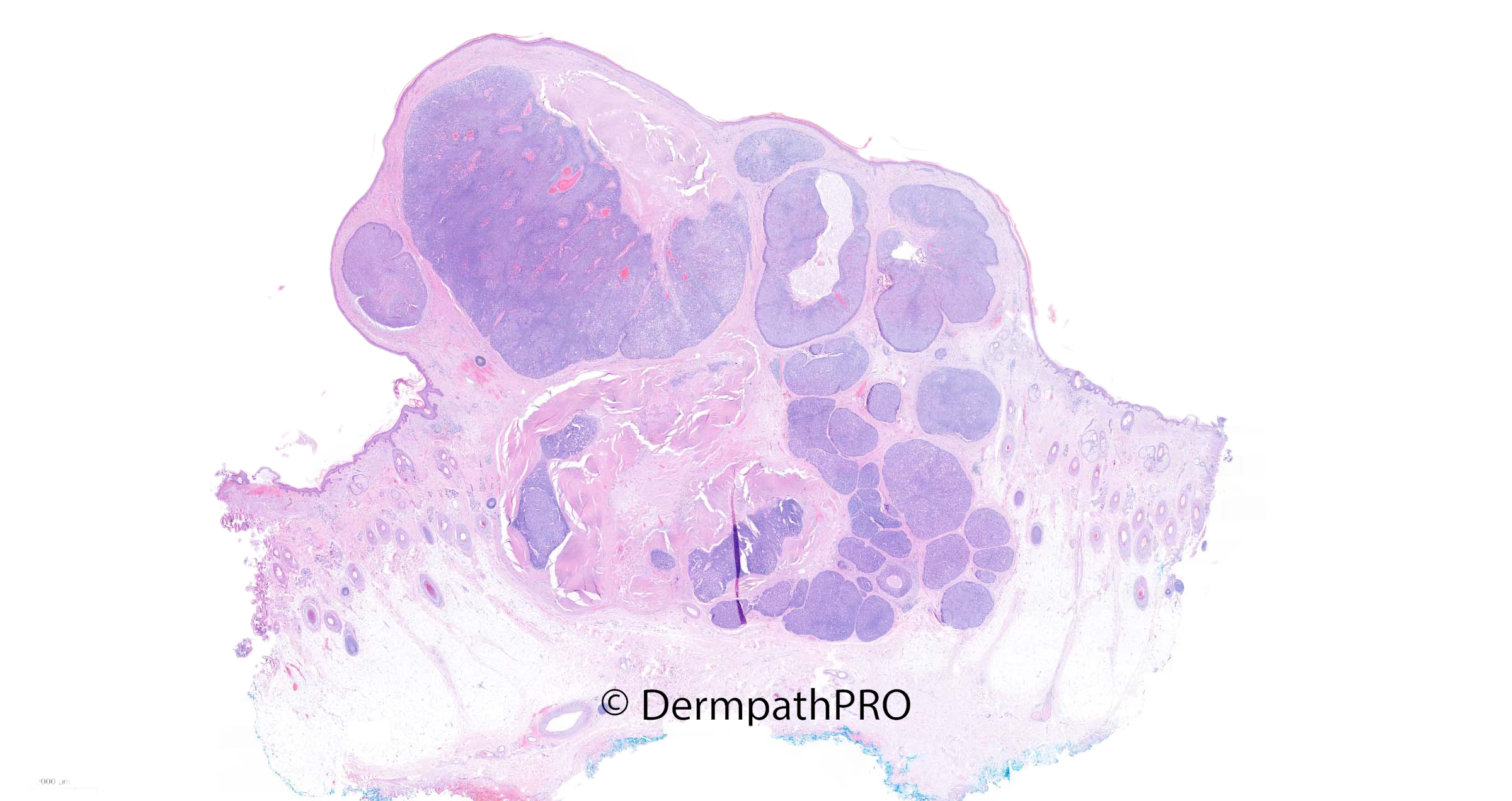
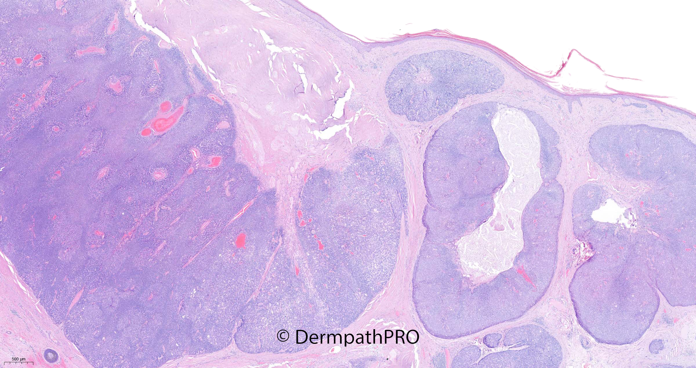
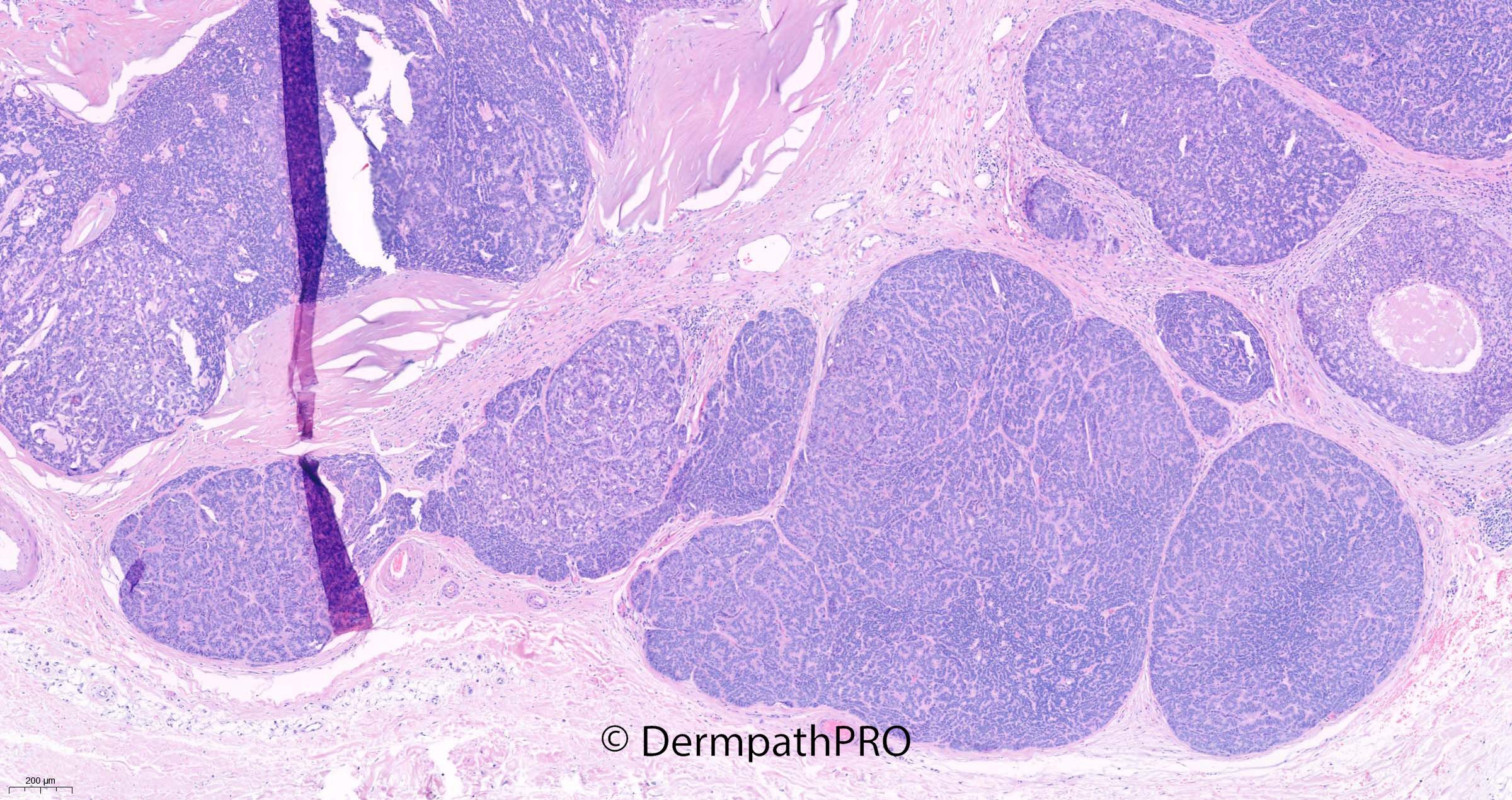
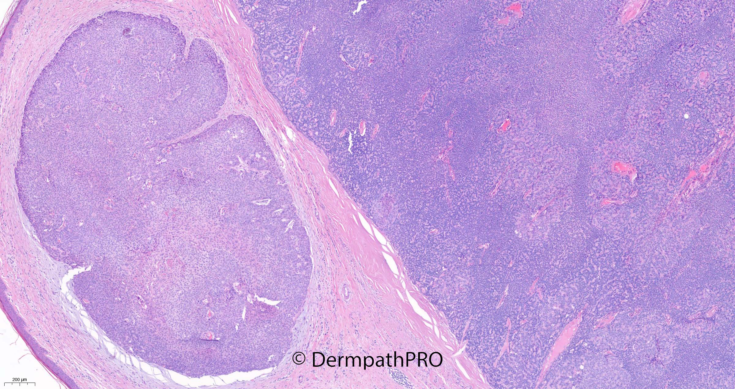
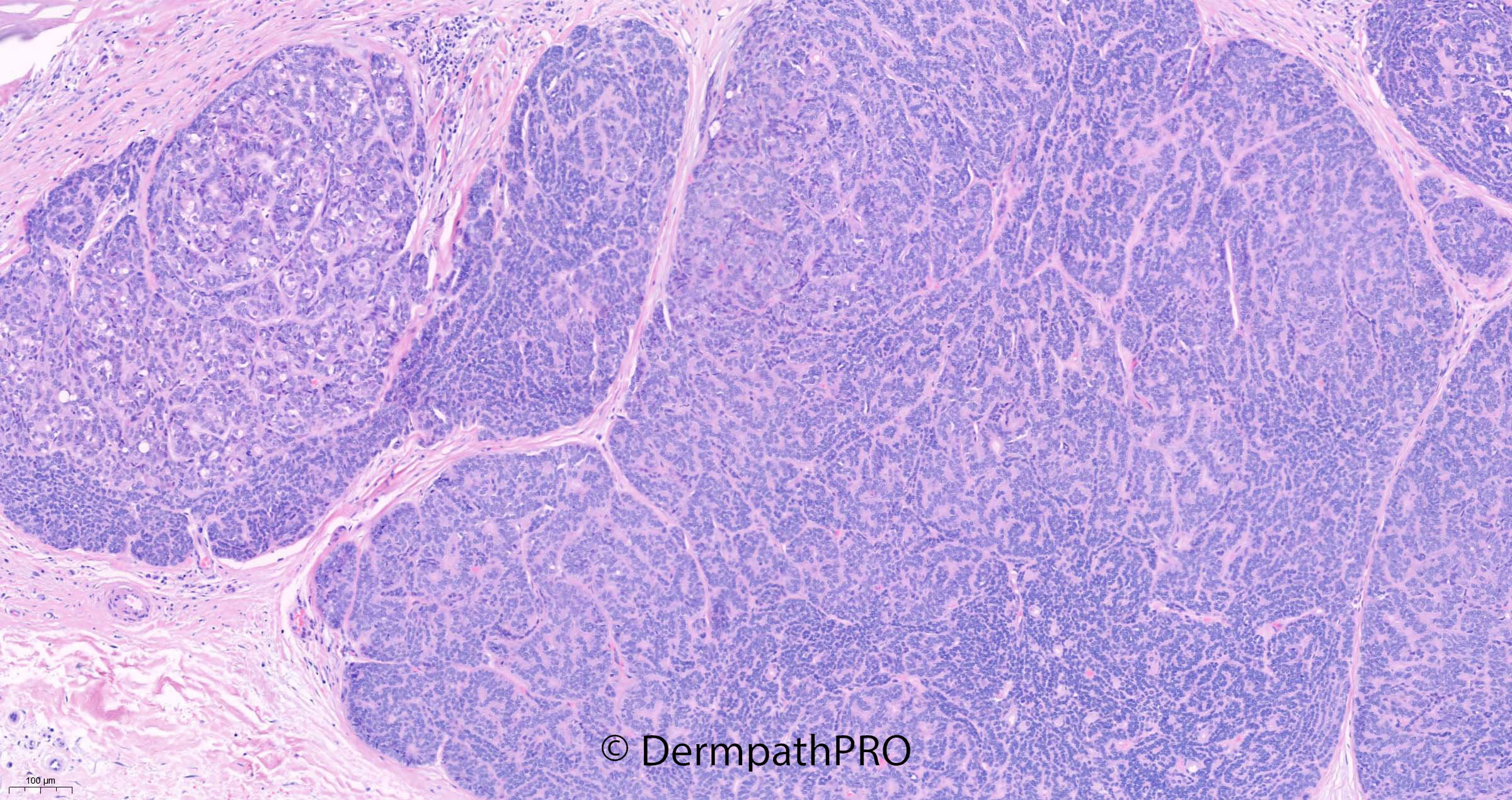
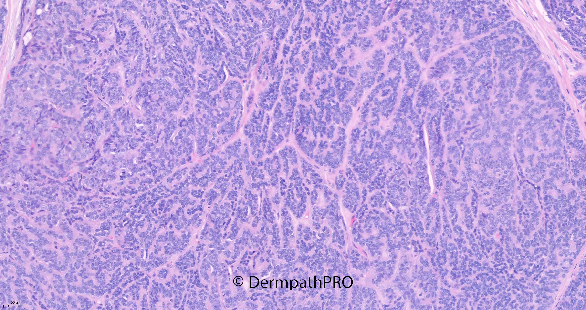
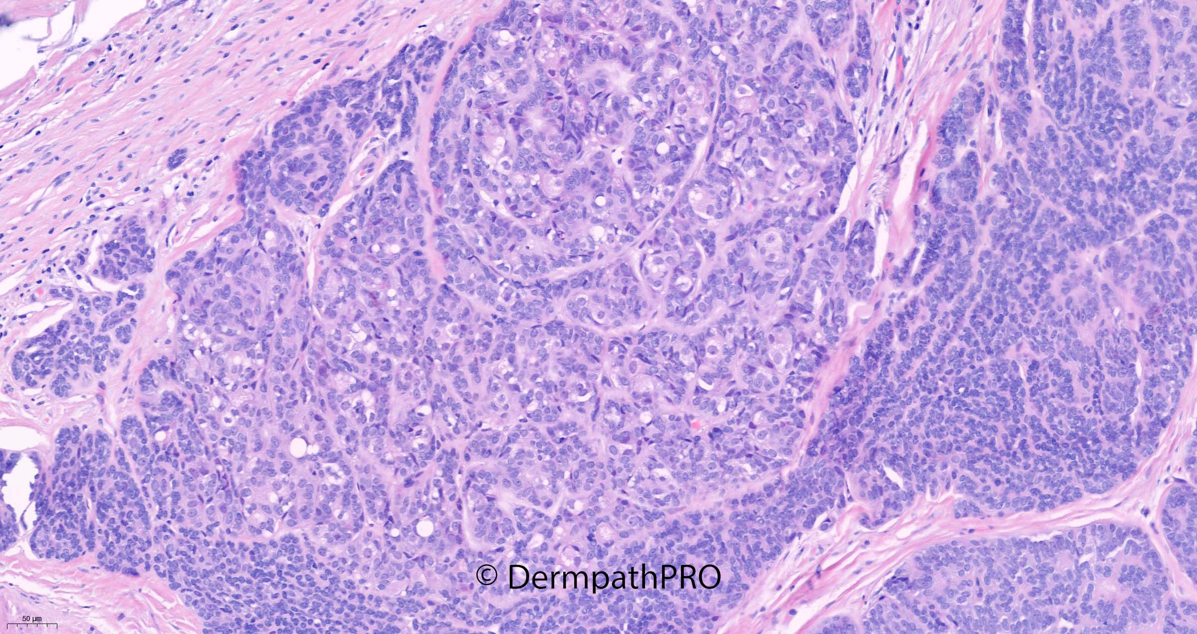
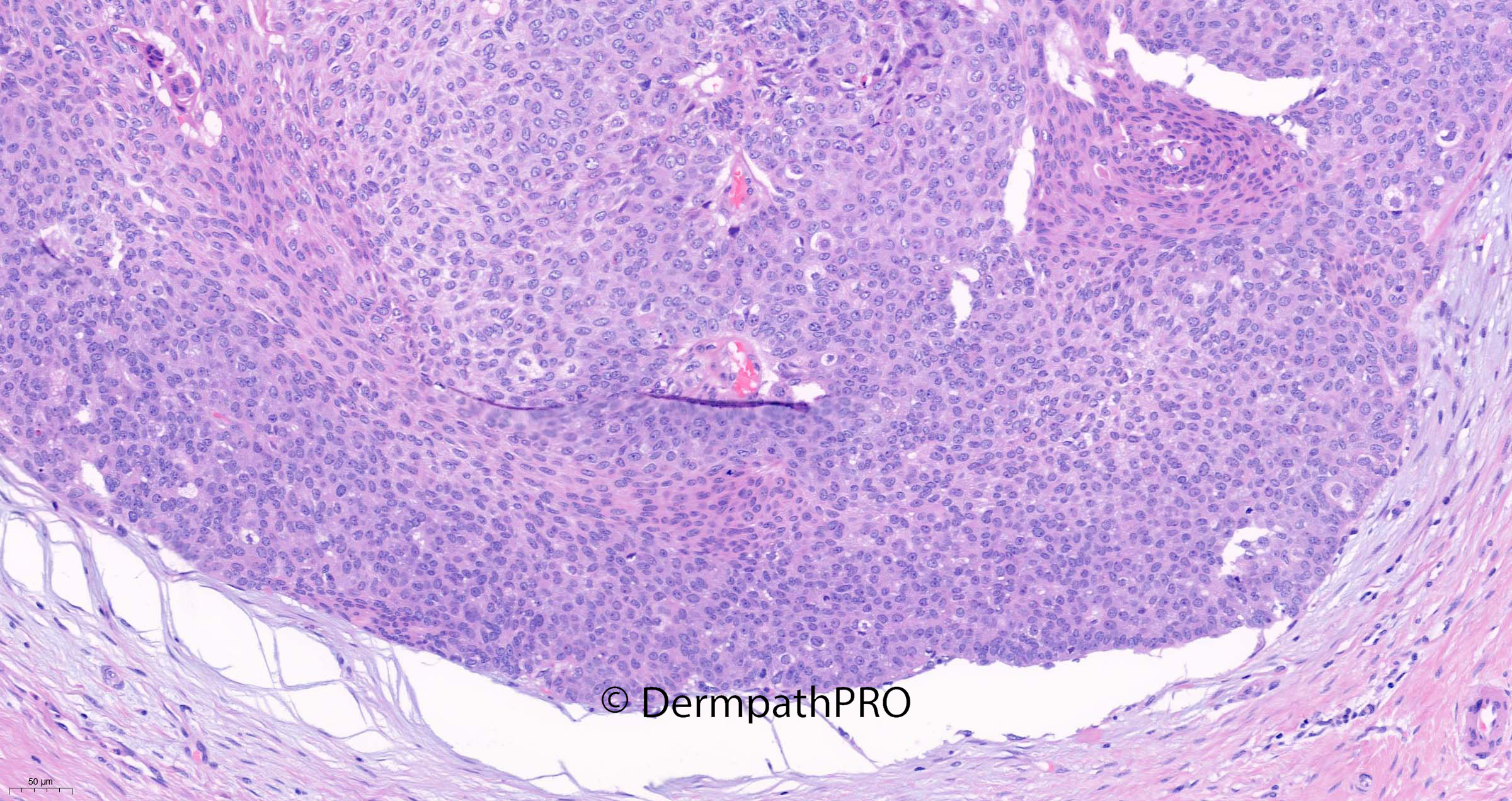
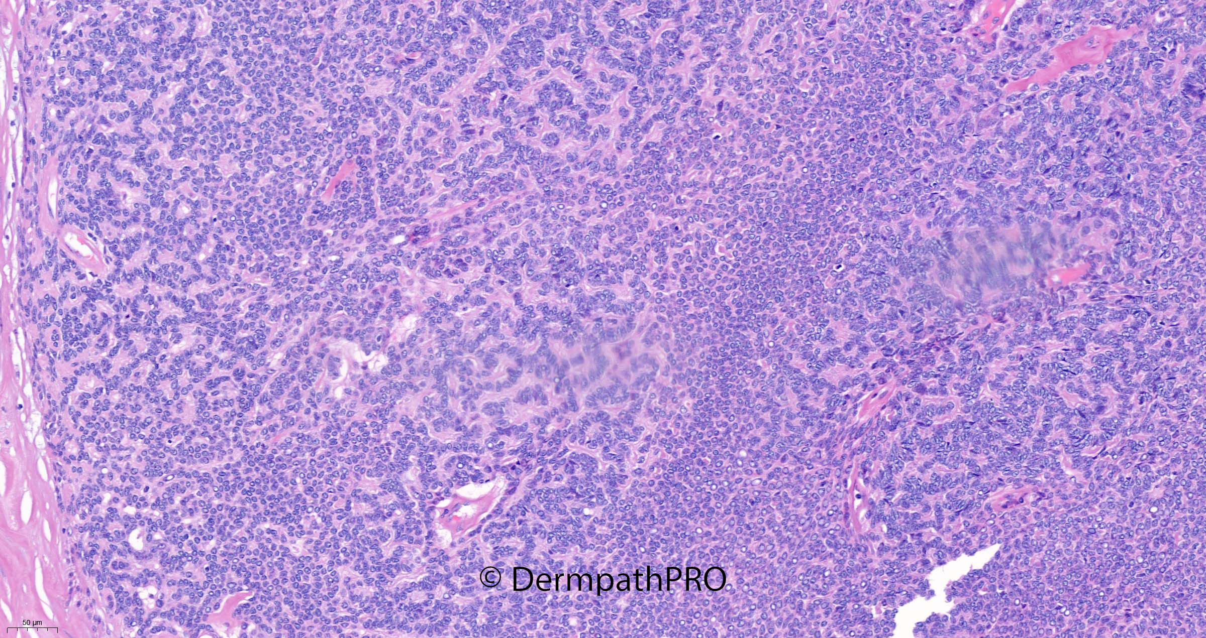
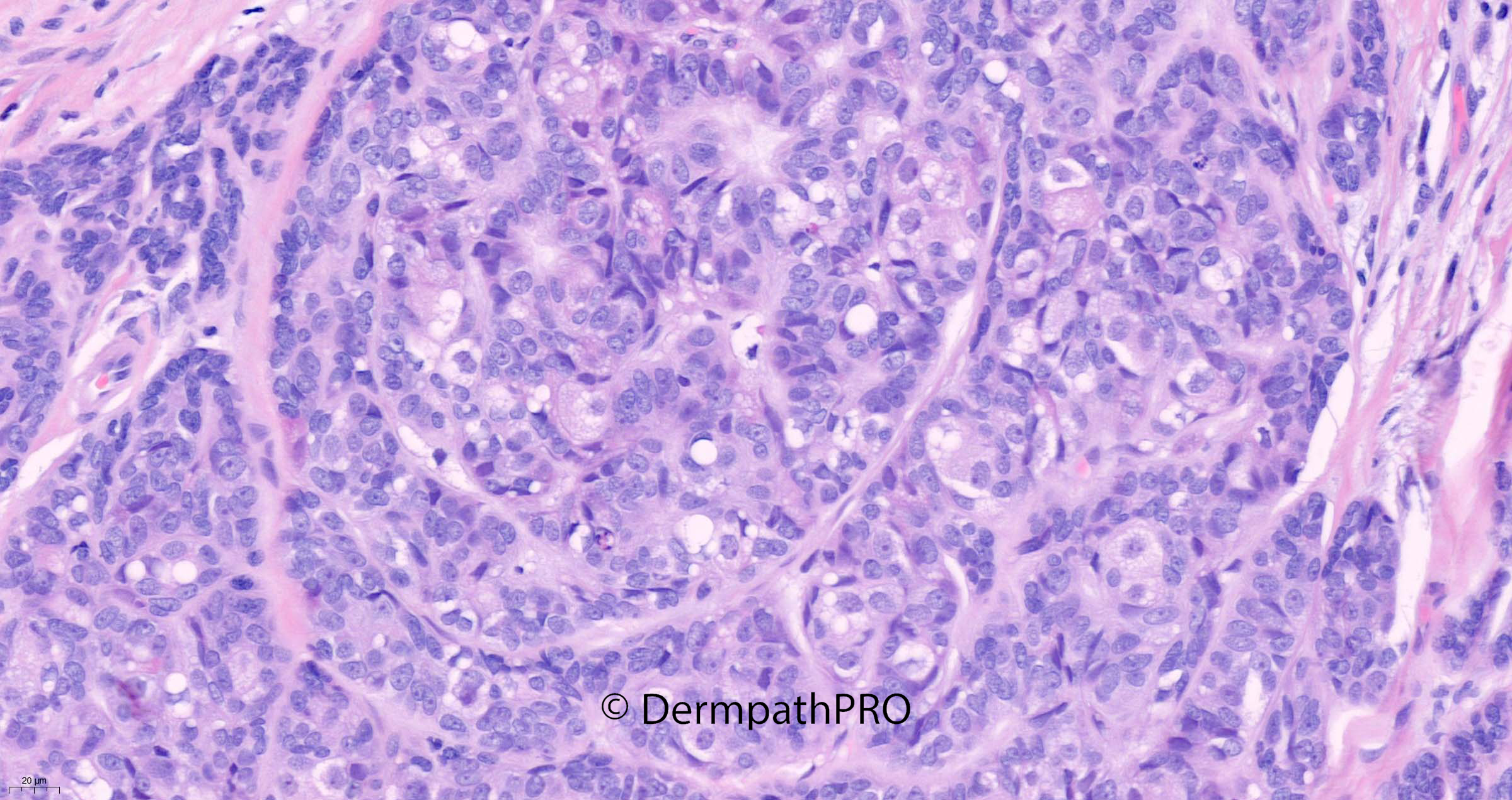
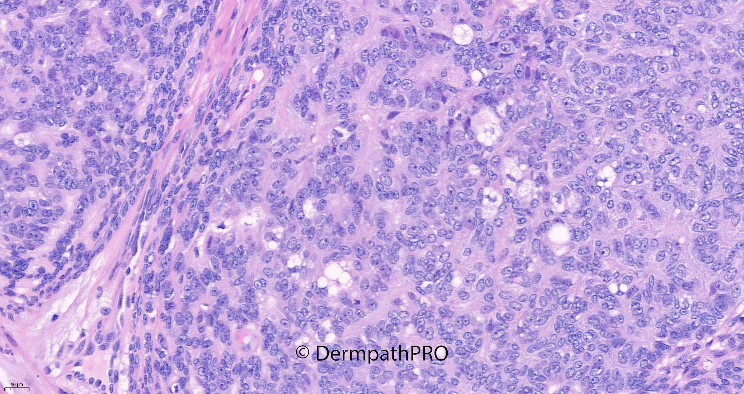
80M Excision scalp
Join the conversation
You can post now and register later. If you have an account, sign in now to post with your account.