Jump to content
In this section we have spot diagnoses posted on a daily basis since June 2010, now over 4000! You can review the archived cases and read the suggested diagnoses by users and the final comment by the contributors.
Case are uploaded each week day by 10 am UK time with the correct diagnosis will generally be posted at 8 pm UK time. Why not view the most recent spot diagnosis and proffer a diagnosis?

Case Number : Case 2988 - 20 December 2021 Posted By: Dr. Mona Abdel-Halim

Please read the clinical history and view the images by clicking on them before you proffer your diagnosis.
Submitted Date :
   (0 reviews)

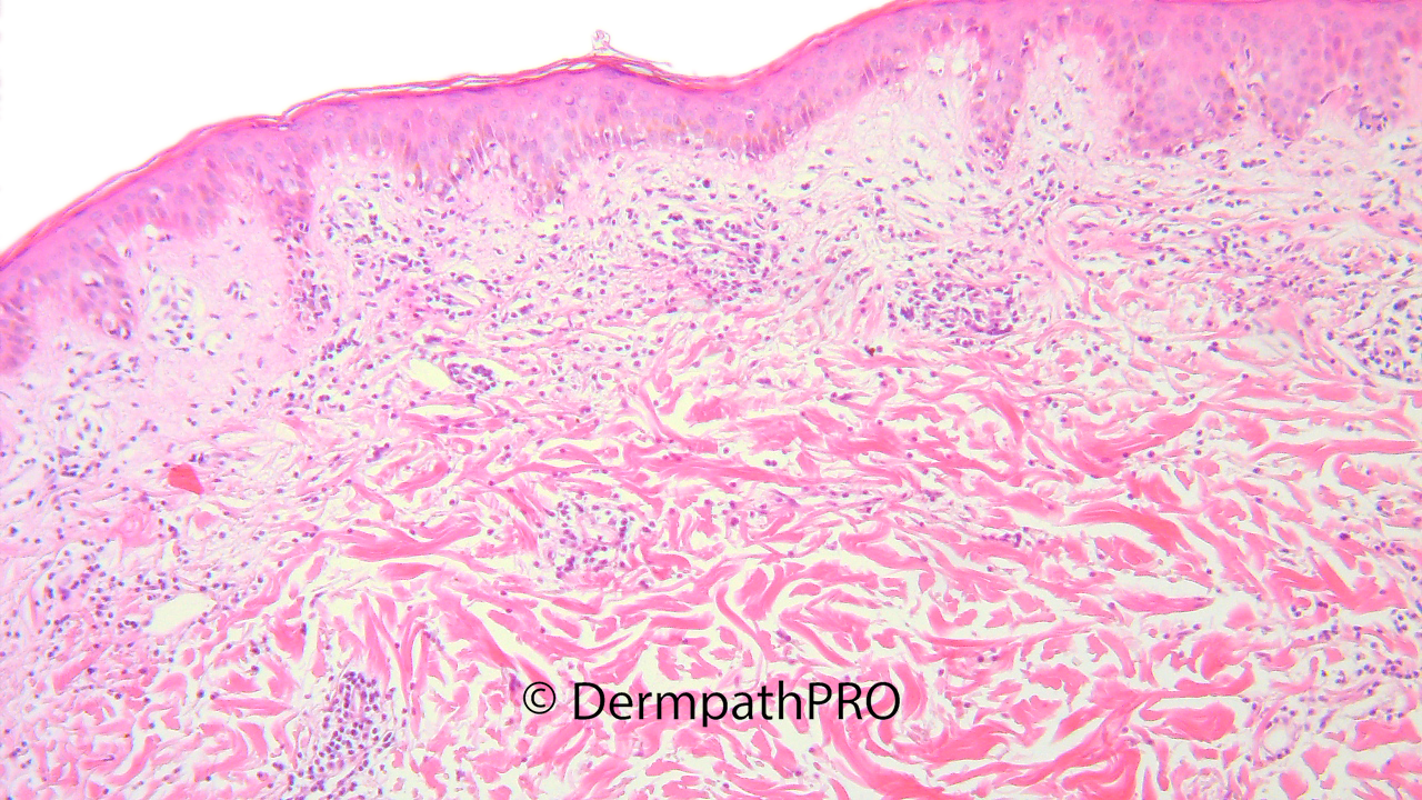
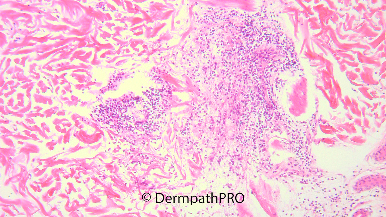
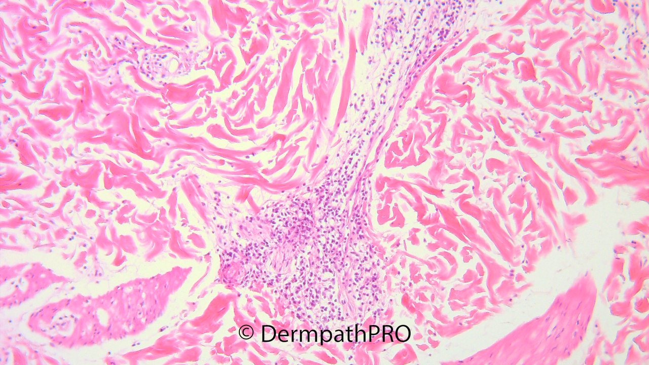
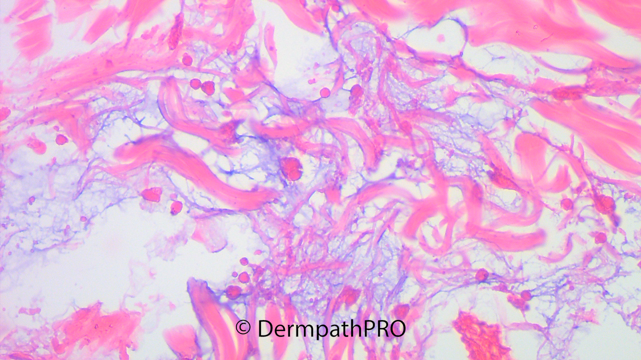
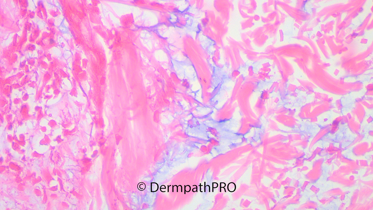
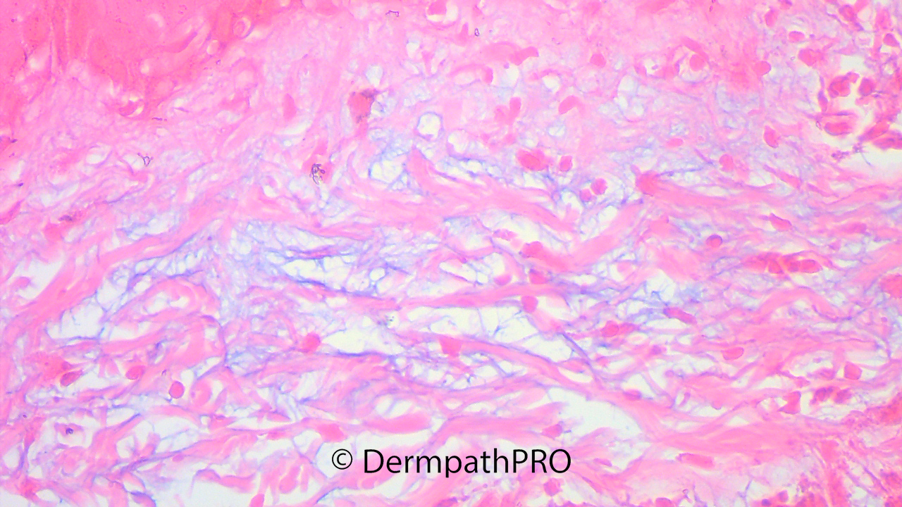
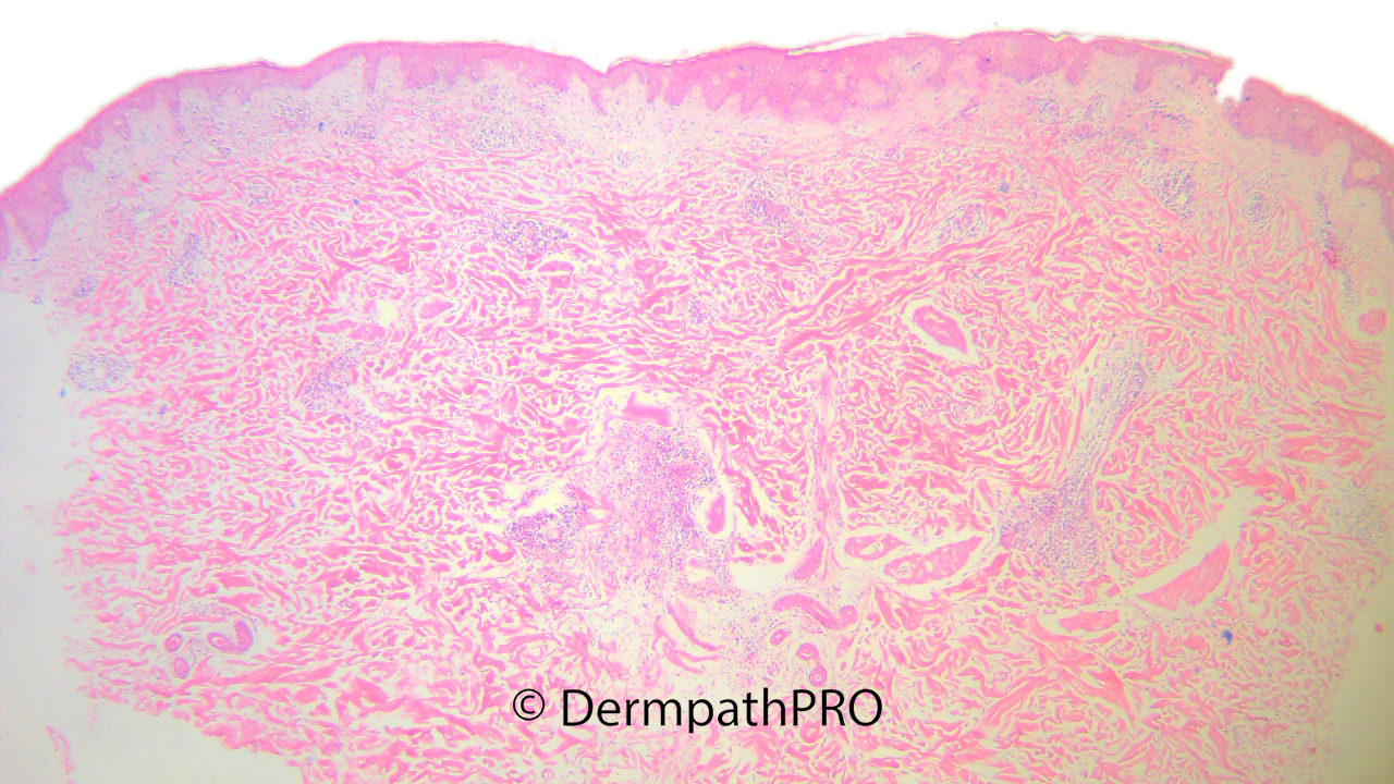
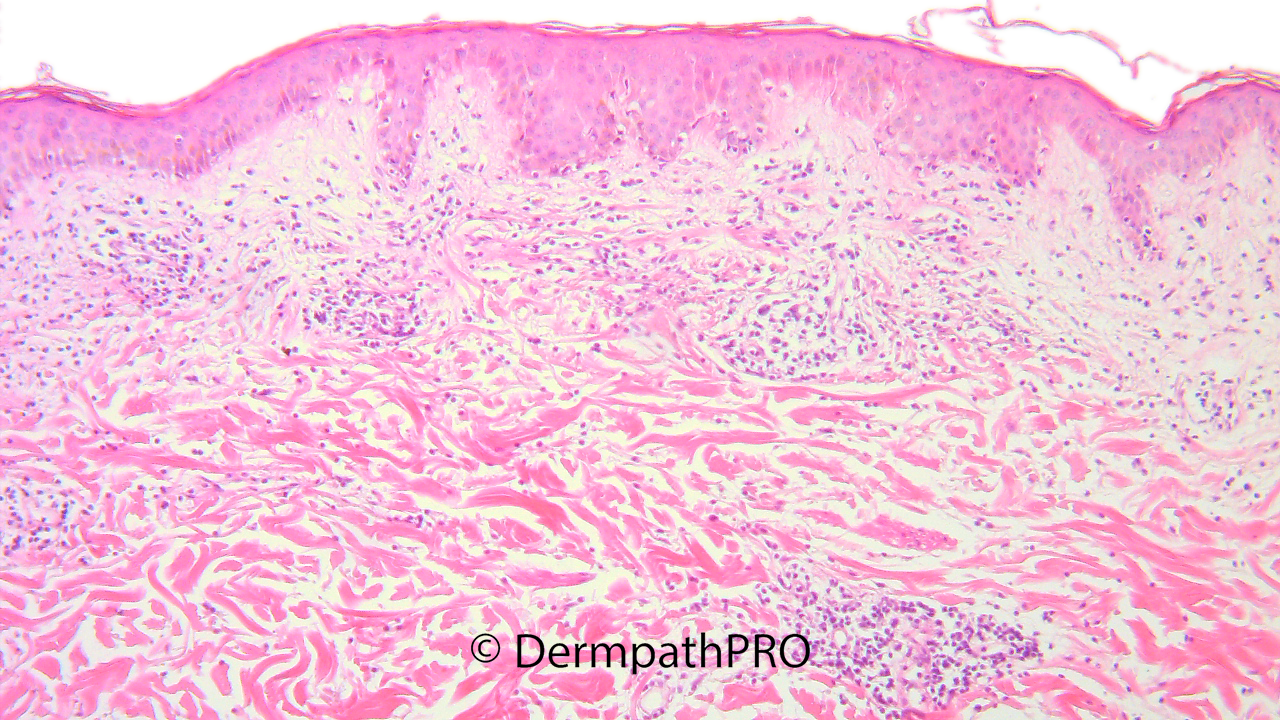
M, 30, Edematous erythematous plaques over the face, chest, upper back and arms. Last three images are Alcian Blue stain.

  • Thanks 1
  Report Record

User Feedback


Thickened, edematous collagen fibres separated by mucin = scleredema.

Maybe post- streptococcal infection or associated with diabetes melitus 

Share this comment


Link to comment
share_externally

Meenakshi Batrani

Posted

Superficial and deep perivascular infiltrate with mucin, there also appears to be subtle interface pathology. Makes me think of tumid LE. 

Share this comment


Link to comment
share_externally

Agree with Meenakshi. Tumid LE is an important differential. Clinical correlation is needed.

Share this comment


Link to comment
share_externally

Krishnakumar subramanian

Posted

usually in tumid lupus there must be no vacuolar interface changes and we see superficial and mid dermal perivascular and peri eccrine lymphocytic infiltrates with a few plasma cells. since some amount of vacuolar interface changes are seen could be SLE.

However i am not against Tumid LE

Share this comment


Link to comment
share_externally

Richard Logan

Posted

I agree with tumid LE. 

In scleredema of Bushcke the skin changes are confluent and uniform rather than forming discrete plaques as in this case.

Share this comment


Link to comment
share_externally

Sorry. I don’t know what this is. Very interesting case  Waiting for the answer. 

Share this comment


Link to comment
share_externally

Eman El-Nabarawy

Posted

What about Reticular erythematous mucinosis (REM), ‘midline mucinosis’ or ‘plaque-like cutaneous mucinosis’?

Share this comment


Link to comment
share_externally

I don’t know the lepromatous follicular mucinosis...but could this be the case? I’m stumbling around in the dark. 

Share this comment


Link to comment
share_externally

Richard Logan

Posted

19 hours ago, vincenzo said:

I don’t know the lepromatous follicular mucinosis...but could this be the case? I’m stumbling around in the dark. 

The pseudo-lepromatous form of follicular mucinosis is nearly always seen in older patients with CTCL-associated follicular mucinosis.  I think the inflammation and distribution of mucin in this case is rather too diffuse to be called follicular mucinosis, which is why I am leaning more towards lupus.

Share this comment


Link to comment
share_externally

Dr. Mona Abdel-Halim

Posted

Thank you all.

Clinicopathological correlation was consistent with tumid LE. 

Share this comment


Link to comment
share_externally


Join the conversation

You can post now and register later. If you have an account, sign in now to post with your account.

Guest
Add a comment...

×   Pasted as rich text.   Paste as plain text instead

  Only 75 emoji are allowed.

×   Your link has been automatically embedded.   Display as a link instead

×   Your previous content has been restored.   Clear editor

×   You cannot paste images directly. Upload or insert images from URL.

Loading...

×
×
  • Create New...