Jump to content
In this section we have spot diagnoses posted on a daily basis since June 2010, now over 4000! You can review the archived cases and read the suggested diagnoses by users and the final comment by the contributors.
Case are uploaded each week day by 10 am UK time with the correct diagnosis will generally be posted at 8 pm UK time. Why not view the most recent spot diagnosis and proffer a diagnosis?

Case Number : Case 2996 - 30 December 2021 Posted By: Saleem Taibjee

Please read the clinical history and view the images by clicking on them before you proffer your diagnosis.
Submitted Date :
   (0 reviews)

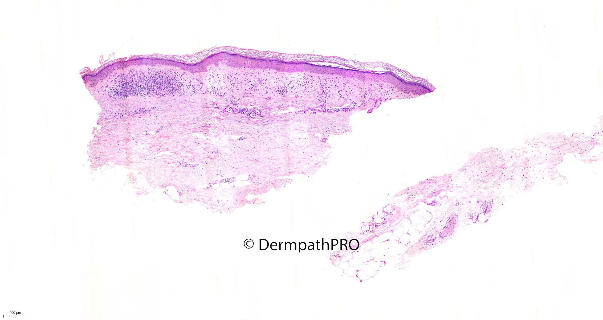
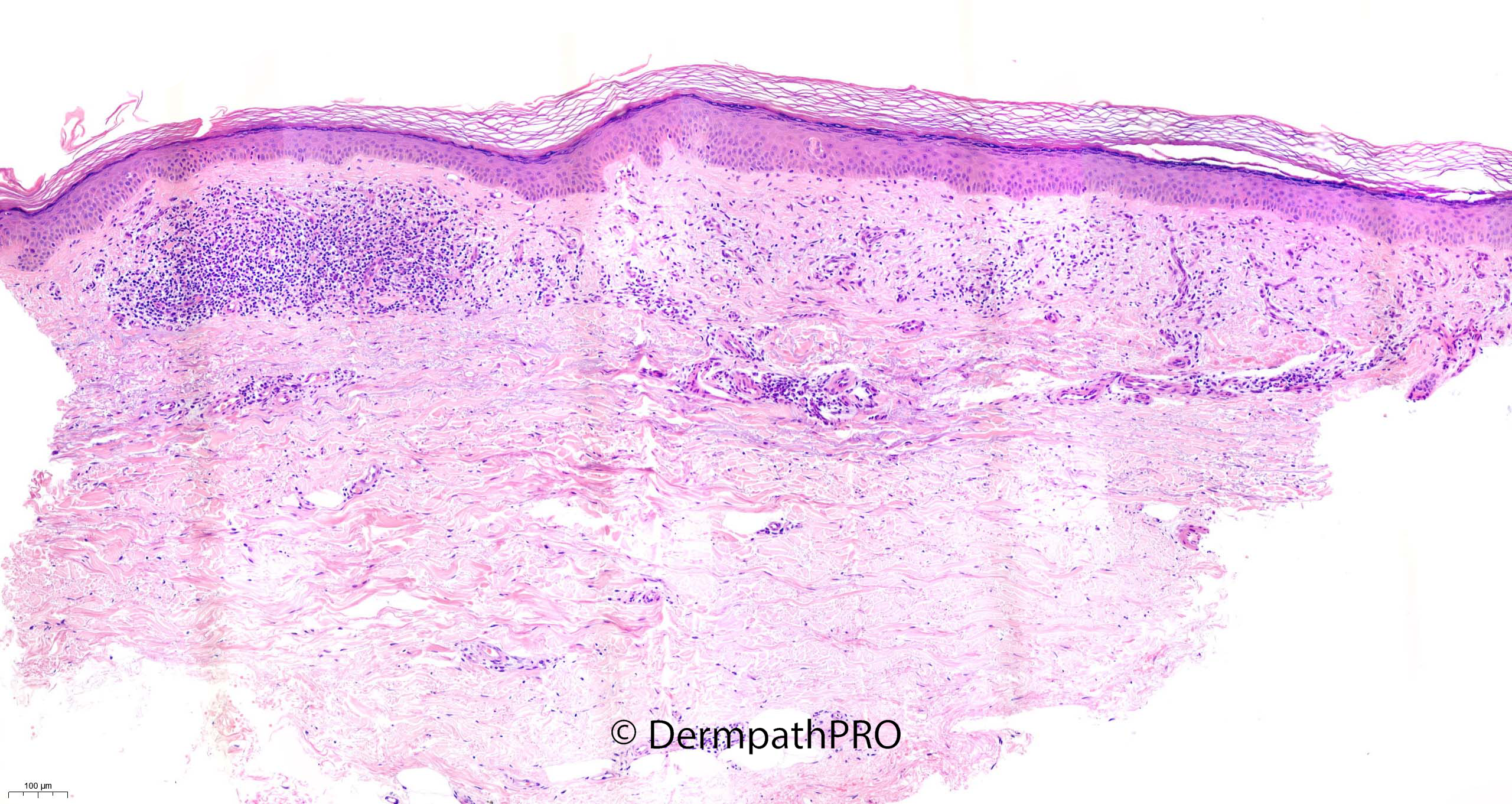
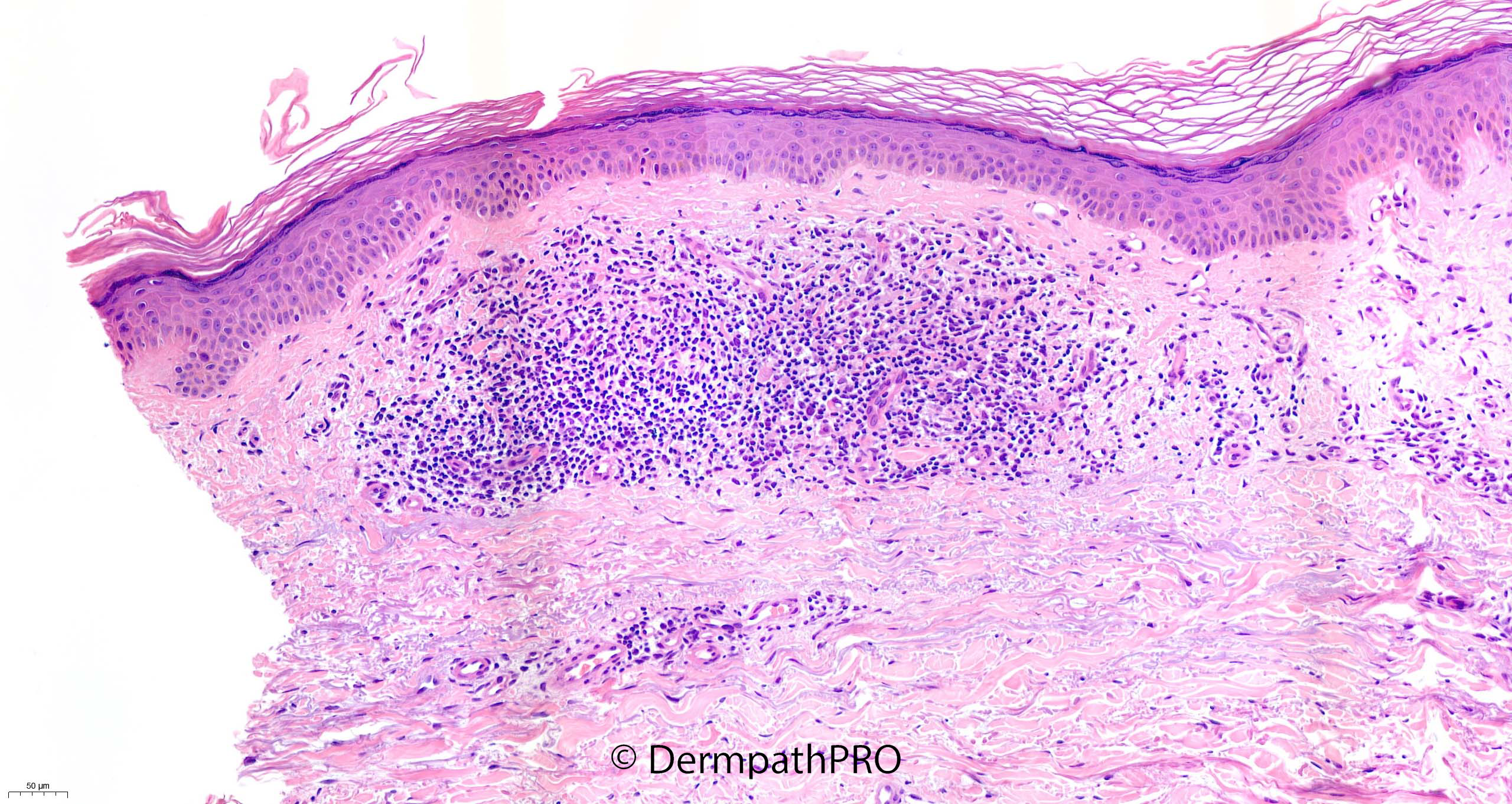
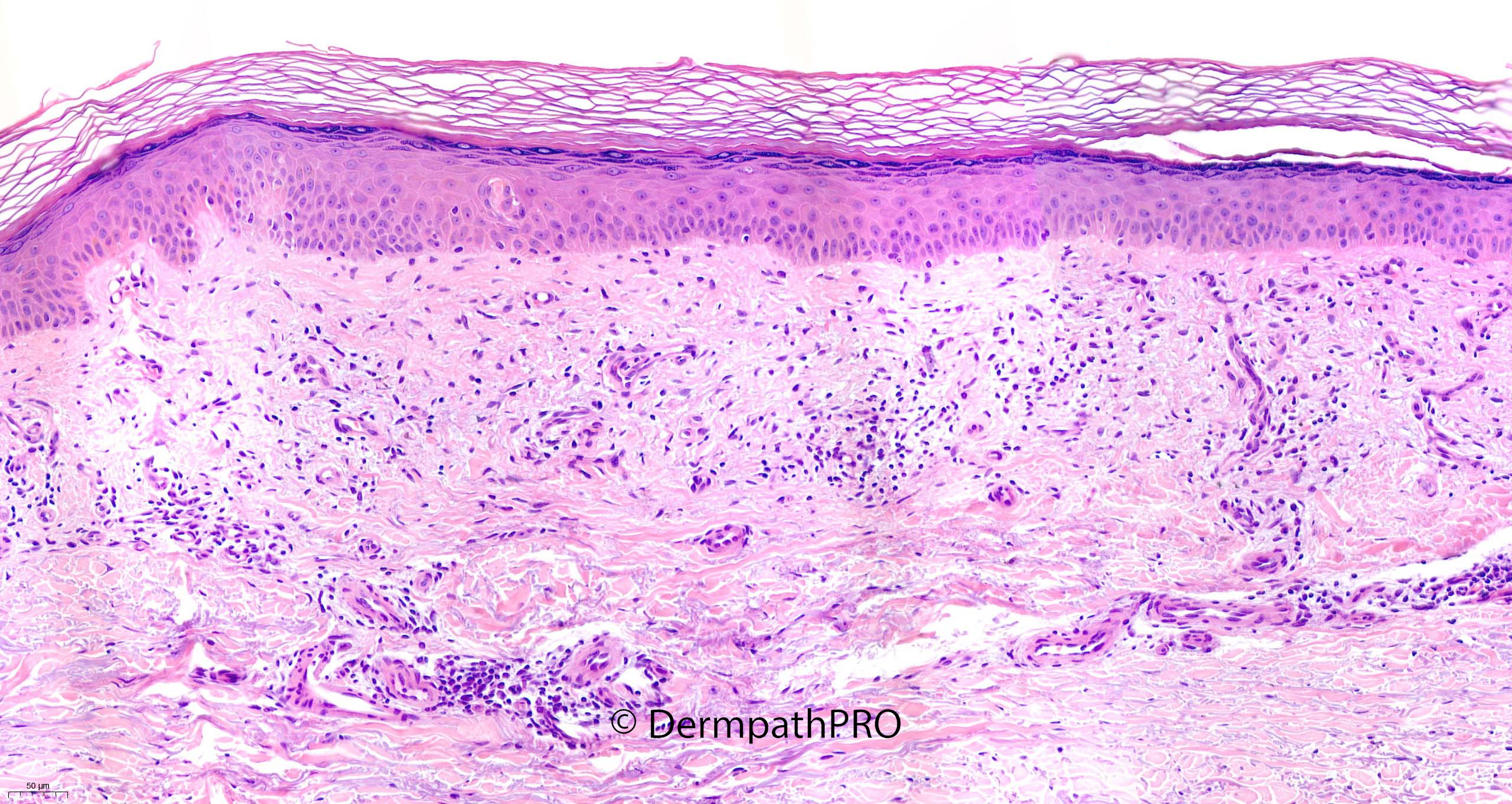
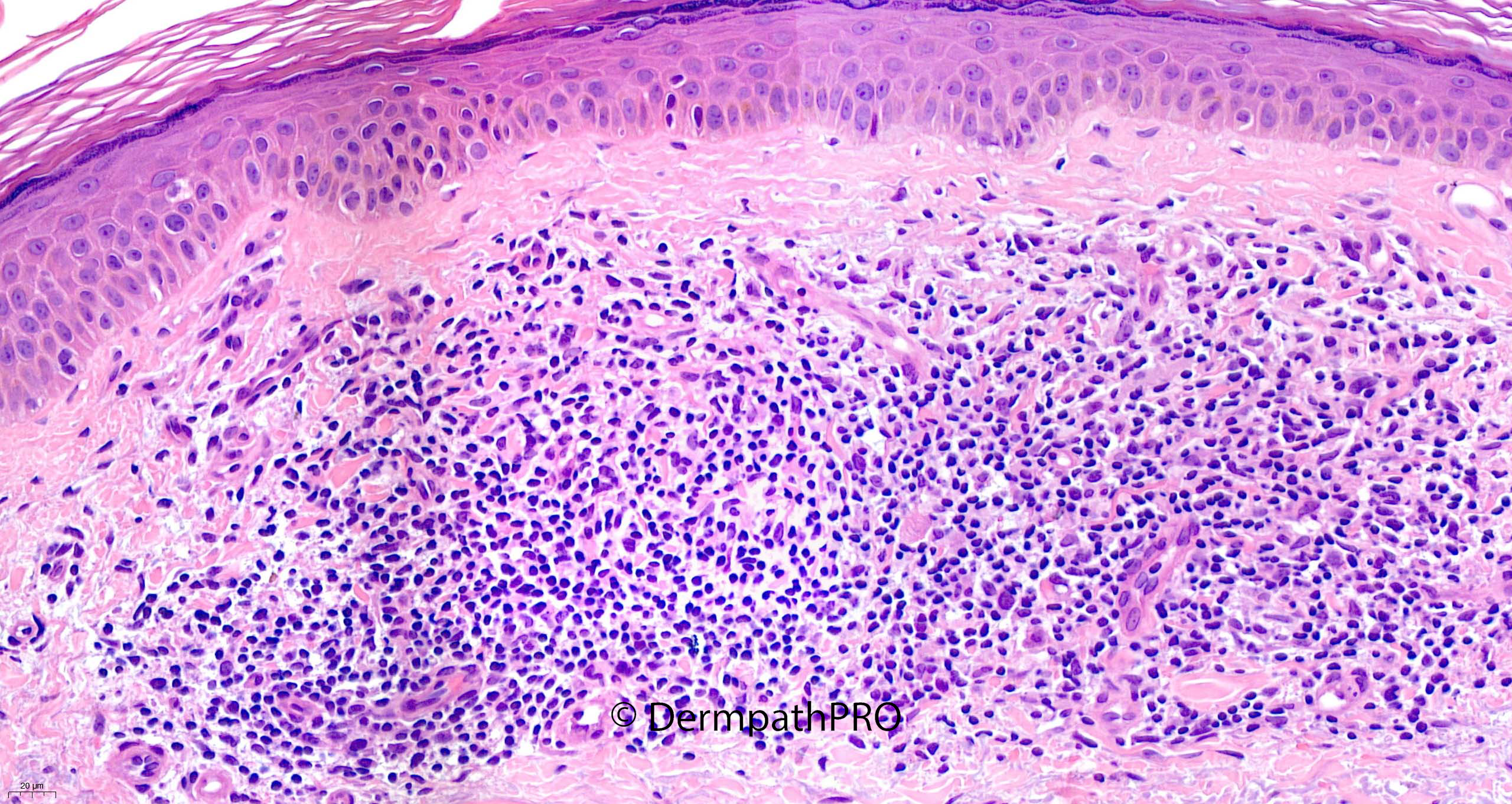
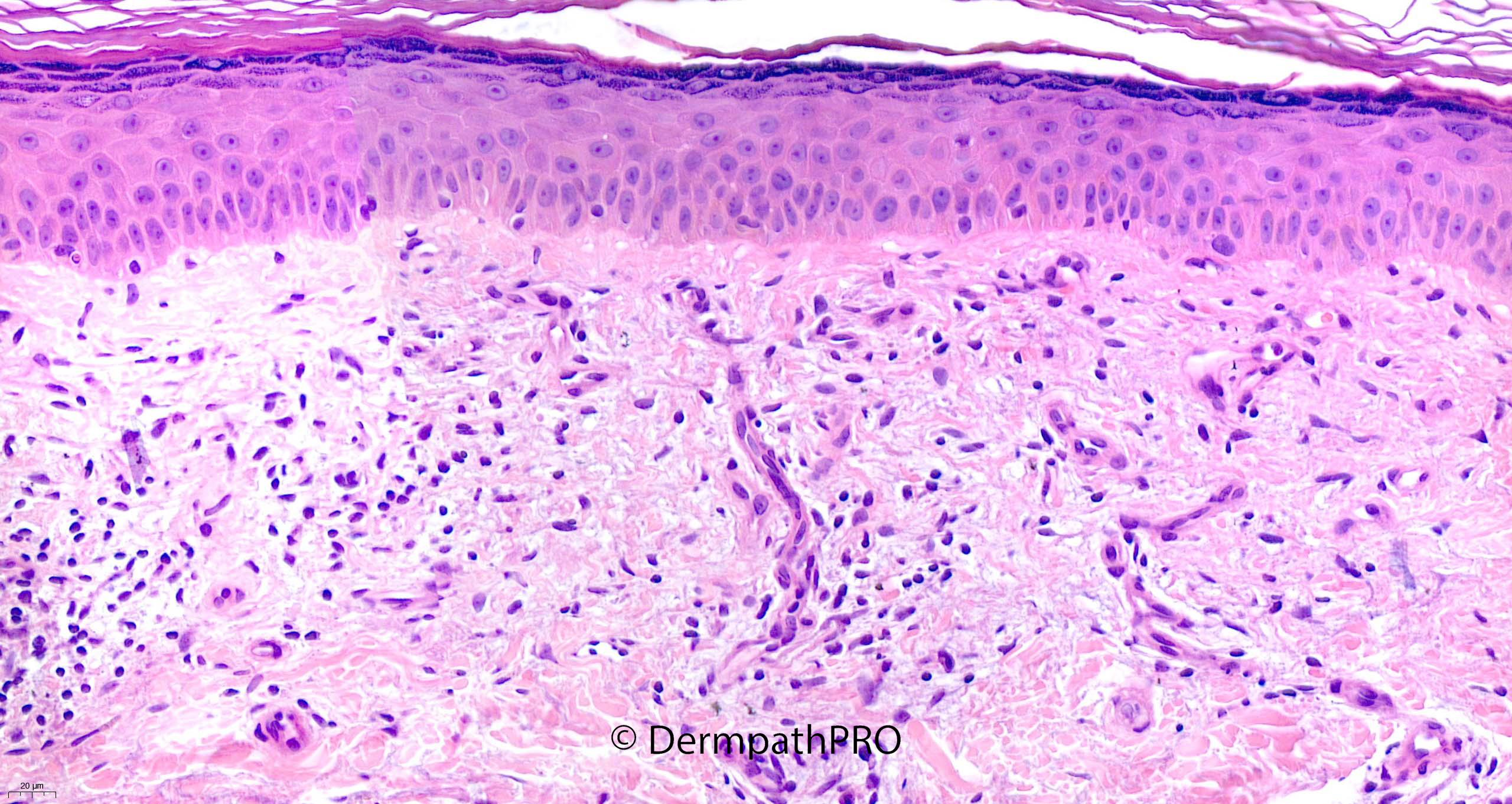
58F Punch biopsy right leg ?seborrhoeic keratosis ?BCC


  Report Record

User Feedback


Krishnakumar subramanian

Posted

lymphocytic vasculitis

some sort of dermatological purpura

Share this comment


Link to comment
share_externally

Meenakshi Batrani

Posted (edited)

To me there appears to be some vascular proliferation with plump endothelial cells and lymphoid infiltrate. Makes me think of Acral pseudolymphomatous angiokeratoma, which was originally described in children (APACHE) but there have been reports in adults. 

Edited by Meenakshi Batrani

Share this comment


Link to comment
share_externally

A regressive lesion. But canestrated ortokeratosis should be in keeping with some benign or recent lesion. No parakeratosis typical of a chronic process. 

Share this comment


Link to comment
share_externally

Meenakshi Batrani

Posted

12 hours ago, vincenzo said:

A regressive lesion. But canestrated ortokeratosis should be in keeping with some benign or recent lesion. No parakeratosis typical of a chronic process. 

Yes. May be some regressed lesion

Share this comment


Link to comment
share_externally

Anil Patki

Posted

Meenakshi's suggestion is plausible and will need some IHC for confirmation. 

Another possibility is a regressing lesion of lymphomatoid papulosis type E which is usually paucilesional.

Will be interesting to see the findings of IHC.

Share this comment


Link to comment
share_externally

Saleem Taibjee

Posted

Happy New Year!

This is a common scenario. Yes, the pattern of superficial dermal fibrosis, vascular proliferation and chronic inflammation is very suggestive of a regressed / previously treated lesion, and this possibility should be raised in the report. In such cases it can be important to remind the clinician that the biopsy may not be fully representative i.e. that if the biopsy has been taken from a much larger lesion, to consider further biopsy.

In the absence of significant alteration of the keratin layer or frank lichenoid damage, the most common scenario seems to be a regressed superficial BCC.

In this instance the punch biopsy was processed on end with 3 initial levels by the laboratory. In this situation I have a very low threshold for requesting additional levels because I see many examples where a clearer diagnosis manifests with deeper levels and presumably the biopsy was not adequately 'cut-in' on the initial sections. Lo-and-behold, the deeper levels did reveal superficial BCC in this case (see images below). When the biopsy is bisected from the outset, the initial sections tend to be more reliably fully representative. Even in that situation I might still consider requesting additional levels. But I would certainly encourage bisecting of punch biopsy specimens from the outset whenever possible.

BW
Saleem

64820_10.0x.jpg

64820_20.0x.jpg

Share this comment


Link to comment
share_externally


Join the conversation

You can post now and register later. If you have an account, sign in now to post with your account.

Guest
Add a comment...

×   Pasted as rich text.   Paste as plain text instead

  Only 75 emoji are allowed.

×   Your link has been automatically embedded.   Display as a link instead

×   Your previous content has been restored.   Clear editor

×   You cannot paste images directly. Upload or insert images from URL.

Loading...

×
×
  • Create New...