Jump to content
In this section we have spot diagnoses posted on a daily basis since June 2010, now over 4000! You can review the archived cases and read the suggested diagnoses by users and the final comment by the contributors.
Case are uploaded each week day by 10 am UK time with the correct diagnosis will generally be posted at 8 pm UK time. Why not view the most recent spot diagnosis and proffer a diagnosis?

Case Number : Case 2997 - 31 December 2021 Posted By: Dr. Richard Carr

Please read the clinical history and view the images by clicking on them before you proffer your diagnosis.
Submitted Date :
   (0 reviews)

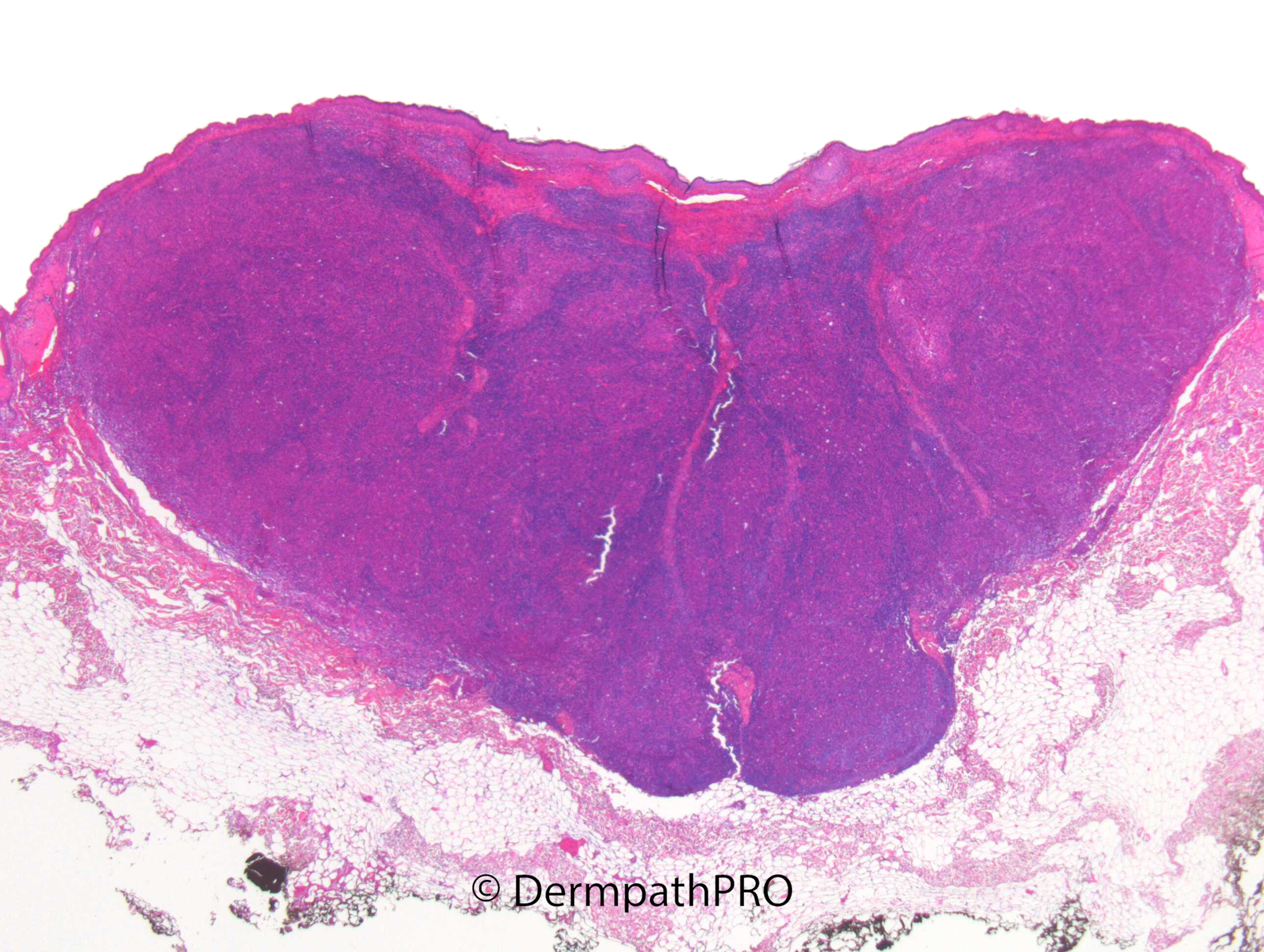
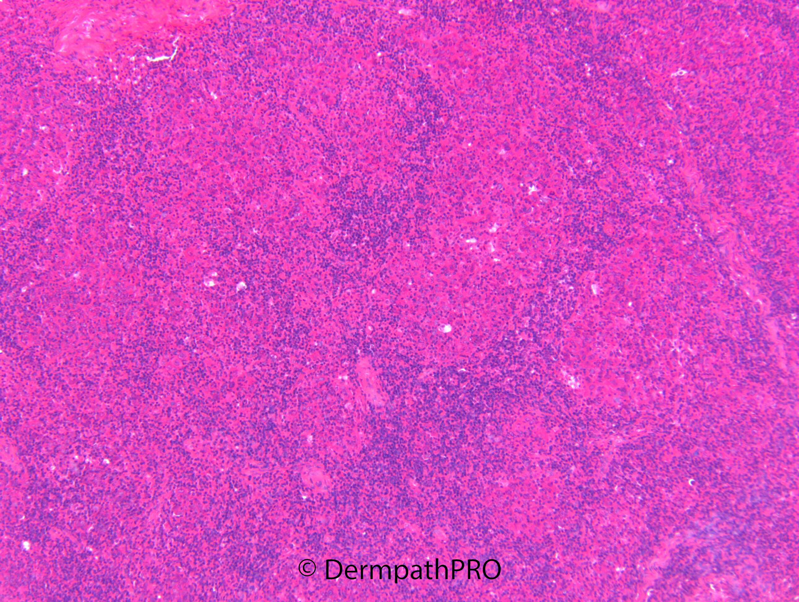
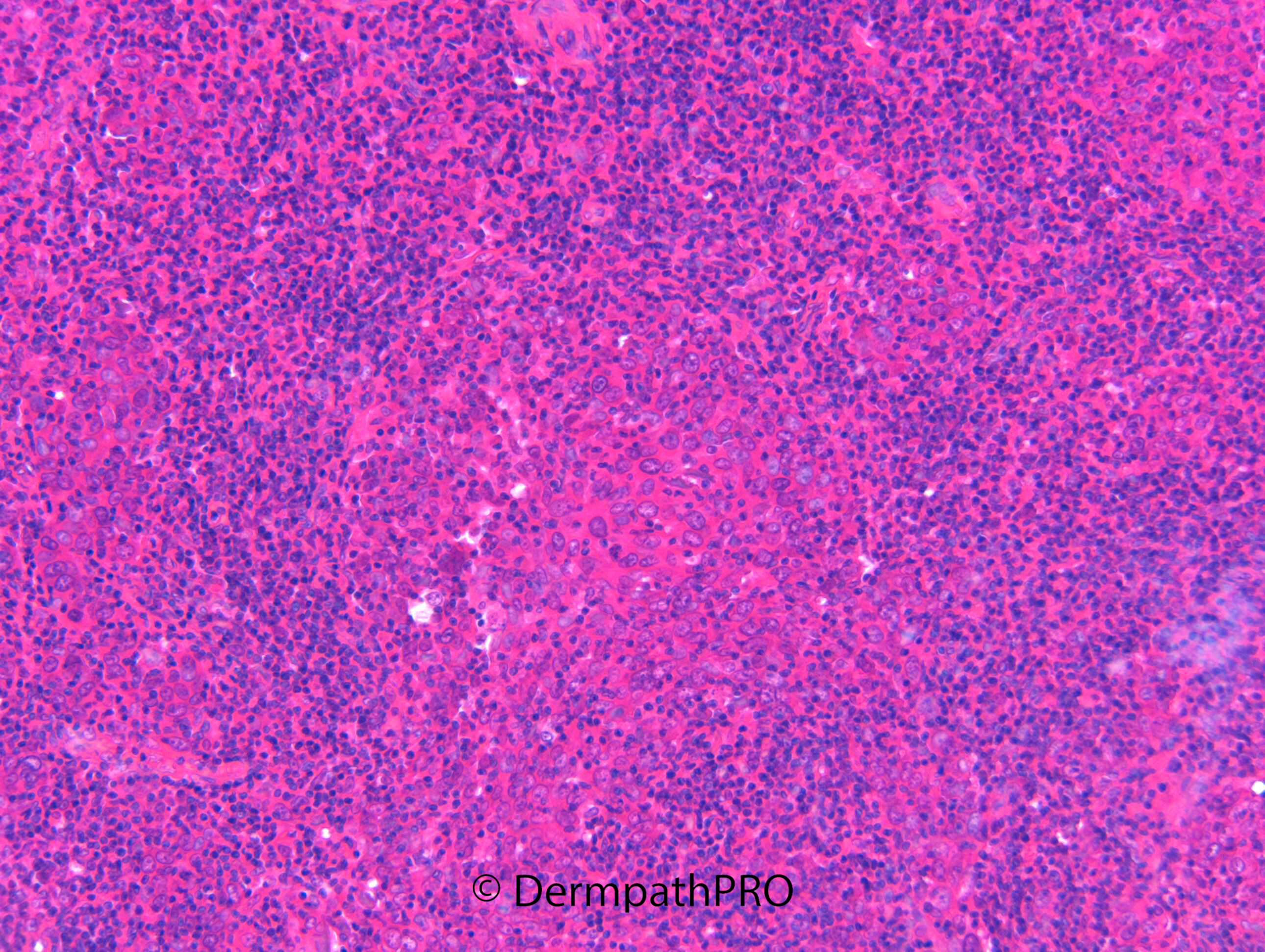
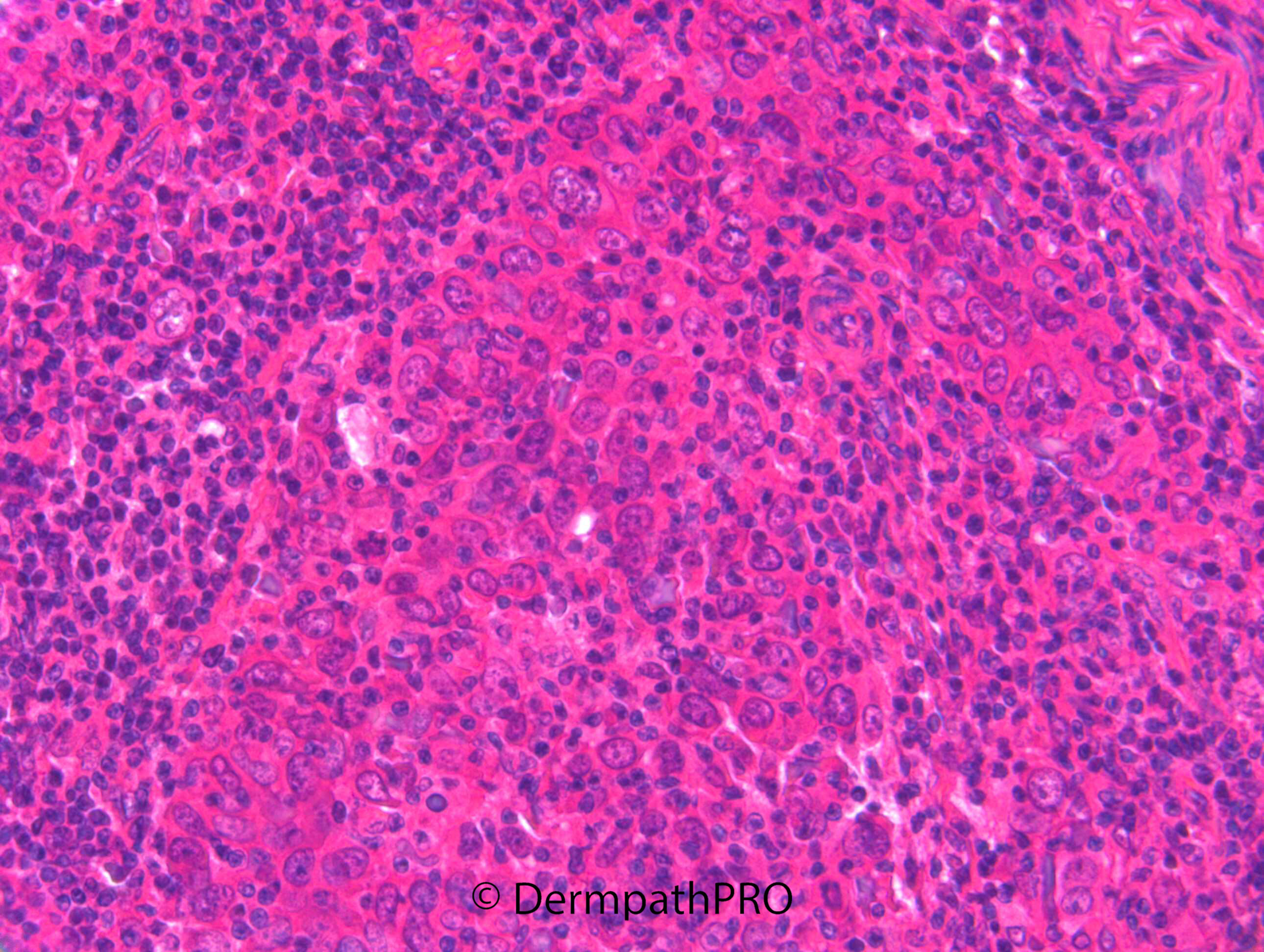
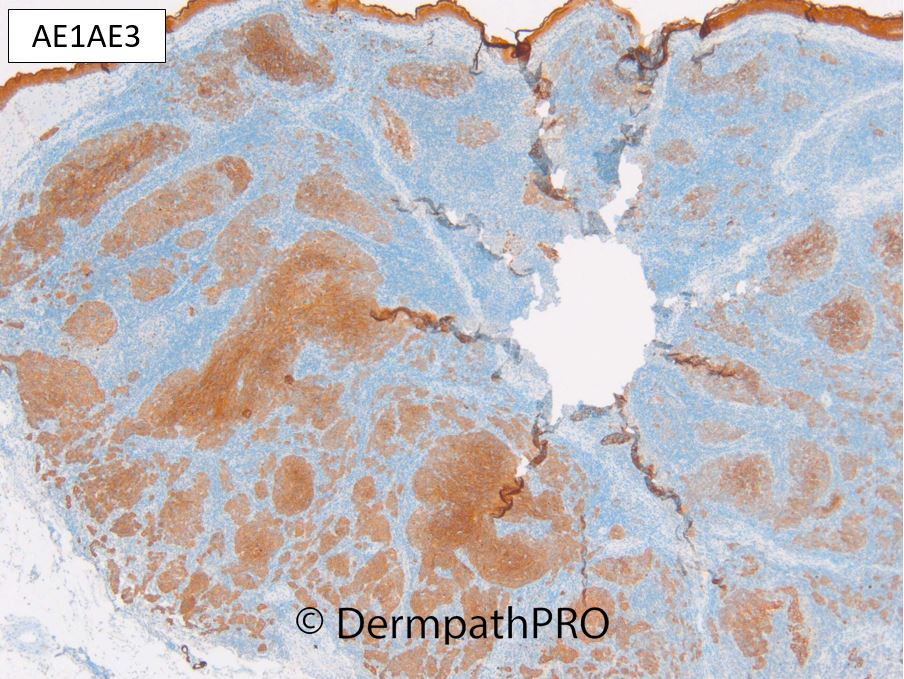
F80. Cheek. BCC 10 x 8mm


  Report Record

User Feedback


Krishnakumar subramanian

Posted

lobules of atypical squamoid cells surrounded by dense lymphocytic cells or vague granuloma with surrounding lymphocytes

Not clear on morphology

may be lymphoepithelial tumor

Share this comment


Link to comment
share_externally

I was thinking of a lymphoepithelial carcinoma, too, ?primary.

Share this comment


Link to comment
share_externally

Perhaps lymphoepithelial-like carcinoma would be a more appropriate definition. 

Share this comment


Link to comment
share_externally

msofopoulos

Posted

Happy new year everybody!

Looks like lymphoepithelioid Ca. I would like to see an EBER stain

Share this comment


Link to comment
share_externally

Krishnakumar subramanian

Posted

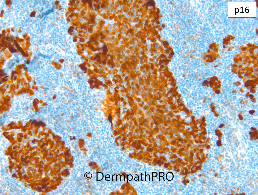
p16 immunohistochemistry is usually negative in lymphoepithelial carcinoma since P 16 is positive should we do testing for HPV where 16 is positive

Share this comment


Link to comment
share_externally

Dr. Richard Carr

Posted

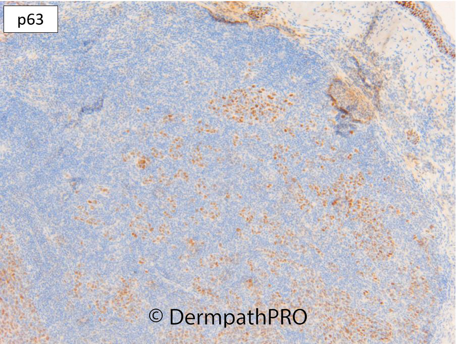
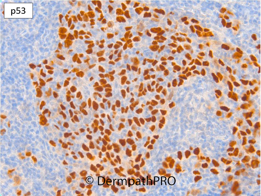
Yes this is a lymphoepithelioma-like carcinoma. I suspect many are from the adnexa (probably follicular stem cells mainly) because, as is typical,  there no surface tumour. Do you have a reference regarding p16 and LELC as I'm not familiar with any studies off the top my head although I didn't google it specifically. I do know that p16 diffuse positive >90% of the cells is quite uncommon generally but often seen in Bowenoid epidermal dysplasia (whether HPV or non-HPV associated) and very uncommon in follicular SCC and non-bowens associated SCC. My understanding is that high p16 in the setting of high risk HPV is accumulation of wild type protein. We allow staining up to 90% that we suspect is wild type and reactive in KA but nearly always retains more variation in intensity mild/moderate/strong mosaic which might even be the case in this tumour. p16 can also appear high when there is a clear cut highly aberrant (null or diffuse moderate/strong) p53 as in this case. So my semi-educated guess of the molecular in a case like this is a mutation of a p53 that has abnormally sequestered due to longer half-like and a preserved but up-regulated p16 as a secondary response to the loss of p53 function. We don't know if the presumed mutations in p53 are bi-allelic, theoretically a normal allele could compensate but I'd guess it is likely, judging by the p16 over-expression p53 function has been lost i.e. the 2nd allele lost, mutated or methylated, or the 1st allele with a dominant mutation that stops the normal p53 function from the other allele. Sadly IHC and genetics are rather complex to correlate without very detailed studies. However like most carcinomas on the severely sun-damaged skin we do have highly aberrant patterns for one if not both IHC markers in this case. 

Share this comment


Link to comment
share_externally


Join the conversation

You can post now and register later. If you have an account, sign in now to post with your account.

Guest
Add a comment...

×   Pasted as rich text.   Paste as plain text instead

  Only 75 emoji are allowed.

×   Your link has been automatically embedded.   Display as a link instead

×   Your previous content has been restored.   Clear editor

×   You cannot paste images directly. Upload or insert images from URL.

Loading...

×
×
  • Create New...